49 Circulatory Systems Chapter 49 Key Concepts 49


- Slides: 160
49 Circulatory Systems
Chapter 49 Key Concepts 49. 1 Circulatory Systems Serve Many Functions 49. 2 Vertebrate Circulatory Systems Evolved from Single to Double Circuits 49. 3 Heart Function Depends on Properties of Cardiac Muscle
Chapter 49 Key Concepts 49. 4 Circulatory System Functions Depend on Blood and Blood Vessels 49. 5 The Circulation Is Controlled by Hormonal and Neural Signals
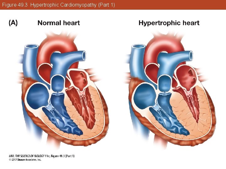
Investigating Life: Athletes with Big Hearts Many young, fit athletes have died from cardiac arrest during physical exertion, caused, in most cases, by hypertrophic cardiomyopathy (HCM). Gene mutations affect the contractile proteins of the heart, the heart has to work harder, and it enlarges.
Investigating Life: Athletes with Big Hearts In athletes, the heart walls get so thick that they impede blood flow from the heart and interfere with the signals that coordinate heart contractions. The result is cardiac arrest. Are there potential therapies that could treat HCM?
Key Concept 49. 1 Focus Your Learning • A circulatory system functions to transport materials around the body. • Small or thin aquatic organisms may not have circulatory systems, and the needs of their cells are served by direct exchange with the environment.
Key Concept 49. 1 Focus Your Learning • Circulatory systems use a muscular heart to distribute extracellular fluid throughout the body to transport nutrients, respiratory gases, wastes, and other materials. • Closed circulatory systems allow more rapid flow and differential control of blood flow to different tissues.
49. 1 Circulatory Systems Serve Many Functions A circulatory system consists of: • Muscular pump—the heart • Fluid—blood • Series of conduits—blood vessels Also known as the cardiovascular system.
49. 1 Circulatory Systems Serve Many Functions Some animals do not need circulatory systems. • Single-celled organisms exchange materials directly with the environment. • Some aquatic multicellular organisms are small or thin, so that all cells are close to the environment to allow exchange.
49. 1 Circulatory Systems Serve Many Functions • Other aquatic organisms have branched gastrovascular systems that bring the external environment inside the animal (e. g. , sponges). • Large animals without a circulatory system tend to be inactive or sessile.
49. 1 Circulatory Systems Serve Many Functions Larger animals must use circulatory systems to deliver nutrients to all cells, and remove wastes. Cells exchange nutrients and wastes with the extracellular fluid. A circulatory system has a muscular pump to move the extracellular fluid around the body.
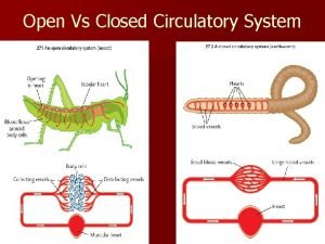
49. 1 Circulatory Systems Serve Many Functions Open circulatory system: Fluid leaves the circulatory system and moves between the cells. Closed systems: Fluid is contained in a continuous system of vessels.
49. 1 Circulatory Systems Serve Many Functions In a closed system, extracellular fluid refers to the fluid in the circulatory system (blood plasma) plus the fluid around cells (interstitial fluid).
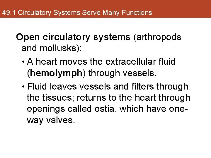
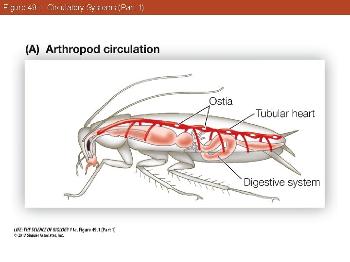
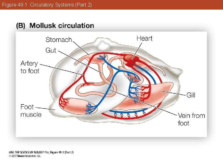
49. 1 Circulatory Systems Serve Many Functions Open circulatory systems (arthropods and mollusks): • A heart moves the extracellular fluid (hemolymph) through vessels. • Fluid leaves vessels and filters through the tissues; returns to the heart through openings called ostia, which have oneway valves.
Figure 49. 1 Circulatory Systems (Part 1)
Figure 49. 1 Circulatory Systems (Part 2)
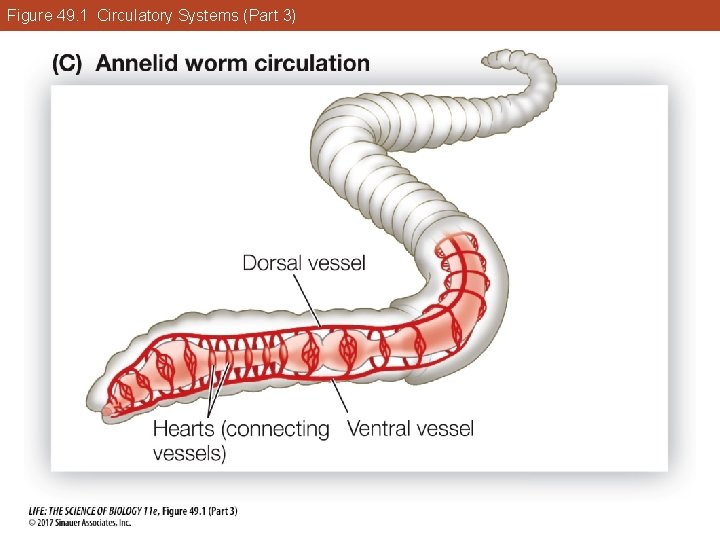
49. 1 Circulatory Systems Serve Many Functions Closed circulatory systems (vertebrates, annelids): • Blood is kept separate from the interstitial fluid; pumped through the vascular system by one or more hearts. • Cellular and macromolecular components of the blood never leave the vessels.
Figure 49. 1 Circulatory Systems (Part 3)
49. 1 Circulatory Systems Serve Many Functions Advantages of closed systems: • Faster transport of materials through vessels. • Blood can be directed to specific tissues by varying resistance (diameter of blood vessel). • Specialized carriers can travel in the vessels and transport hormones or nutrients to specific sites.
Key Concept 49. 1 Learning Outcomes • Describe the diverse functions of circulatory systems. • Explain why some animals can function without a circulatory system. • Describe the differences among hemolymph, blood plasma, and interstitial fluid.
Key Concept 49. 1 Learning Outcomes • Identify features that make a closed circulatory system more efficient than an open circulatory system.
Key Concept 49. 2 Focus Your Learning • The circulatory system in fishes is organized as a single circuit in which blood is pumped by a four-chambered heart through the gills and then to the other organs of the body. • The lungfish is a transitional form in the evolution of a single blood-flow circuit into separate pulmonary and systemic circuits in other vertebrates.
Key Concept 49. 2 Focus Your Learning • Ectothermic reptiles have the ability to control blood flow to the pulmonary circuit, enabling them to conserve energy when they are not breathing. • Separation of pulmonary and systemic circulations maximizes oxygen transport to body cells and gas exchange in respiratory tissues, and allows the two circuits to operate at different pressures.
49. 2 Vertebrate Circulatory Systems Evolved from Single to Double Circuits A general evolutionary theme: • As circulatory systems become more complex, the blood that flows to the gas exchange organs (gills or lungs) is increasingly separated from the blood that flows to the rest of the body.
49. 2 Vertebrate Circulatory Systems Evolved from Single to Double Circuits In fish, the heart pumps blood to the gills and then the rest of the body in a single circuit. In birds and mammals, blood is pumped from heart to lungs and back in a pulmonary circuit, and from the heart to the rest of the body in a systemic circuit.
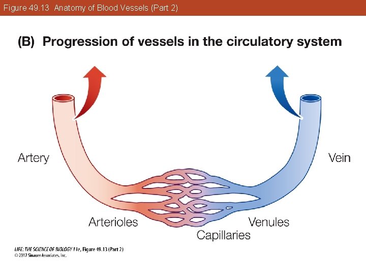
49. 2 Vertebrate Circulatory Systems Evolved from Single to Double Circuits Arteries carry blood away from the heart and branch into arterioles that feed into capillary beds. Capillaries: Tiny, thin-walled vessels; site of exchange between blood and interstitial fluid. Venules drain the capillary beds and form veins, which deliver blood back to the heart.
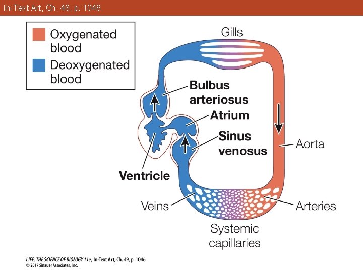
49. 2 Vertebrate Circulatory Systems Evolved from Single to Double Circuits Fish circulatory system • Heart: Four chambers connected in series. • Arterial blood leaves bulbus arteriosus and moves through gills, then to the aorta and on to body tissues. • Unidirectional flow of blood is enabled by one-way valves between the heart chambers.
In-Text Art, Ch. 48, p. 1046
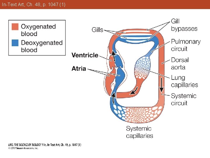
49. 2 Vertebrate Circulatory Systems Evolved from Single to Double Circuits Lungfish show the transition from breathing water to breathing air. An outpocketing of the gut forms a lung. Blood flowing through vessels around the lung can pick up O 2 from air gulped into the lung. Some vessels in the gill arches are modified to carry blood.
In-Text Art, Ch. 48, p. 1047 (1)
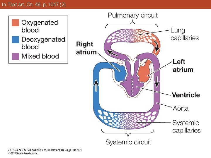
49. 2 Vertebrate Circulatory Systems Evolved from Single to Double Circuits The atrium is partially divided: The left side receives oxygenated blood from the lung; the right side receives deoxygenated blood from the body. The lung evolved to supplement oxygen uptake from the gills. In oxygen-depleted water, the lungfish can rely on the lung.
49. 2 Vertebrate Circulatory Systems Evolved from Single to Double Circuits Amphibian circulatory system • Heart has three chambers; ventricle pumps blood to the lungs and body. • One atrium receives oxygenated blood from the lungs, a second atrium receives blood from the body. • Ventricle directs blood flow to either the pulmonary or systemic circuit.
In-Text Art, Ch. 48, p. 1047 (2)
49. 2 Vertebrate Circulatory Systems Evolved from Single to Double Circuits Partial separation of pulmonary and systemic circulation has the advantage of allowing blood going to the tissues to sidestep the large pressure drop that occurs in the gas exchange organ. Amphibians can also pick up oxygen in blood flowing through small vessels in their skin.
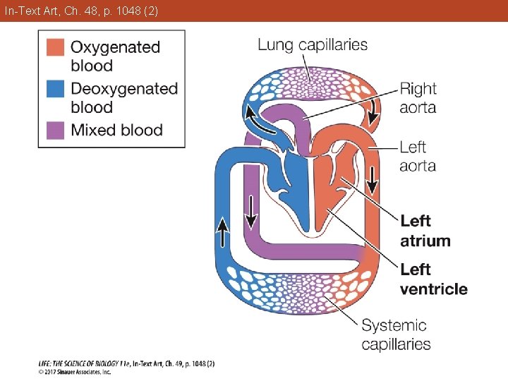
49. 2 Vertebrate Circulatory Systems Evolved from Single to Double Circuits Reptile circulatory systems • Turtles, snakes, and lizards have threechambered hearts. • Ectothermic reptiles can pump blood to the lungs and the rest of the body when they are active and breathing, but when inactive and not breathing, they can bypass the pulmonary circuit.
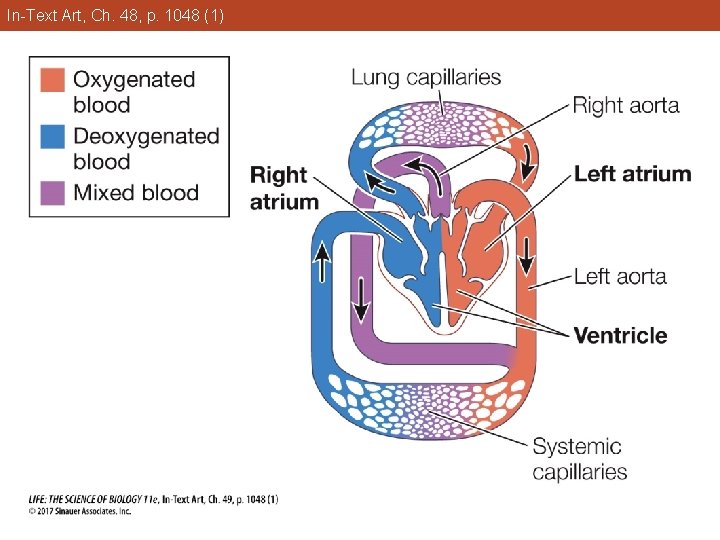
49. 2 Vertebrate Circulatory Systems Evolved from Single to Double Circuits • Ventricle is partially divided; two aortas. The left aorta receives oxygenated blood from the left side of the ventricle and delivers it to the body. § The right aorta can receive blood from either side of the ventricle. §
49. 2 Vertebrate Circulatory Systems Evolved from Single to Double Circuits When breathing, resistance in the pulmonary circuit is lower than in the systemic circuit, so blood from right side of the ventricle flows into the pulmonary artery. § When not breathing, pulmonary vessels constrict, and blood from right side of ventricle flows into the right aorta. §
In-Text Art, Ch. 48, p. 1048 (1)
49. 2 Vertebrate Circulatory Systems Evolved from Single to Double Circuits • Crocodiles have four separate chambers and two aortas. § The aortas are connected and the amount of blood going to either circuit can be altered. § Blood is directed to a circuit by changes in resistance and one-way valves.
In-Text Art, Ch. 48, p. 1048 (2)
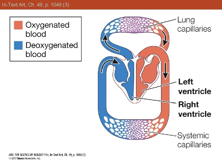
49. 2 Vertebrate Circulatory Systems Evolved from Single to Double Circuits Bird and mammal systems • Four-chamber hearts and completely separate pulmonary and systemic circuits. • Advantages: § Systemic circuit always receives blood with higher O 2 content. § Gas exchange is maximized. § Circuits can operate at different pressures.
In-Text Art, Ch. 48, p. 1048 (3)
Key Concept 49. 2 Learning Outcomes • Trace the flow of blood through the circulatory system of a fish. • Describe how a lungfish can distribute oxygen to its tissues in both water and air environments.
Key Concept 49. 2 Learning Outcomes • Explain how and why ectothermic reptiles shunt blood from the pulmonary circuit when not breathing. • Apply knowledge about four-chambered hearts and fully separate pulmonary and systemic circulatory systems to explain the consequences of heart defects.
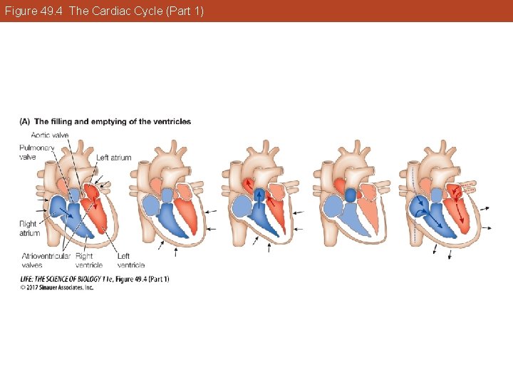
Key Concept 49. 3 Focus Your Learning • Blood flow in mammalian hearts goes from right atrium to right ventricle to lungs and from left atrium to left ventricle to the systemic circulation. Valves prevent backflow. • In each cardiac cycle, both atria contract first, followed by both ventricles contracting, which is then followed by relaxation.
Key Concept 49. 3 Focus Your Learning • Action potentials in cardiac muscle cells depend on the properties of the ion channels in their membranes.
49. 3 Heart Function Depends on Properties of Cardiac Muscle Human heart has two atria and two ventricles. • Right ventricle pumps blood through the pulmonary circuit. • Left ventricle pumps blood through the systemic circuit.
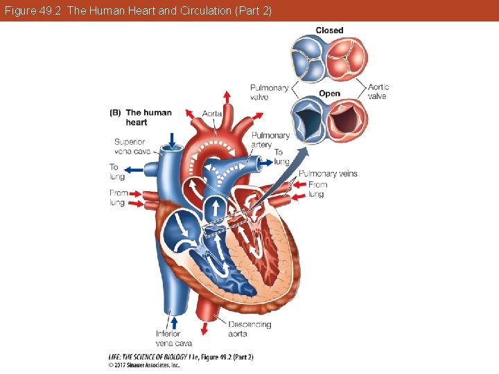
49. 3 Heart Function Depends on Properties of Cardiac Muscle One-way valves prevent backflow of blood: • Atrioventricular (AV) valves between atria and ventricles prevent backflow when ventricles contract. § Right: Tricuspid valve § Left: Bicuspid or mitral valve
49. 3 Heart Function Depends on Properties of Cardiac Muscle • Pulmonary valve goes to the lungs. • Aortic valve goes to the aorta. § They prevent backflow when ventricles relax.
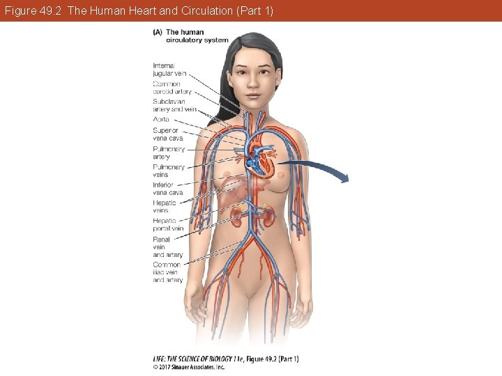
Figure 49. 2 The Human Heart and Circulation (Part 1)
Figure 49. 2 The Human Heart and Circulation (Part 2)
49. 3 Heart Function Depends on Properties of Cardiac Muscle The right atrium receives deoxygenated blood from the body through two large veins: • Superior vena cava—blood from upper body • Inferior vena cava—blood from lower body
49. 3 Heart Function Depends on Properties of Cardiac Muscle Blood passes from the right atrium through the tricuspid valve into the right ventricle. The atrium contracts, then the ventricle contracts. The AV valve closes and blood is pumped through the pulmonary artery to the lungs.
49. 3 Heart Function Depends on Properties of Cardiac Muscle Pulmonary veins return oxygenated blood to the left atrium. Oxygenated blood moves into the left ventricle through the bicuspid valve. The left atrium contracts, then the left ventricle. The aortic valve opens and blood flows into the aorta.
49. 3 Heart Function Depends on Properties of Cardiac Muscle Ventricle walls are powerful muscles that contract with a wringing motion. Left ventricle walls are thicker—it has to propel blood through many kilometers of blood vessels and must therefore push against more resistance than the right ventricle.
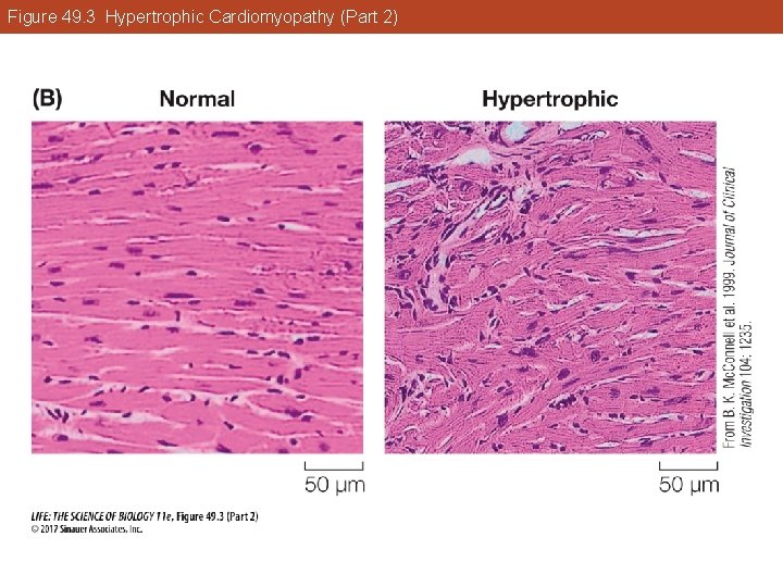
49. 3 Heart Function Depends on Properties of Cardiac Muscle In hypertrophic cardiomyopathy (HCM), mutations that reduce efficiency of contraction increase the work load of the left ventricle, which compensates by getting bigger (hypertrophy). This can block blood flowing into the aorta and disrupt signals that coordinate contractions.
Figure 49. 3 Hypertrophic Cardiomyopathy (Part 1)
Figure 49. 3 Hypertrophic Cardiomyopathy (Part 2)
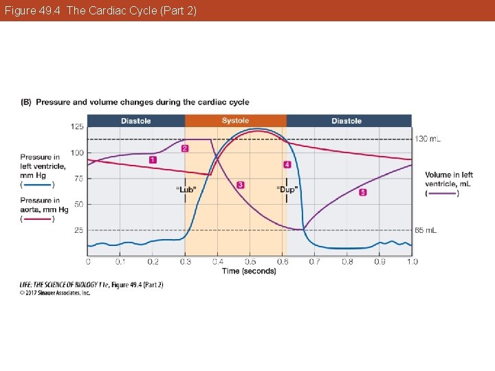
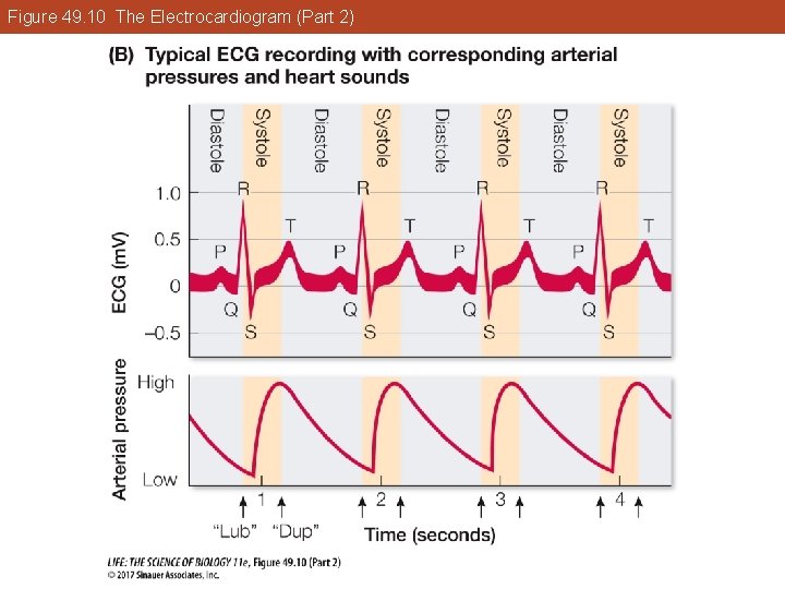
49. 3 Heart Function Depends on Properties of Cardiac Muscle Cardiac cycle: Both sides contract at the same time—first the atria, then the ventricles. Phases: • Systole—when ventricles contract • Diastole—when ventricles relax
Figure 49. 4 The Cardiac Cycle (Part 1)
Figure 49. 4 The Cardiac Cycle (Part 2)
49. 3 Heart Function Depends on Properties of Cardiac Muscle Sounds of the cardiac cycle heard through a stethoscope are created by the heart valves slamming shut. Closing and opening of the valves result from pressure differences on the two sides of the valves. Defective valves that do not close properly lead to sounds called heart murmurs.
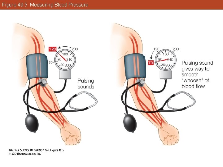
49. 3 Heart Function Depends on Properties of Cardiac Muscle Blood pressure changes are measured with a sphygmomanometer and a stethoscope. • Systolic value—pressure needed to compress an artery so blood does not flow. • Diastolic value—pressure needed to allow intermittent flow though the artery.
Figure 49. 5 Measuring Blood Pressure
49. 3 Heart Function Depends on Properties of Cardiac Muscle Cardiac cells are in electrical contact with each other through gap junctions—action potentials can spread rapidly. Large groups of cardiac muscle contract in unison—essential for pumping blood efficiently. Pacemaker cells initiate action potentials without input from the nervous system.
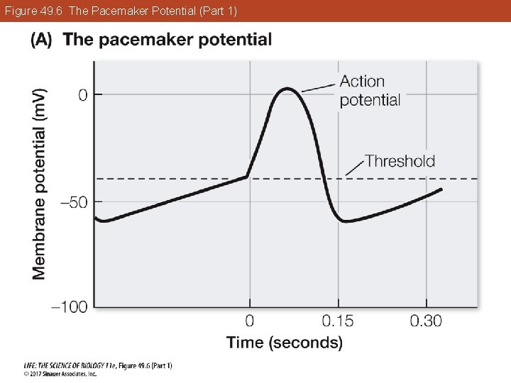
49. 3 Heart Function Depends on Properties of Cardiac Muscle The primary pacemaker is the sinoatrial node. Resting membrane potential of these cells is less negative and unstable so that cells gradually reach threshold. The action potentials are slower to rise, broader, and slower to return to resting potential than those of neurons and other muscle cells.
Figure 49. 6 The Pacemaker Potential (Part 1)
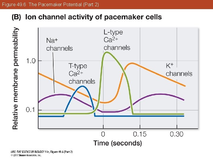
49. 3 Heart Function Depends on Properties of Cardiac Muscle Ion channels in pacemaker cells are different from other cardiac cells: • Na+ channels are open more, so resting potential is less negative than other cardiac cells. • Action potential is due to voltage-gated Ca 2+ channels, which open more slowly than Na+ channels.
Figure 49. 6 The Pacemaker Potential (Part 2)
49. 3 Heart Function Depends on Properties of Cardiac Muscle The unstable resting potential of pacemaker cells is due to activity of cation channels. Pacemaker cells have channels that are more permeable to Na+ than to K+. Na+ enters cells more easily than K+ leaves, so resting membrane potential becomes less negative.
49. 3 Heart Function Depends on Properties of Cardiac Muscle As the membrane becomes less negative, some Ca 2+ channels open: membrane potential continues to rise. These Ca 2+ channels are called T-types because they open transiently. Membrane potential reaches threshold for L-type (long-lasting) Ca 2+ channels to open, which generates the action potential.
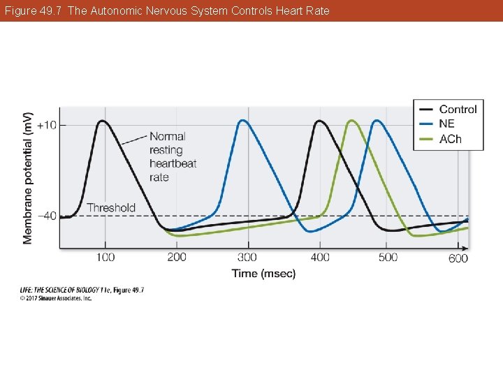
49. 3 Heart Function Depends on Properties of Cardiac Muscle Autonomic nervous system controls heart rate by influencing rate at which resting potentials of pacemaker cells drift upward. • Norepinephrine from sympathetic nerves increases permeability of Na+/K+ and Ca 2+ channels. § Resting potential rises more rapidly and action potentials are closer together.
49. 3 Heart Function Depends on Properties of Cardiac Muscle • Acetylcholine from parasympathetic nerves has the opposite effect. § It increases permeability of K+ and decreases that of Ca 2+ channels. o Resting potential rises more slowly and action potentials are farther apart.
Figure 49. 7 The Autonomic Nervous System Controls Heart Rate
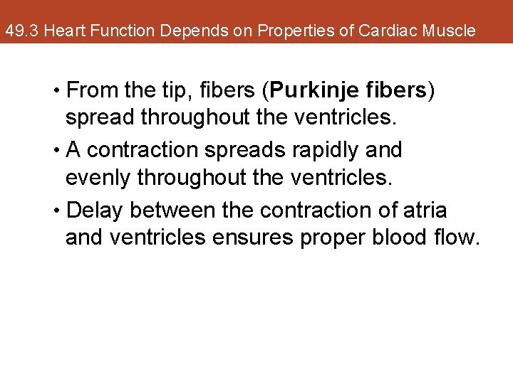
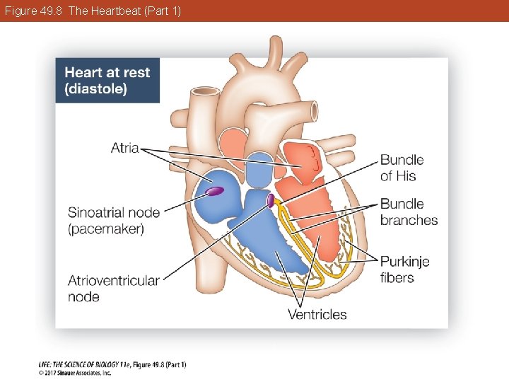
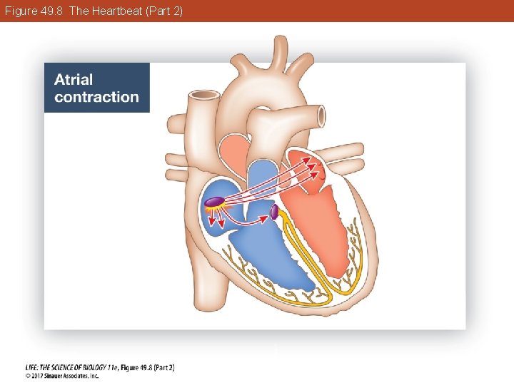
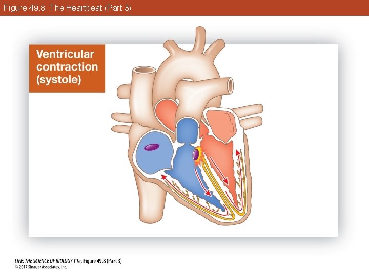
49. 3 Heart Function Depends on Properties of Cardiac Muscle Heart muscle contraction is coordinated: • An action potential is generated in the sinoatrial node. • It spreads through gap junctions in the atria and they contract together, but it does not spread to the ventricles.
49. 3 Heart Function Depends on Properties of Cardiac Muscle • The action potential in the atria stimulates the atrioventricular node. • The node consists of cells that do not contract, but do conduct action potentials to the ventricles via the bundle of His. § The bundle divides into right and left bundle branches that run to the tips of the ventricles.
49. 3 Heart Function Depends on Properties of Cardiac Muscle • From the tip, fibers (Purkinje fibers) spread throughout the ventricles. • A contraction spreads rapidly and evenly throughout the ventricles. • Delay between the contraction of atria and ventricles ensures proper blood flow.
Figure 49. 8 The Heartbeat (Part 1)
Figure 49. 8 The Heartbeat (Part 2)
Figure 49. 8 The Heartbeat (Part 3)
49. 3 Heart Function Depends on Properties of Cardiac Muscle HCM impairs function of the left ventricle: • Septum between ventricles is modified, which disrupts the conducting fibers. • Thickening of the septum can block the entrance to the aorta and impede blood flow.
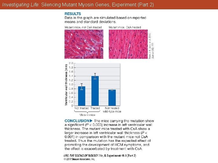
Investigating Life: Silencing Mutant Myosin Genes Hypotheses: 1. Mice genetically engineered to carry a mutation in the myosin gene most commonly responsible for HCM in humans will show the HCM phenotype, and it will be exacerbated by treatment with Cs. A. 2. Silencing expression of the mutant gene will prevent the development of the HCM phenotype.
Investigating Life: Silencing Mutant Myosin Genes Method: Generate a mouse model with a mutation in a myosin gene. Measure left ventricular wall thickness in mutant mice with and without treatment with Cs. A (cyclosporine A) to exacerbate development hyperthrophy.
Investigating Life: Silencing Mutant Myosin Genes Develop an interference RNA (RNAi) that will silence expression of the mutant gene in vitro. Transfect mice heterozygous for the mutation with the RNAi and later, assess them for hypertrophy.
Investigating Life: Silencing Mutant Myosin Genes, Experiment (Part 2)
Investigating Life: Silencing Mutant Myosin Genes Conclusion: Mice carrying the mutation show a significant (P < 0. 003) increase in left ventricular wall thickness. Mutant mice treated with Cs. A show a larger increase (P < 0. 001) in comparison with mutant mice not Cs. A treated. Thus the mutation has the expected effect of promoting development of HCM symptoms, and the effect is exacerbated by treatment with Cs. A.
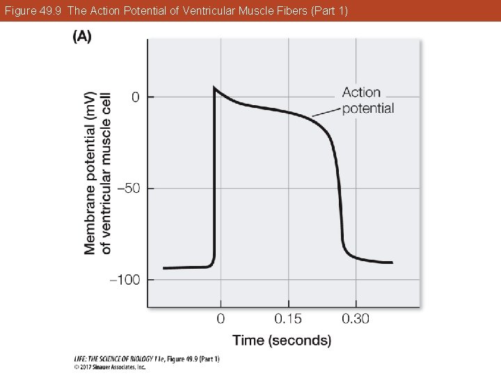
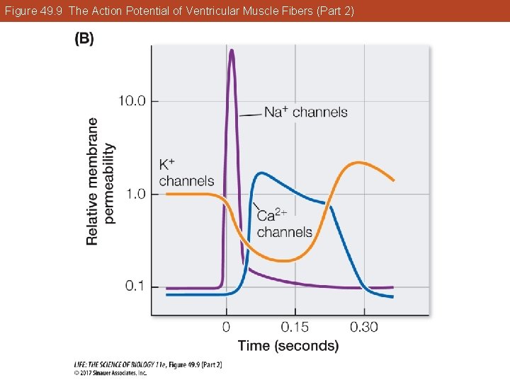
49. 3 Heart Function Depends on Properties of Cardiac Muscle Ventricular muscle fibers contract for a much longer time than skeletal muscle fibers. The extended action potential is due to longer opening of voltage-gated Ca 2+ channels and increased availability of Ca 2+ to stimulate contraction.
Figure 49. 9 The Action Potential of Ventricular Muscle Fibers (Part 1)
Figure 49. 9 The Action Potential of Ventricular Muscle Fibers (Part 2)
49. 3 Heart Function Depends on Properties of Cardiac Muscle To terminate systole, Ca 2+ must be rapidly cleared from the sarcoplasm. Ca 2+ pumps in the sarcoplasmic reticulum (SR) membrane actively transport Ca 2+ ions out of the sarcoplasm and into the SR where they are sequestered until the next action potential arrives.
49. 3 Heart Function Depends on Properties of Cardiac Muscle Thus the rate of cycling of Ca 2+ puts limits on heart rate and strength of ventricular contraction.
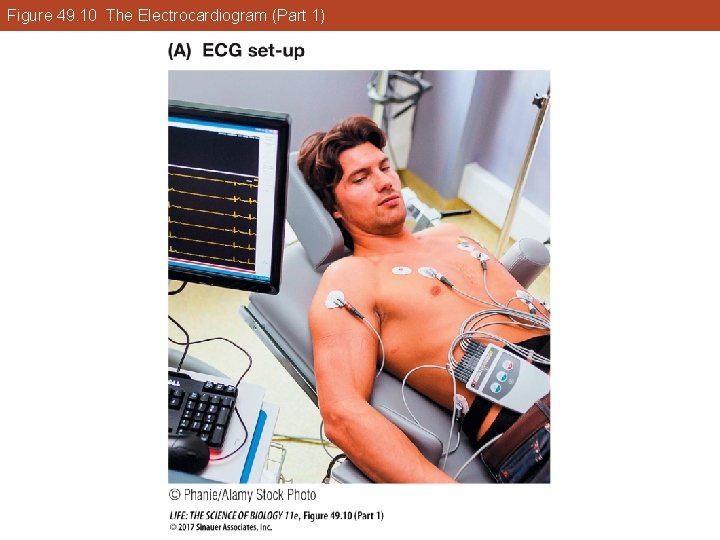
49. 3 Heart Function Depends on Properties of Cardiac Muscle An electrocardiogram (ECG or EKG) uses electrodes to record events in the cardiac cycle. Large action potentials in the heart cause electrical current to flow outward to all parts of the body. Electrodes register the voltage difference at different times.
Figure 49. 10 The Electrocardiogram (Part 1)
Figure 49. 10 The Electrocardiogram (Part 2)
Key Concept 49. 3 Learning Outcomes • Trace the path of blood through both sides of the heart, naming the major blood vessels and heart valves. • Describe how heart valves function. • Describe the cardiac cycle in terms of left ventricular volume and pressure, aortic pressure, and heart sounds.
Key Concept 49. 3 Learning Outcomes • Explain how calcium ion movement into and out of the sarcoplasm of cardiac muscle cells influences the rate and strength of contraction.
Key Concept 49. 4 Focus Your Learning • Red blood cells are generated in bone marrow and, until they are destroyed in the spleen, they transport respiratory gases. • Platelets and blood proteins are responsible for blood clotting. • Smooth muscle cells in arteries and arterioles control blood flow to specific tissues and also control blood pressure.
Key Concept 49. 4 Focus Your Learning • Extracellular fluid exchange between the blood and the interstitial space occurs in capillary beds and depends on hydrostatic pressure pushing water out of the capillaries and osmotic pressure pulling water back into the capillaries. • One-way valves and muscle contraction help return venous blood to the heart.
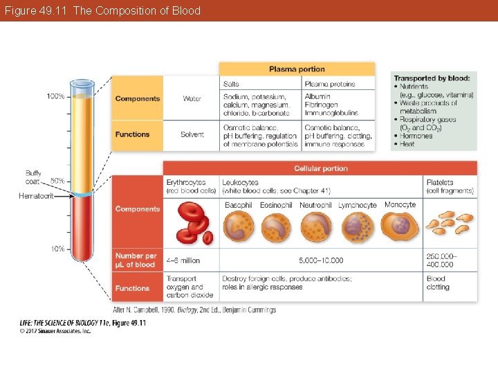
49. 4 Circulatory System Functions Depend on Blood and Blood Vessels Blood is a connective tissue made of cells in an extracellular fluid matrix, the blood plasma. The hematocrit is the percent of blood that is red blood cells. Normal hematocrit is 42– 46%; higher in people who live at high altitudes because low O 2 stimulates production of more red blood cells.
Figure 49. 11 The Composition of Blood
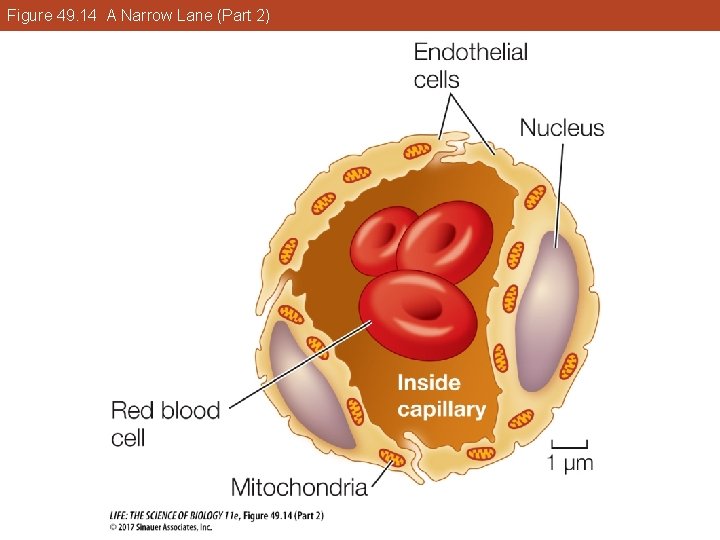
49. 4 Circulatory System Functions Depend on Blood and Blood Vessels Mature erythrocytes, or red blood cells, are biconcave, flexible discs packed with hemoglobin. They transport respiratory gases; the shape gives them a large surface area for gas exchange, and flexibility enables them to squeeze through narrow capillaries.
49. 4 Circulatory System Functions Depend on Blood and Blood Vessels Red blood cells are generated in the bone marrow. Erythropoietin (EPO), a hormone released in the kidney in response to hypoxia, controls red blood cell production. Hypoxia-inducible factor 1 (HIF-1) is a transcription factor—in the kidney it activates the gene for erythropoietin.
49. 4 Circulatory System Functions Depend on Blood and Blood Vessels Immature red blood cells divide many times and produce hemoglobin while in the bone marrow. When hemoglobin content reaches 25%, organelles break down and the cells enter the circulation. Cells circulate about 120 days before rupturing as they pass through narrow capillaries, as in the spleen.
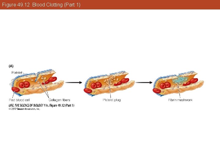
49. 4 Circulatory System Functions Depend on Blood and Blood Vessels Bone marrow stem cells also produce megakaryocytes that break off cell fragments called platelets. Platelets initiate blood clotting when activated by collagen exposed in damaged blood vessels. They release chemical clotting factors that activate other platelets and also form a sticky plug at the damaged site.
Figure 49. 12 Blood Clotting (Part 1)
Figure 49. 12 Blood Clotting (Part 2)
Figure 49. 12 Blood Clotting (Part 3)
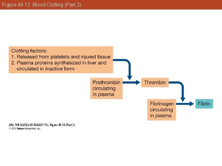
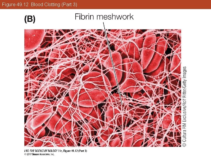
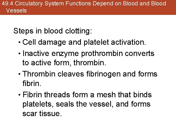
49. 4 Circulatory System Functions Depend on Blood and Blood Vessels Steps in blood clotting: • Cell damage and platelet activation. • Inactive enzyme prothrombin converts to active form, thrombin. • Thrombin cleaves fibrinogen and forms fibrin. • Fibrin threads form a mesh that binds platelets, seals the vessel, and forms scar tissue.
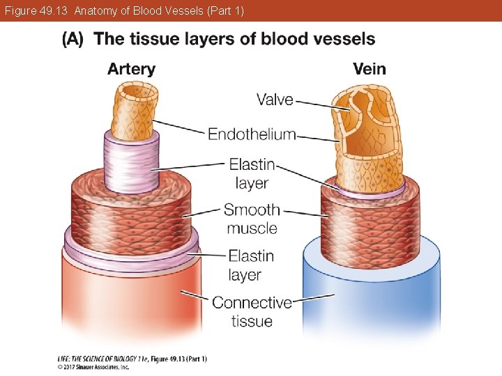
49. 4 Circulatory System Functions Depend on Blood and Blood Vessels Anatomy of different vessel types reflects function. Walls of large arteries have collagen and elastin fibers, which enable them to withstand high blood pressures. They are stretched during systole, storing some of the heart contraction energy. Elastic recoil during diastole squeezes the blood and pushes it forward.
Figure 49. 13 Anatomy of Blood Vessels (Part 1)
Figure 49. 13 Anatomy of Blood Vessels (Part 2)
49. 4 Circulatory System Functions Depend on Blood and Blood Vessels Smooth muscle cells in artery and arteriole walls constrict or dilate those vessels. When the diameter of an artery changes, so does its resistance—blood flow changes as a result. Neuronal and hormonal mechanisms control the resistance by influencing the smooth muscle cells.
49. 4 Circulatory System Functions Depend on Blood and Blood Vessels Arterioles are called resistance vessels because their resistance can vary to control the blood flow to specific tissues.
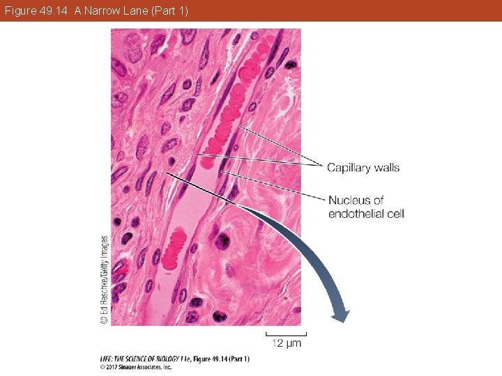
49. 4 Circulatory System Functions Depend on Blood and Blood Vessels Capillary beds lie between arterioles and venules. Capillary walls are thin and permeable to water and many solutes. Blood flows slowly through capillaries, allowing time for exchange.
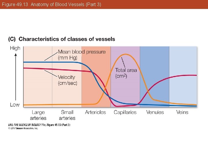
49. 4 Circulatory System Functions Depend on Blood and Blood Vessels Blood flows through large arteries rapidly at high pressures, but when it reaches the capillaries, pressure and rate of flow decrease. The sum total cross-sectional area of the capillaries is much greater than that of any other class of vessels.
Figure 49. 13 Anatomy of Blood Vessels (Part 3)
49. 4 Circulatory System Functions Depend on Blood and Blood Vessels Capillary walls are a single layer of endothelial cells and are permeable to water, ions, and small molecules, but not to large proteins and red blood cells.
Figure 49. 14 A Narrow Lane (Part 1)
Figure 49. 14 A Narrow Lane (Part 2)
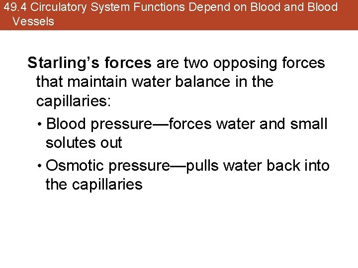
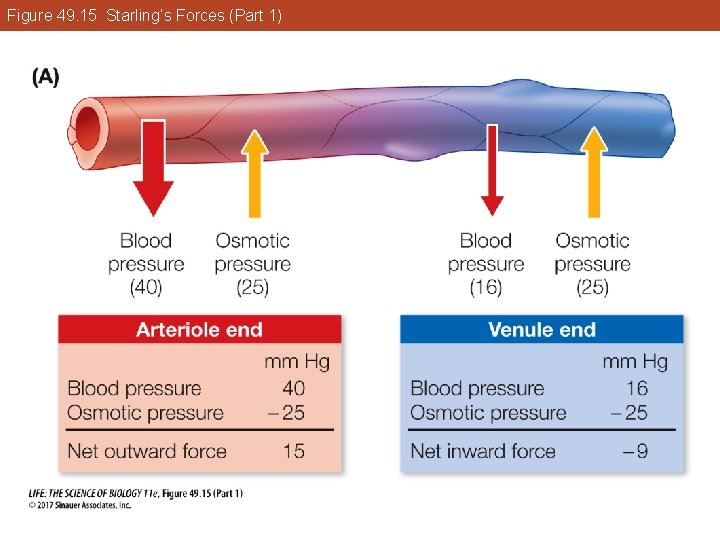
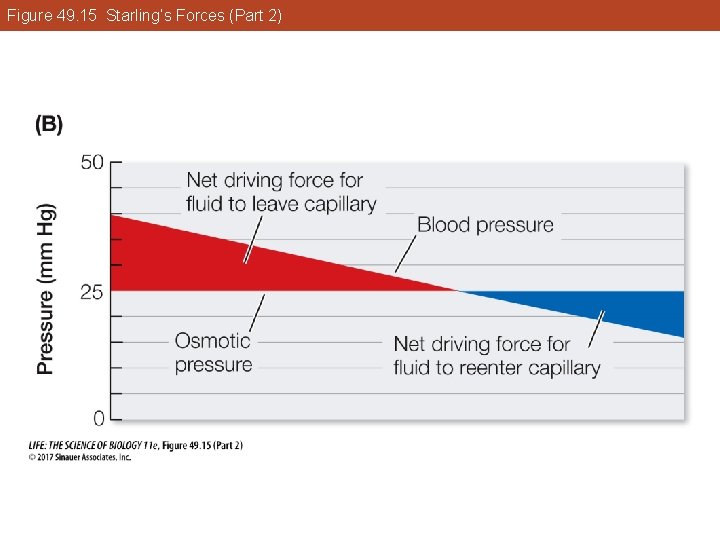
49. 4 Circulatory System Functions Depend on Blood and Blood Vessels Starling’s forces are two opposing forces that maintain water balance in the capillaries: • Blood pressure—forces water and small solutes out • Osmotic pressure—pulls water back into the capillaries
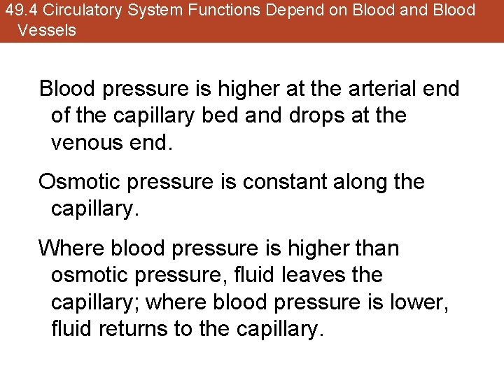
49. 4 Circulatory System Functions Depend on Blood and Blood Vessels Blood pressure is higher at the arterial end of the capillary bed and drops at the venous end. Osmotic pressure is constant along the capillary. Where blood pressure is higher than osmotic pressure, fluid leaves the capillary; where blood pressure is lower, fluid returns to the capillary.
Figure 49. 15 Starling’s Forces (Part 1)
Figure 49. 15 Starling’s Forces (Part 2)
49. 4 Circulatory System Functions Depend on Blood and Blood Vessels Bicarbonate ions (HCO 3–) in blood plasma contribute to the osmotic force that draws water back into capillaries. CO 2 from cellular metabolism diffuses into endothelial cells, is converted to HCO 3– and released into the plasma. Increase in HCO 3– ions at the venous end raises the osmotic pressure, especially during exercise.
49. 4 Circulatory System Functions Depend on Blood and Blood Vessels Permeability varies in different capillary beds. All are permeable to O 2, CO 2, and small ions. Water and small solutes pass between cells in the capillary wall, and through holes called fenestrations in the endothelial cells.
49. 4 Circulatory System Functions Depend on Blood and Blood Vessels Less selective capillaries are found in the digestive tract and kidneys. The endothelial cells also contain transporters for specific molecules, such as glucose and lactate.
49. 4 Circulatory System Functions Depend on Blood and Blood Vessels Capillaries in the brain are not very permeable and are wrapped by glia. Only lipid-soluble substances (e. g. , alcohol and anesthetics) can pass through. This high selectivity of brain capillaries is known as the blood–brain barrier.
49. 4 Circulatory System Functions Depend on Blood and Blood Vessels Blood pressue in veins is low; the walls are expandable and blood tends to accumulate. Because of their capacity to stretch and store blood, veins are called capacitance vessels.
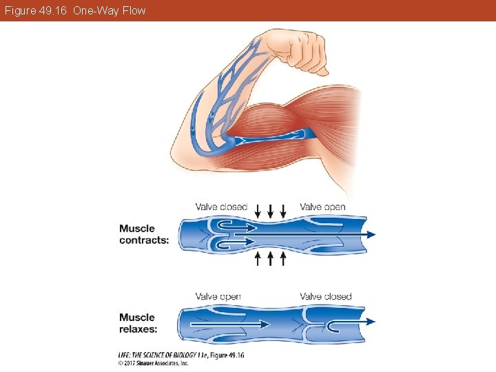
49. 4 Circulatory System Functions Depend on Blood and Blood Vessels Blood returning from the upper body is assisted by gravity. Blood returning from below the heart is assisted by skeletal muscle contractions that squeeze the veins. One-way valves in the veins prevent backflow.
Figure 49. 16 One-Way Flow
49. 4 Circulatory System Functions Depend on Blood and Blood Vessels Frank–Starling law: Cardiac cells contract more forcefully if stretched by increased volume of returning blood. • Both leg muscle contractions and breathing actions help return venous blood to the heart.
49. 4 Circulatory System Functions Depend on Blood and Blood Vessels Lymphatic system returns interstitial fluid to the blood. Capillary beds contain at least one blind-ended lymph capillary. When the fluid enters the vessels it is called lymph. Lymphatic capillaries ultimately merge into two thoracic ducts that empty into veins in the neck.
49. 4 Circulatory System Functions Depend on Blood and Blood Vessels Lymphatic vessels have one-way valves to keep lymph flowing toward the thoracic ducts. Lymph nodes along the vessels are the site of lymphocyte production. Lymphocytes remove microorganisms and foreign materials by phagocytosis.
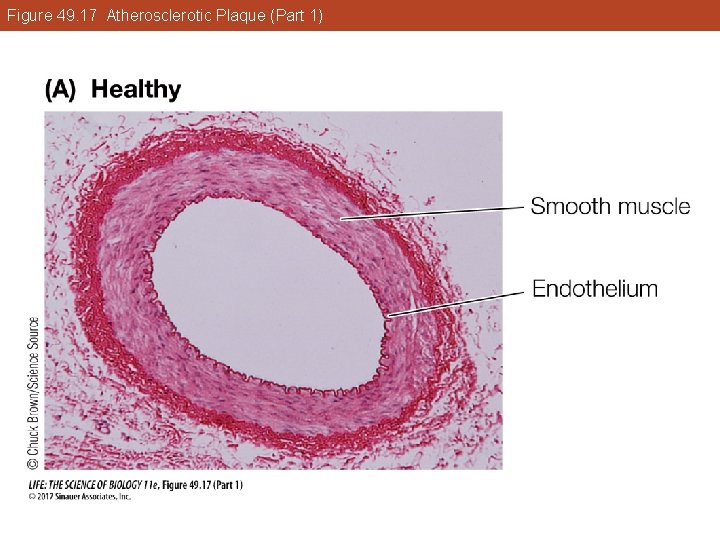
49. 4 Circulatory System Functions Depend on Blood and Blood Vessels Vascular disease • Atherosclerosis is “hardening of the arteries. ” § The endothelial lining of arteries is damaged by high blood pressure, smoking, high-fat diet, or microorganisms.
49. 4 Circulatory System Functions Depend on Blood and Blood Vessels § Plaque forms at damage sites: o Damaged cells attract white blood cells and smooth muscle cells. o Cholesterol is deposited, and the plaque becomes fatty. o Fibrous connective tissue made by the invading smooth muscle cells, plus calcium deposits, make the artery wall less elastic.
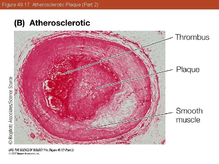
49. 4 Circulatory System Functions Depend on Blood and Blood Vessels The growing plaque deposit narrows the artery and causes turbulence in the blood flow. § Platelets may stick to the plaque and form a thrombus, or blood clot, which blocks the artery. §
Figure 49. 17 Atherosclerotic Plaque (Part 1)
Figure 49. 17 Atherosclerotic Plaque (Part 2)
49. 4 Circulatory System Functions Depend on Blood and Blood Vessels § Coronary arteries supply blood to the heart muscle. § Atherosclerosis in coronary arteries reduces blood flow; marked by chest pain and shortness of breath. • Coronary thrombosis—a thrombus that forms in a coronary artery can lead to a heart attack, or myocardial infarction.
49. 4 Circulatory System Functions Depend on Blood and Blood Vessels • An embolus is a piece of a thrombus that breaks loose. § It can cause an embolism (blockage) if it lodges in a smaller blood vessel. § Arteries narrowed by plaque are likely places for an embolism. § If the embolism is in the brain, cells fed by that artery will die, causing a stroke.
49. 4 Circulatory System Functions Depend on Blood and Blood Vessels Risk factors for atherosclerosis: • Genetic predisposition • Age • Environmental risk factors—high-fat diet, smoking, sedentary lifestyle • Medical conditions—hypertension, obesity, diabetes
Key Concept 49. 4 Learning Outcomes • Describe the features, life cycle, and function of a red blood cell. • Describe the blood clotting process. • Explain the role of arterioles in the control of blood flow. • Explain the forces that cause water to leave and reenter the capillaries. • Describe the factors that affect the return of blood in veins to the heart.
Key Concept 49. 5 Focus Your Learning • Local blood flow is controlled to meet local metabolic need by autoregulatory responses of arteriole smooth muscles. • Mean arterial pressure is regulated through neural and hormonal control of smooth muscles in arterioles.
Key Concept 49. 5 Focus Your Learning • Baroreceptors in the walls of large arteries continuously convey blood pressure information to the cardiovascular regulatory center in the brain, which controls the neural and hormonal mechanisms that influence mean arterial pressure.
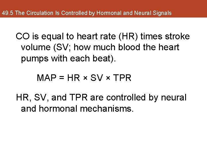
49. 5 The Circulation Is Controlled by Hormonal and Neural Signals Blood flow depends on pressure. Mean arterial pressure (MAP) is determined by cardiac output (CO) and total peripheral resistance (TPR). MAP = CO × TPR
49. 5 The Circulation Is Controlled by Hormonal and Neural Signals CO is equal to heart rate (HR) times stroke volume (SV; how much blood the heart pumps with each beat). MAP = HR × SV × TPR HR, SV, and TPR are controlled by neural and hormonal mechanisms.
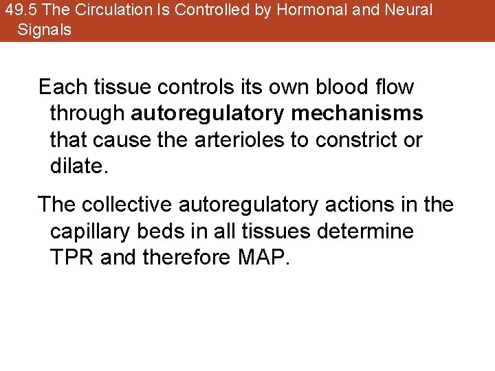
49. 5 The Circulation Is Controlled by Hormonal and Neural Signals Each tissue controls its own blood flow through autoregulatory mechanisms that cause the arterioles to constrict or dilate. The collective autoregulatory actions in the capillary beds in all tissues determine TPR and therefore MAP.
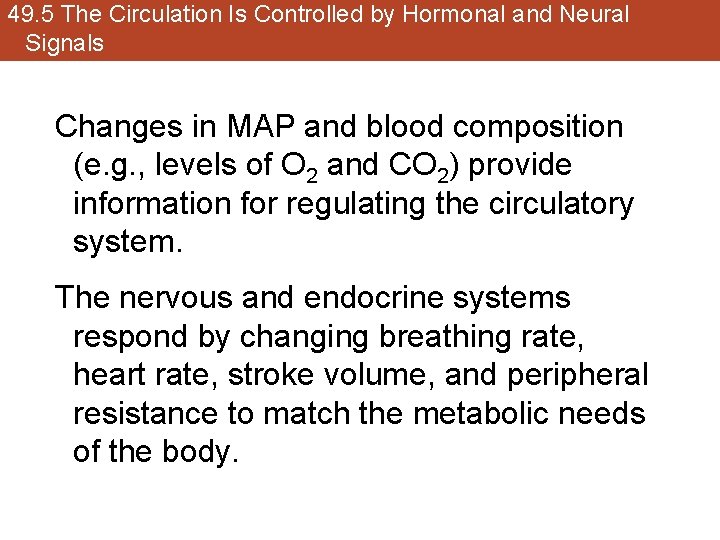
49. 5 The Circulation Is Controlled by Hormonal and Neural Signals Changes in MAP and blood composition (e. g. , levels of O 2 and CO 2) provide information for regulating the circulatory system. The nervous and endocrine systems respond by changing breathing rate, heart rate, stroke volume, and peripheral resistance to match the metabolic needs of the body.
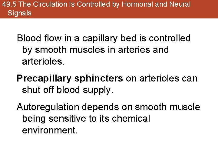
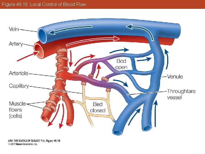
49. 5 The Circulation Is Controlled by Hormonal and Neural Signals Blood flow in a capillary bed is controlled by smooth muscles in arteries and arterioles. Precapillary sphincters on arterioles can shut off blood supply. Autoregulation depends on smooth muscle being sensitive to its chemical environment.
Figure 49. 18 Local Control of Blood Flow
49. 5 The Circulation Is Controlled by Hormonal and Neural Signals Hyperemia: Low O 2 and high CO 2 levels cause smooth muscle to relax, increasing blood supply to capillary bed. Other metabolic by-products produced during exercise, such as lactic acid, also promote hyperemia.
49. 5 The Circulation Is Controlled by Hormonal and Neural Signals Arteries and arterioles are innervated by the sympathetic nervous system. Norepinephrine causes arterioles to constrict in tissues not needed for “fight or flight” response. • This reduces blood flow to these tissues and increases MAP. Sympathetic input also increases HR and SV.
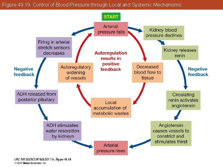
49. 5 The Circulation Is Controlled by Hormonal and Neural Signals Hormones also regulate arterial pressure: • Epinephrine is released from the adrenal medulla in response to a fall in arterial pressure or the “fight-or-flight” response; arterioles constrict. • Angiotensin is produced when blood pressure in the kidneys drops; reduces flow to peripheral tissues and directs it to essential organs.
Figure 49. 19 Control of Blood Pressure through Local and Systemic Mechanisms
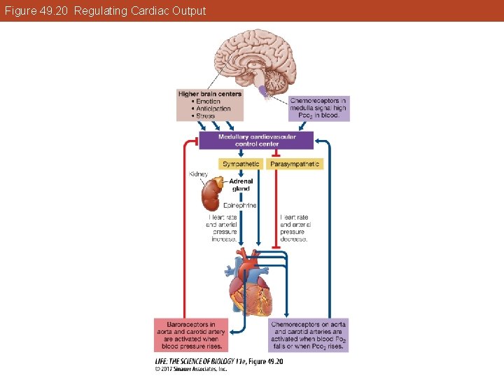
49. 5 The Circulation Is Controlled by Hormonal and Neural Signals The cardiovascular control center in the medulla controls heart rate and vessel constriction. Information on blood composition and pressure comes from baroreceptors (stretch receptors) and chemoreceptors in the aorta and carotid arteries.
Figure 49. 20 Regulating Cardiac Output
49. 5 The Circulation Is Controlled by Hormonal and Neural Signals Increased activity of the stretch receptors signals rising blood pressure: • Response—inhibition of sympathetic nervous system and increased parasympathetic input • Result—heart slows and arterioles dilate, blood pressure falls
49. 5 The Circulation Is Controlled by Hormonal and Neural Signals Antidiuretic hormone (ADH), or vasopressin, is released in response to low baroreceptor activity. The kidneys absorb more water and increase blood pressure. Increased baroreceptor activity inhibits the release of ADH and kidneys excrete more water.
49. 5 The Circulation Is Controlled by Hormonal and Neural Signals Other chemoreceptors send feedback to the cardiovascular and respiratory regulatory control centers: • In the medulla, chemoreceptors are activated by rising arterial CO 2 levels. • In the aorta and carotid arteries chemoreceptors respond to falling arterial O 2 levels.
Investigating Life: Athletes with Big Hearts Are there potential therapies that could treat HCM? Physical exams and genetic screening may allow carriers of the mutated genes that result in HCM to make decisions about safe levels of exercise. Gene therapy may be possible in the future.