What is molecular pathology Molecular pathology is the


- Slides: 23


What is molecular pathology? Molecular pathology: is the study of diseases at molecular level and includes detection and diagnosis of abnormalities at the level of DNA of the cell.

Functional components of the cell Cells are the smallest functional unit of the body. They contain structures that are strikingly similar to those needed to maintain total body function. There are three major components of the eukaryotic cell: 1 - Cell (Plasma) membrane 2 -Nucleus 3 -Cytoplasm: The bulk of the cytoplasm is water, in which inorganic and organic chemicals are dissolved. This fluid suspension is called the cytosol. The cytosol contains membrane-enclosed compartments or organelles that perform distinctive functions.

Cell membrane Structure

Cell membrane Structure A main structural component of the cell membrane is its lipid bilayer, that consists primarily of phospholipids, with glycolipids and cholesterol. Phospholipids, compose of a hydrophilic (water-soluble) head and a hydrophobic (waterinsoluble) tail. The integral proteins span the entire lipid bilayer, and pass directly through the membrane. Also, there are transmembrane proteins. Many integral transmembrane proteins form the ion channels found on the cell surface. Other proteins, called the peripheral proteins, are bound to one or the other side of the membrane and do not pass into the lipid bilayer. Several peripheral proteins serve as receptors or are involved in intracellular signaling systems. The cell surface is surrounded by a layer, called the cell coat or glycocalyx. It consists of, complex carbohydrate chains attached to protein molecules and carbohydrate-binding proteins called lectins. The cell coat participates in cell-tocell recognition and adhesion. It contains tissue transplant antigens that label cells as self or nonself.

Cell membrane Function 1 - The cell membrane encloses the cell. 2 - It acts as a semipermeable structure that separates the intracellular and extracellular environments. 3 - It controls the transport of materials from the extracellular fluids to the interior of the cell. 4 - It maintains the electrical activities that power cell function. 5 - It provides receptors for hormones and other biologically active substances.

Diseases associated with Cell Membrane Disorders • Membrane Structural Defects in human RBC 1 -Hereditary Ovalocytosis 2 -Hereditary Spherocytosis 3 -Hereditary Elliptocytosis • Membrane Permeability Defects in human RBC 1 -Hereditary Xerocytosis 2 -Hereditary Hydrocytosis • Also, Alzheimer's disease as a disorder of the plasma membrane Depending on the molecular pathways and biological signals utilized, the plasma membrane can be the source of both beneficial and detrimental signals to further modulate amyloidogenic, inflammatory or neurotrophic aspects of the Alzheimer’s disease process.

Nucleus Structure • The nucleus is the control center for the cell. It also contains most of the hereditary material. • It is enclosed in a nuclear envelope and contains chromatin, the genetic material of the nucleus, and a distinct region called the nucleolus. The complex structure of DNA and DNA-associated proteins dispersed in the nuclear matrix (chromatin). • The nuclear envelope is formed by an inner and outer nuclear membrane. The doublemembrane envelope is penetrated by pores in which nuclear pore complexes are positioned and continuous with the rough endoplasmic reticulum. The nuclear lamina on the surface of the inner membrane bind to DNA and hold the chromosomes in place.

Nucleus Function • The nucleus can be regarded as the control center for the cell. • It contains the deoxyribonucleic acid (DNA) that is essential to the cell because its genes encode the information necessary for the synthesis of proteins that the cell must produce to stay alive. The genes also represent the individual units of inheritance that transmit information from one generation to another. • The nucleus also is the site for the synthesis of the three types of ribonucleic acid (messenger RNA, ribosomal RNA, and transfer RNA) that move to the cytoplasm and carry out the actual synthesis of proteins.

Mutations in nuclear lamina and DNA damage associated with a diverse set of diseases • Over 200 diseases have been associated with mutations in lamina genes. Most mutations are in lamina and lead to wide variety of phenotypes. How do mutations in one gene lead to such diverse phenotypes? Currently, we don’t know the answer. Mutations in lamina likely affect nuclear structure and possible gene expression (e. g. Atypical progeria syndrome). • DNA damage induces sequential responses to either repair the damage, and defects in this critical response leads to wide array of human pathologies that include : Cancer predisposition, Inflammation responses, Cell Ageing and Neurodegeneration.

The Cytoplasm and Its Organelles • The cytoplasm surrounds the nucleus. Embedded in the cytoplasm are various membrane-enclosed compartments (endoplasmic reticulum, Golgi apparatus, mitochondria, lysosomes). Most organelles are surrounded by one or two lipid membranes, similar to plasma membrane, that separate the organelles from the cytosol. • The ribosomes, endoplasmic reticulum, and Golgi apparatus represent the primary sites of protein synthesis in the cell. Because these organelles lack direct communication with the cytosol, they use transport vesicles to move newly synthesized proteins, membrane components, and soluble molecules from one organelle to another. These transport vesicles bud off from the membrane of one organelle and fuse with another, carrying the transported material.

Ribosomes structure and function • The ribosomes are small particles of nucleoproteins (r. RNA and proteins) that are held together by a strand of m. RNA to form polyribosomes (also called polysomes). Polysomes exist as isolated clusters of free ribosomes within the cytoplasm or attached to the membrane of the endoplasmic reticulum. • Free ribosomes are involved in the synthesis of proteins, mainly enzymes that aid in the control of cell function, whereas those attached to the endoplasmic reticulum translate m. RNAs that code for proteins secreted from the cell or stored within the cell (e. g. , granules in white blood cells).

Ribosomopathies: human disorders of ribosome dysfunction • Ribosomopathies compose a collection of disorders in which genetic abnormalities cause impaired ribosome biogenesis and function, resulting in specific clinical phenotypes. • Congenital mutations in RPS 19 and other genes encoding ribosomal proteins cause Diamond-Blackfan anemia, a rare congenital bone marrow failure syndrome.

Endoplasmic reticulum structure • The endoplasmic reticulum (ER) is an extensive system of paired membranes and flat vesicles. • Between the paired ER membranes is a fluid filled space called the matrix. The matrix connects the space between the two membranes of the nuclear envelope, the cell membrane, and various cytoplasmic organelles. • Two forms of ER exist in cells: rough and smooth. Rough ER is studded with ribosomes attached to specific binding sites on the membrane. • The smooth ER is free of ribosomes and is continuous with the rough ER.

Endoplasmic reticulum function 1. 2. 3. 4. 5. 6. 7. It functions as a tubular communication system for transporting various substances from one part of the cell to another. The multiple enzyme systems attached to the ER membranes provide the machinery for a major share of the cell’s metabolic functions. Proteins produced by the rough ER are used in the generation of lysosomal enzymes. The rough ER segregates these proteins from other components of the cytoplasm and modifies their structure for a specific function. The smooth ER does not participate in protein synthesis; instead, its enzymes are involved in the synthesis of lipid molecules, regulation of intracellular calcium, and metabolism and detoxification of certain hormones and drugs. The smooth ER is the site of lipid, lipoprotein, and steroid hormone synthesis. The smooth ER of the liver is involved in glycogen storage and metabolism of lipid-soluble drugs.

Endoplasmic reticulum dysfunction • Endoplasmic reticulum dysfunction might have an important part to play in a range of neurological disorders, including: 1. Cerebral ischaemia 2. Sleep apnoea 3. Alzheimer's disease 4. Multiple sclerosis 5. Amyotrophic lateral sclerosis 6. The prion diseases 7. And familial encephalopathy with neuroserpin inclusion bodies.

Golgi apparatus structure and function • The Golgi apparatus (Golgi complex) consists of stacks of thin, flattened vesicles or sacs. • These Golgi bodies are found near the nucleus and function in association with the ER. Substances produced in the ER are carried to the Golgi complex in small, membrane-covered transport vesicles. • The Golgi complex modifies the large inactive proteins and packages them into secretory granules or vesicles (e. g. insulin). • The Golgi complex is thought to produce large carbohydrate molecules that combine with proteins produced by the rough ER to form glycoproteins.

Human Diseases Associated with Golgi Disorders

Lysosomes structure and function • Lysosomes are small, membrane-enclosed sacs filled with hydrolytic enzymes. All of the lysosomal enzymes are acid hydrolases, thus the lysosomes provide an acid environment by maintaining a p. H of approximately 5. 0 in their interior. • The lysosomes are the digestive organelles in the cell. • The lysosomal enzymes can break down most proteins, carbohydrates, and lipids to their basic constituents. • Lysosomes are also repositories where cells accumulate abnormal substances that cannot be completely digested or broken down.

Lysosomal storage diseases • Lysosomal storage disorders are caused by lysosomal dysfunction usually as a consequence of deficiency of a single enzyme required for the metabolism of lipids and glycoproteins. • Examples: Niemann–Pick disease, type C. Fabry disease and Hunter syndrome (MPS II).

Mitochondria structure and function • The mitochondria are composed of two membranes: an outer membrane that encloses the periphery of the mitochondrion and an inner membrane that forms shelflike projections, called cristae. The narrow space between the outer and inner membranes is called the intermembrane space, whereas the large space enclosed by the inner membrane is termed the matrix space. The inner membrane contains respiratory chain enzymes and transport proteins needed for the synthesis of ATP. • Mitochondria contain their own DNA and ribosomes and are self-replicating. The DNA is found in the mitochondrial matrix and is distinct from the chromosomal DNA found in the nucleus.

Mitochondria Function • The mitochondria are literally the “power plants” of the cell because they transform organic compounds into energy. They do not make energy, but extract it from organic compounds. Much of this energy is stored in the high-energy phosphate bonds of compounds such as adenosine triphosphate (ATP) that power the various cellular activities. • Mitochondrial DNA directs the synthesis of 13 of the proteins required for mitochondrial function.

Mitochondrial diseases • Mitochondrial diseases are sometimes caused by mutations in the mitochondrial DNA that affect mitochondrial function. • Other mitochondrial diseases are caused by mutations in genes of the nuclear DNA, whose gene products are imported into the mitochondria (mitochondrial proteins) as well as acquired mitochondrial conditions. • Examples of mitochondrial diseases include: 1. Mitochondrial myopathy 2. Diabetes mellitus and deafness (DAD) • this combination at an early age can be due to mitochondrial disease • Diabetes mellitus and deafness can be found together for other reasons 3. Leber's hereditary optic neuropathy (LHON) • visual loss beginning in young adulthood • eye disorder characterized by progressive loss of central vision due to degeneration of the optic nerves and retina
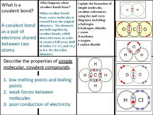
 Giant molecular structure vs simple molecular structure
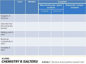
 Giant molecular structure vs simple molecular structure
 Giant molecular structure vs simple molecular structure
 Sự nuôi và dạy con của hổ
 Các châu lục và đại dương trên thế giới
 Dot
Dot Biện pháp chống mỏi cơ
 Bổ thể
 Phản ứng thế ankan
 Thiếu nhi thế giới liên hoan
 Fecboak
 Hát lên người ơi alleluia
 điện thế nghỉ
 Một số thể thơ truyền thống
 Sơ đồ cơ thể người
 Số nguyên tố là gì
 Công thức tính thế năng
 đặc điểm cơ thể của người tối cổ
 Tỉ lệ cơ thể trẻ em
 Các châu lục và đại dương trên thế giới
 ưu thế lai là gì
 Thẻ vin
 Các môn thể thao bắt đầu bằng tiếng chạy
 Tư thế ngồi viết

