Vascular Technology Lecture 22 Venous Gross Anatomy HHHoldorf


 • Formed by confluence of venules • Empties the")
 • Formed by confluence of venules • Empties front")
 between the adductor magnus muscle")
 • Popliteal vein becomes FV when vein passed through adductor hiatus")

 • Common femoral vein becomes EIV when passes through inguinal")


 • Formed by confluence of common iliac veins • Commonly")
 vein (SSV) • Ascends back of calf joining")
 Saphenous Vein (GSV) • Longest vein in the body,")


 to the deep venous system (D)")


 • Formed by confluence of venules • Empties lateral")
 • Formed by confluence of radial and ulnar veins")
 • Formed by confluence of subclavian vein and internal jugular")


 • Formed by confluence of innominate veins •")


- Slides: 53
Vascular Technology Lecture 22 : Venous Gross Anatomy HHHoldorf
Venous Gross Anatomy �Lower Extremity Veins • Note: The anatomical relationship of the veins to the heart is the same as for the arteries. Veins located at the ankle are considered distal: while veins located closer to the heart (e. g. , Femoral) are considered more proximal • Note: Be sure to know the orientation of vessels from medial to lateral and from lateral to medial
The paired, deep veins of the calf (Anterior Tibials, Peroneals, and posterior tibilas, follow the corresponding arteries: are called comitantes (corresponding veins).
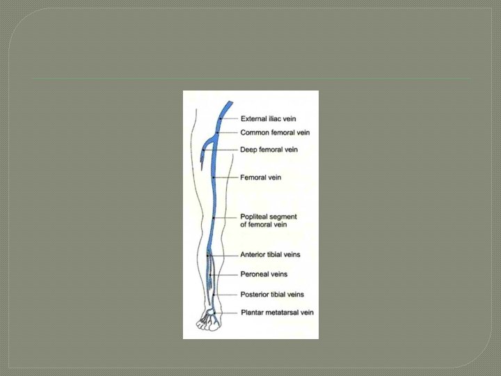
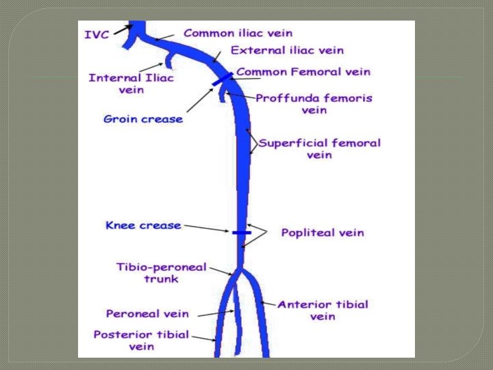
�Paired peroneal veins (Per. V) • Formed by confluence of venules • Empties the lateral leg • Paired veins may form common trunk and carry blood cephalad into tibial-peroneal trunk �Paired posterior tibial veins (PTV) • Formed by confluence of venules • Empties back of leg • Paired veins may form common trunk and carry blood cephalad into tibial-peroneal trunk
�Paired anterior tibial veins (ATV) • Formed by confluence of venules • Empties front of leg �Popliteal vein (Pop. V) • Formed by union of ATV and Tib-Peroneal Trunk • Usually just below the knee • Becomes femoral vein (previously called superficial femoral vein) when passes through adductor hiatus in lower thigh
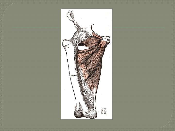
Adductor hiatus The adductor hiatus is a hiatus (gap) between the adductor magnus muscle and the femur that allows the passage of the femoral vessels from the anterior thigh to the posterior thigh and then the popliteal fossa.
�Femoral Vein (FV) • Popliteal vein becomes FV when vein passed through adductor hiatus �Common Femoral Vein (CFV) • Formed by joining of FV & Deep femoral vein
Common Femoral Vein 5 Formed by 6 and 8
�External iliac Vein (EIV) • Common femoral vein becomes EIV when passes through inguinal ligament �Common Iliac Vein (CIV) • Formed by confluence of external and internal iliac veins
The inguinal ligament
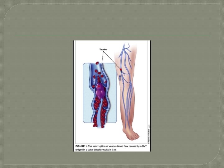
�Because the left common iliac vein passes under the right common iliac artery, extrinsic compression may be evident. �This pressure point may account for left sided DVT; also known as May-Thurner Syndrome
May-Thurner Syndrome
�Inferior Vena Cava (IVC) • Formed by confluence of common iliac veins • Commonly at level of 5 th lumbar vertebra • Carries blood into right atrium of heart
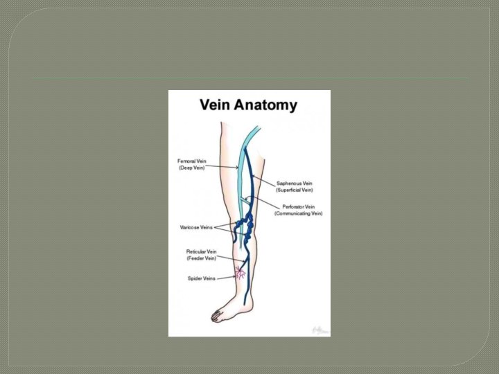
Superficial Veins �Small saphenous (formally lesser) vein (SSV) • Ascends back of calf joining popliteal vein
Superficial Veins �Great (formerly Greater) Saphenous Vein (GSV) • Longest vein in the body, originating on dorsum of foot, traveling medially to saphenofemoral junction in the groin (about level of CFA bifurcation)
Great Saphenous Vein
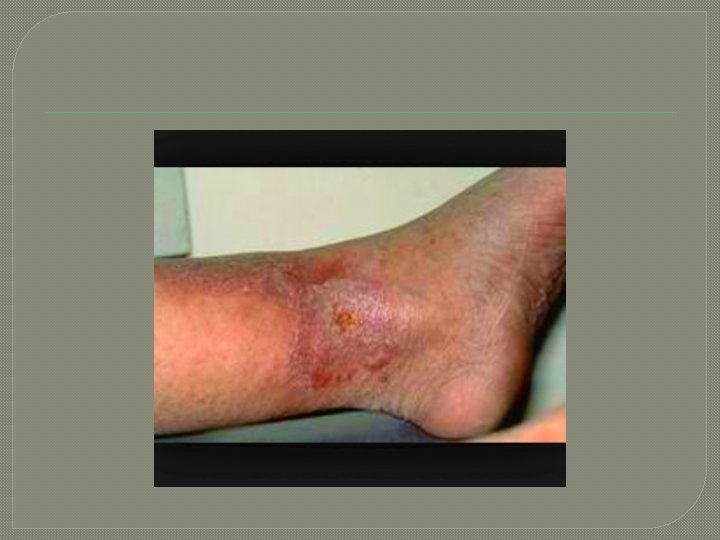
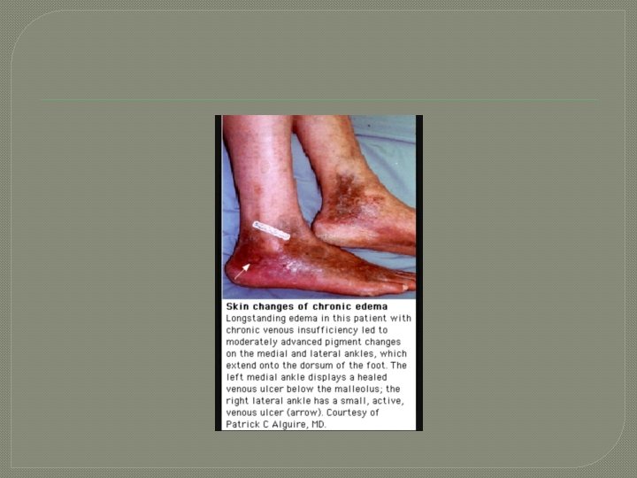
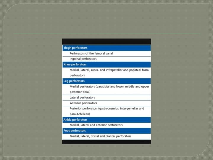
Perforators �Carry blood from superficial veins into deep veins �Posterior arch vein • Has three ankle perforators • Plays major role in development of venous stasis ulcers
Blood Flows from superficial veins (S) to the deep venous system (D)
Important perforators of the Posterior Arch Vein
Posterior arch vein
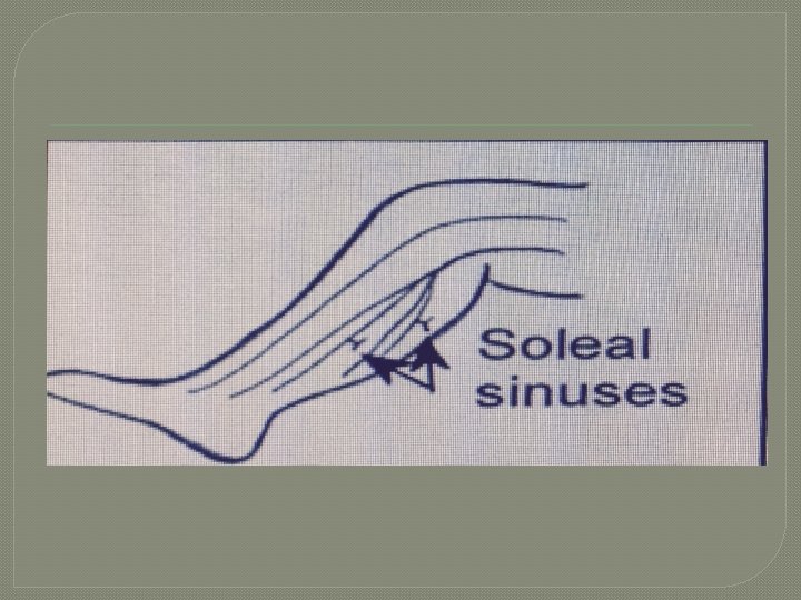
Venous Sinuses �Intracranial: • Spaces between dura mater and periosteum that drain blood into the Internal Jugular Vein �Lower extremity: • Dilated channels in soleal and gastrocnemius muscles • Drains blood into the posterior tibial vein and Peroneal Vein • Major part of calf muscle pump
Upper Extremity Veins �Deep Veins • The paired deep veins of the arm follow the corresponding arteries; called venae comitantes (corresponding veins) �Radial veins �Ulnar veins �Brachial veins
Deep Veins of the upper extremity
�Paired radial veins (Rad. V) • Formed by confluence of venules • Empties lateral hand forearm �Paired ulnar veins (Uln. V) • Formed by confluence of venules • Empties medial hand forearm
�Paired brachial veins (Bra. V) • Formed by confluence of radial and ulnar veins �Axillary veins (Ax. V) • Formed by confluence of brachial vein and basilic vein (basilic vein is in the superficial system) �Subclavian vein (Sub. V) • Formed by the confluence of axillary vein and cephalic vein (cephalic vein is superficial system
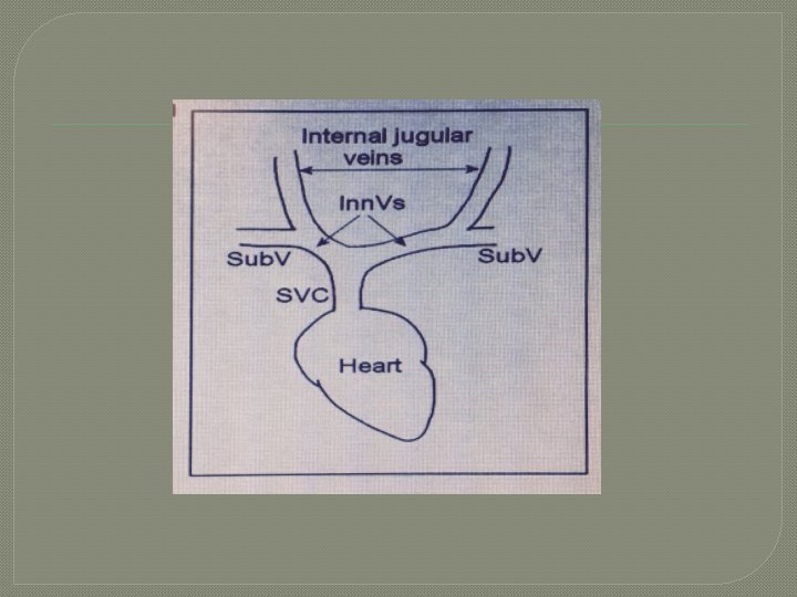
�Innominate vein (Inn. V) • Formed by confluence of subclavian vein and internal jugular (Also called brachiocephalic) �Superior vena cava (SVC) • Formed by confluence of right and left innominate veins • Carries blood into right atrium
Superficial Veins �Basilic • • Vein: Formed by the digital veins Empties medial aspect of arm Joins brachial vein to form axillary vein Can be harvested for arterial bypass conduit
Cephalic vein �Formed by digital veins �Empties lateral aspect �Joins axillary vein to form subclavian vein �Can be harvested for arterial bypass conduit
Central veins �Superior Vena Cava (SVC) • Formed by confluence of innominate veins • Drains head and upper extremity veins • Terminates in right atrium �Inferior Vena Cava (IVC) • Formed by confluence of common iliac veins • Drains lower half of body • Terminates in right atrium
Portal System �Portal vein • Formed by superior mesenteric and splenic veins • Drains abdominal part of digestive tract, pancreas, spleen, and gallbladder • Carries blood into sinusoids of liver (hepato-petal flow) • Carries approximately 80% of blood flow to the liver
�Hepatic Veins • Carries blood from the liver into IVC (flow away from the liver called hepato-fugal) �Renal veins – empties into the IVC
Structural Anatomy of Veins �Function • Thin walled, collapsible tubes that transport blood from capillaries toward heart • Carry away waste products of cellular activity • Not completely passive structures; have some element of reactivity, which may be referred to as veno-motor tone; contraction of smooth muscle cells can occur in response to stimulation of sympathetic nervous system, i. e. , temperature, exercise, stress, traume
Anatomy �Same three layers as arteries, e. g. , intima, media, adventitia (Media layer very thin) �Venous system starts at capillary level with progressive increase in size (venules smallest – vena cava – largest)
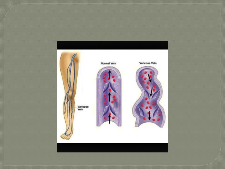
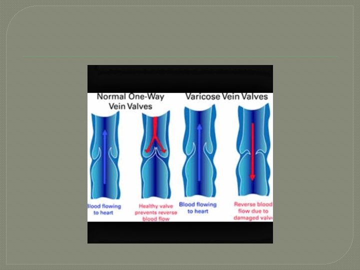
Venous Valves �Extensions of intimal layer �Bicuspid structures providing unidirectional flow �Valves of lower extremities more susceptible to disease secondary to the effects of venous thrombosis, increased ambulatory venous pressure from gravity, increased intraabdominal pressure and or venous obstruction.
Venous Valves
Veins without Valves �Soleal sinuses �External iliac vein: contains valves approximately 25% of the time �Common Iliac and internal iliac �Innominate �Superior and Inferior vena cava
Veins With Valves � Great saphenous vein has approximately 12 valves: most are below the knee � Small Saphenous: 6 -12 valves � Perforators: each contains a valve � Intrapopliteal (deep veins: 7 – 12 valves each � Popliteal and femoral: 1 -3 valves each � External iliac vein: contains valves approximately 25% of the time � Common femoral: 1 valve � Jugular Vein: 1 valve � Basilica and cephalic: 1 valve each � Variable number of UE deep veins has valves
Additional Notes: Lower extremity venous �Peroneal veins are paired �Posterior tibial veins par paired �Take note if May-Thurner Syndrome Upper extremity veins �Know medial to lateral and lateral to medial
Hepatic blood flow direction � Hepato-petal flow is blood flow into the liver � Hepato-fugal flow is blood flow away from the liver Veins with valves � Where the most of the valves are: the further away from the heart!! � The closer to the heart, the least likely for a vein to have a valve.
Homework �Textbook: Chapter 24 • Gross Anatomy of the Central Peripheral Venous systems �Pages 265 – 276 �SDMS Assignments