Venous drainage and Lymphatics of the upper limb

Venous drainage and Lymphatics of the upper limb Dr. Qudsia sultana

Why veins are important to us ?

What is this?

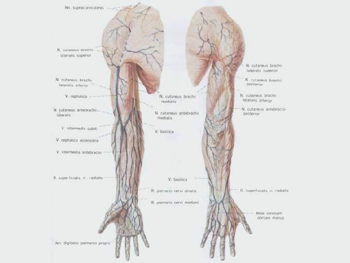
The veins of upper limb They are divided into two sets, • superficial and • deep which anastomose freely with each other.

Deep veins of the upper limb • Follow the course of arteries. • Arranged in pairs on either side of the arteries of upper limb and are called as venae commitants (except the axillary Artery).

The superficial veins of upper limb • placed immediately under the skin, in the superficial fascia. • Communicate with deep veins and finally drain in axillary vein. • They are Ø Cephalic, Ø Basilic and Ø Median cubital vein.

Dorsal venous arch • It drains the blood from back of the hand. • Formed by the union of three dorsal metacarpal veins. Which in return formed by the union of dorsal digital veins. • On its lateral side it receives, dorsal digital veins from the radial side of index finger and both the sides of the thumb.

• On its medial side , it receives dorsal digital vein from the ulnar side of the little finger.

Cephalic vein • Formed in the anatomical snuff box. • Begins –lateral side of dorsal venous arch. • winds upwards round the radial border of the forearm to its anterior surface in the cubital fossa, where it is connected to basilic vein through median cubital vein.

Cephalic vein • it then ascends subcutaneously along the lateral side of the biceps in arm.

• It lies in the groove between the pectoralis major and deltoid • It pierces the clavipectoral fascia with right angled bend to end in the axillary vein.

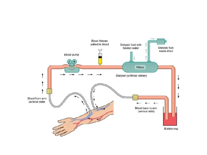
Applied anatomy • Internal arterio-venous fistulas for haemodialysis in chronic renal failure is created between cephalic vein and Radial artery.

Basilic vein • Begins in the ulnar side of the dorsal venous arch of the hand. • It runs up for some distance on the posterior surface of the ulnar side of the forearm. • Inclines forwards to the anterior surface of medial epicondyle where it is connected to the cephalic vein by median cubital vein

• It then ascends medial to biceps • perforates the deep fascia a little below the middle of the arm and • join with the venae commitantes of the brachial artery to form the axillary vein.

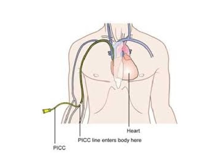
Applied • The basilic vein is used for cardiac catheterization.

Median cubital vein • It is given off from the cephalic vein about 2. 5 cms below and front of the elbow , and passes medially to join the basilic vein about 2. 5 cms above the elbow. • It rests on bicipital aponeurosis which seperates it from brachial artery and median nerve. • Receives tributaries from forearm - median antebrachial vein

As it is fixed to deep veins with perforators it is used: • Blood sampling • Blood transfusion • Intravenous injection in general, are often performed at the bend of the elbow( the median cubital vein). • Cardiac catheterization

Lymph nodes of upper limb

Introduction • Lymphatics • Lymph nodes • Applied anatomy

Lymphatics of upperlimb. • Superficial Lymphatics. • Deep Lymphatics.

Superficial Lymphatics – Collect lymph from skin and subcutaneous tissue and accompany superficial veins. – Drain into axillary nodes • Medial three fingers, medial side of the arm and forearm –along the basilic vein- Supratrochlear lymphnodes - lateral group of axillary lymph nodes. • Index and thumb finger, Lateral side of the arm and forearm – along the cephalic veininfraclavicular nodes-apical group of axillary nodes.

Deep Lymphatics • Follow the arteries – Less numerous. – Drains structures deeper to deep fascia. – End in the lateral group of axillary nodes.

. – Scattered in the fibro-fatty tissue")
Lymph nodes • Axillary Lymph Node (Pectoralis group). – Scattered in the fibro-fatty tissue of the axilla. – Five groups Ø Anterior group ØPosterior group ØLateral group ØCentral group ØApical group

1. Anterior Group: • Along the lateral thoracic vessels. (lower border of the pectoralis minor). • Receives lymph from the upper half of the anterior wall of the trunk and from the major part of the breast.
. – Along the subscapular vessels, on the posterior fold")
2. Posterior Group (Scapular group). – Along the subscapular vessels, on the posterior fold of the axilla. – Receive lymph from the posterior wall of the trunk upto the iliac crest.

3. Lateral Group: – Lateral wall of axilla, medial to the axillary vein. – Receive lymph from the medial side of upper limb.

4. Central group: – base of the axilla embedded in fat. – Receives lymph from other groups and drains into the apical group.

Applied • Intercosto brachial nerve passes through the central nodes. • If central nodes are enlarged due to ca breast – compresses the nerve – referred pain along the medial side of arm.

5. Apical/ Infraclavicular: q – Lie deep to the clavipectoral fascia along the axillary vessels. – Receive lymph from all the groups of axillary lymphnodes, – Lmphatics along the cephalic vein(thumb and its web) – upper part of the breast. – Drains into subclavian trunk

Lymph nodes • Deltopectoral nodes: – Lies in the deltopectoral groove along the cephalic vein. • Superficial cubital / supratrochlear nodes. – Lie above the medial epicondyle. – Drains the ulnar side of the hand forearm.

Lymph nodes • Deep lymph nodes: 1. Medial side of the brachial artery. 2. At the bifurcation of the brachial artery. 3. Occasionally along the arteries of the forearm.

Applied Anatomy • Supratrochlear lymphnodes are enlarged in Syphilis.

Grouping according to the location of Pectoralis minor

Rotter’s nodes

• Lymphangitis. • Lymphadenitis. • Lymphedema.

- Slides: 40