21 7 The Systemic Circuit Veins of the


- Slides: 51


21 -7 The Systemic Circuit • Veins of the Hand – Digital veins • Empty into superficial and deep palmar veins • Which interconnect to form palmar venous arches

21 -7 The Systemic Circuit • Veins of the Hand – Superficial arch empties into: • Cephalic vein • Median antebrachial vein • Basilic vein • Median cubital vein – Deep palmar veins drain into: • Radial and ulnar veins • Which fuse above elbow to form brachial vein

Figure 21 -30 The Venous Drainage of the Abdomen and Chest Median cubital Cephalic Anterior crural interosseous Basilic Ulnar Palmar venous arches Digital veins KEY Superficial veins Deep veins Radial Median antebrachial

21 -7 The Systemic Circuit • The Brachial Vein – Merges with basilic vein – To become axillary vein • Cephalic vein joins axillary vein – To form subclavian vein – Merges with external and internal jugular veins » To form brachiocephalic vein » Which enters thoracic cavity

Figure 21 -30 The Venous Drainage of the Abdomen and Chest Vertebral Internal jugular External jugular Subclavian Highest intercostal Brachiocephalic Axillary Cephalic Accessory hemiazygos Hemiazygos Brachial Intercostal veins INFERIOR VENA CAVA Basilic Phrenic veins Adrenal veins KEY Superficial veins Deep veins

21 -7 The Systemic Circuit • Veins of the Thoracic Cavity – Brachiocephalic vein receives blood from: • Vertebral vein • Internal thoracic vein • The Left and Right Brachiocephalic Veins – Merge to form the superior vena cava (SVC)

21 -7 The Systemic Circuit • Tributaries of the Superior Vena Cava – Azygos vein and hemiazygos vein, which receive blood from: 1. Intercostal veins 2. Esophageal veins 3. Veins of other mediastinal structures

Figure 21 -30 The Venous Drainage of the Abdomen and Chest SUPERIOR VENA CAVA Mediastinal veins Esophageal veins Azygos Internal thoracic Hepatic veins Renal veins Gonadal veins Lumbar veins Common iliac KEY Superficial veins Deep veins Internal iliac External iliac Medial sacral

Figure 21 -31 a Flowcharts of Circulation to the Superior and Inferior Venae Cavae Left vertebral Right internal jugular Right external jugular Right subclavian Right brachiocephalic Left internal jugular Left and right internal thoracic veins Collects blood from cranium, face, and neck Collect blood from structures of anterior thoracic wall Right axillary Mediastinal veins Veins of the right upper limb Right intercostal veins Collect blood from vertebrae and body wall KEY Superficial veins Deep veins Collect blood from the mediastinum Collects blood from cranium, spinal cord, vertebrae SUPERIOR VENA CAVA Left brachiocephalic Through highest intercostal vein RIGHT ATRIUM Azygos Esophageal veins Hemiazygos Collect blood from the eophagus Tributaries of the superior vena cava Left intercostal veins Collect blood from vertebrae and body wall

Figure 21 -31 a Flowcharts of Circulation to the Superior and Inferior Venae Cavae Left external jugular Left brachiocephalic Collects blood from neck, face, salivary glands, scalp KEY Left subclavian Left axillary Left cephalic Left basilic Collects blood from lateral surface of upper limb Collects blood from medial surface of upper limb Interconnected by median cubital vein and median antebrachial network Superficial veins Deep veins Left brachial Left radial Collects blood from forearm, wrist, and hand Radial side of forearm Left ulnar Ulnar side of forearm Venous network of wrist and hand Tributaries of the superior vena cava

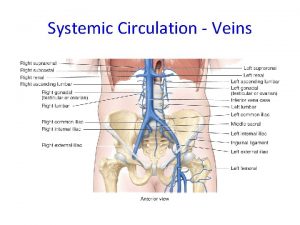
21 -7 The Systemic Circuit • The Inferior Vena Cava (IVC) – Collects blood from organs inferior to the diaphragm

21 -7 The Systemic Circuit • Veins of the Foot – Capillaries of the sole • Drain into a network of plantar veins • Which supply the plantar venous arch • Drain into deep veins of leg: – Anterior tibial vein – Posterior tibial vein – Fibular vein » All three join to become popliteal vein

21 -7 The Systemic Circuit • The Dorsal Venous Arch – Collects blood from: • Superior surface of foot • Digital veins – Drains into two superficial veins 1. Great saphenous vein (drains into femoral vein) 2. Small saphenous vein (drains into popliteal vein)

21 -7 The Systemic Circuit • The Popliteal Vein – Becomes the femoral vein • Before entering abdominal wall, receives blood from: – Great saphenous vein – Deep femoral vein – Femoral circumflex vein • Inside the pelvic cavity – Becomes the external iliac vein

21 -7 The Systemic Circuit • The External Iliac Veins – Are joined by internal iliac veins • To form right and left common iliac veins – The right and left common iliac veins » Merge to form the inferior vena cava

Figure 21 -32 a Venous Drainage from the Lower Limb External iliac Common iliac Internal iliac Gluteal Internal pudendal Lateral sacral Obturator Femoral circumflex Deep femoral Femoral Great saphenous Popliteal Small saphenous Anterior tibial Posterior tibial Fibular Dorsal venous arch Plantar venous arch Digital An anterior view

Figure 21 -32 b Venous Drainage from the Lower Limb External iliac Gluteal Internal pudendal Obturator Femoral circumflex Deep femoral Femoral Great saphenous Popliteal Small saphenous Anterior tibial Posterior tibial Fibular A posterior view

Figure 21 -32 c Venous Drainage from the Lower Limb EXTERNAL ILIAC Deep femoral Small saphenous Collects blood from the thigh Femoral Collects blood from superficial veins of the leg and foot Great saphenous KEY Superficial veins Deep veins Popliteal Fibular Collects blood from the superficial veins of the lower limb Posterior tibial Anterior tibial Extensive anastomoses interconnect veins of the ankle and foot A flowchart of venous circulation from a lower limb

21 -7 The Systemic Circuit • Major Tributaries of the Abdominal Inferior Vena Cava 1. Lumbar veins 2. Gonadal veins 3. Hepatic veins 4. Renal veins 5. Adrenal veins 6. Phrenic veins

21 -7 The Systemic Circuit • The Hepatic Portal System – Connects two capillary beds – Delivers nutrient-laden blood • From capillaries of digestive organs • To liver sinusoids for processing

21 -7 The Systemic Circuit Tributaries of the Hepatic Portal Vein 1. Inferior mesenteric vein • Drains part of large intestine 2. Splenic vein • Drains spleen, part of stomach, and pancreas 3. Superior mesenteric vein • Drains part of stomach, small intestine, and part of large intestine 4. Left and right gastric veins • Drain part of stomach 5. Cystic vein • Drains gallbladder

21 -7 The Systemic Circuit • Blood Processed in Liver – After processing in liver sinusoids (exchange vessels): • Blood collects in hepatic veins and empties into inferior vena cava

Figure 21 -33 The Hepatic Portal System Inferior vena cava Hepatic Liver Cystic Hepatic portal Superior Mesenteric Vein and Its Tributaries Pancreaticoduodenal Middle colic (from transverse colon) Right colic (ascending colon) Ileocolic (Ileum and ascending colon) Intestinal (small intestine) Pancreas

Figure 21 -33 The Hepatic Portal System Left gastric Right gastric Splenic Vein and Its Tributaries Stomach Spleen Pancreas Left gastroepiploic (stomach) Right gastroepiploic (stomach) Pancreatic Descending colon Inferior Mesenteric Vein and Its Tributaries Left colic (descending colon) Sigmoid (sigmoid colon) Superior rectal (rectum)

Figure 21 -31 b Flowcharts of Circulation to the Superior and Inferior Venae Cavae KEY RIGHT ATRIUM Superficial veins Deep veins INFERIOR VENA CAVA Hepatic veins Collect blood from the liver Gonadal veins Collect blood from the gonads (testes or ovaries) Lumbar veins Collect blood from the spinal cord and body wall Right common iliac Right external iliac Right internal iliac Blood from veins in right lower limb Phrenic veins Collect blood from the diaphragm Adrenal veins Collect blood from the adrenal glands Renal veins Collect blood from the kidneys Left common iliac Collect blood from the pelvic muscles, skin, urinary and reproductive organs of pelvic cavity Superior gluteal veins Internal pudendal veins Left external iliac Left internal iliac Obturator veins Blood from veins in left lower limb Lateral sacral veins Tributaries of the inferior vena cava

21 -8 Fetal and Maternal Circulation • Fetal and Maternal Cardiovascular Systems Promote the Exchange of Materials – Embryonic lungs and digestive tract nonfunctional – Respiratory functions and nutrition provided by placenta

21 -8 Fetal and Maternal Circulation • Placental Blood Supply – Blood flows to the placenta • Through a pair of umbilical arteries that arise from internal iliac arteries • Enters umbilical cord – Blood returns from placenta • In a single umbilical vein that drains into ductus venosus – Ductus venosus • Empties into inferior vena cava

21 -8 Fetal and Maternal Circulation • Before Birth – Fetal lungs are collapsed – O 2 provided by placental circulation

21 -8 Fetal and Maternal Circulation • Fetal Pulmonary Circulation Bypasses – Foramen ovale • Interatrial opening • Covered by valve-like flap • Directs blood from right to left atrium – Ductus arteriosus • Short vessel • Connects pulmonary and aortic trunks

21 -8 Fetal and Maternal Circulation • Cardiovascular Changes at Birth – Newborn breathes air – Lungs expand • Pulmonary vessels expand • Reduced resistance allows blood flow • Rising O 2 causes ductus arteriosus constriction • Rising left atrium pressure closes foramen ovale – Pulmonary circulation provides O 2

Figure 21 -34 a Fetal Circulation Aorta Foramen ovale (open) Ductus arteriosus (open) Pulmonary trunk Umbilical vein Liver Placenta Umbilical cord Inferior vena cava Ductus venosus Umbilical arteries Blood flow to and from the placenta in full-term fetus (before birth)

Figure 21 -34 b Fetal Circulation Ductus arteriosus (closed) Pulmonary trunk Left atrium Foramen ovale (closed) Right atrium Left ventricle Inferior vena cava Blood flow through the neonatal (newborn) heart after delivery Right ventricle

Figure 21 -35 Congenital Heart Problems Normal Heart Structure Most heart problems reflect deviations from the normal formation of the heart and its connections to the great vessels.

21 -8 Fetal and Maternal Circulation • Patent Foramen Ovale and Patent Ductus Arteriosus – In patent (open) foramen ovale blood recirculates through pulmonary circuit instead of entering left ventricle • The movement, driven by relatively high systemic pressure, is a “left-to-right shunt” • Arterial oxygen content is normal, but left ventricle must work much harder than usual to provide adequate blood flow through systemic circuit

21 -8 Fetal and Maternal Circulation • Patent Foramen Ovale and Patent Ductus Arteriosus – Pressures rise in the pulmonary circuit • If pulmonary pressures rise enough, they may force blood into systemic circuit through ductus arteriosus • A patent ductus arteriosus creates a “right-to-left shunt” • Because circulating blood is not adequately oxygenated, it develops deep red color • Skin develops blue tones typical of cyanosis and infant is known as a “blue baby”

Figure 21 -35 Congenital Heart Problems Patent ductus arteriosus Patent foramen ovale Patent Foramen Ovale and Patent Ductus Arteriosus If the foramen ovale remains open, or patent, blood recirculates through the pulmonary circuit instead of entering the left ventricle. The movement, driven by the relatively high systemic pressure, is called a “left-to-right shunt. ” Arterial oxygen content is normal, but the left ventricle must work much harder than usual to provide adequate blood flow through the systemic circuit. Hence, pressures rise in the pulmonary circuit. If the pulmonary pressures rise enough, they may force blood into the systemic circuit through the ductus arteriosus. This condition—a patent ductus arteriosus— creates a “right-to-left shunt. ” Because the circulating blood is not adequately oxygenated, it develops a deep red color. The skin then develops the blue tones typical of cyanosis and the infant is known as a “blue baby. ”

21 -8 Fetal and Maternal Circulation • Tetralogy of Fallot – Complex group of heart and circulatory defects that affect 0. 10 percent of newborn infants 1. Pulmonary trunk is abnormally narrow (pulmonary stenosis) 2. Interventricular septum is incomplete 3. Aorta originates where interventricular septum normally ends 4. Right ventricle is enlarged and both ventricles thicken in response to increased workload

Figure 21 -35 Congenital Heart Problems Patent ductus arteriosus Pulmonary stenosis Ventricular septal defect Enlarged right ventricle Tetralogy of Fallot The tetralogy of Fallot (fa-LO) is ¯a complex group of heart and circulatory defects that affect 0. 10 percent of newborn infants. In this condition, (1) the pulmonary trunk is abnormally narrow (pulmonary stenosis), (2) the interventricular septum is incomplete, (3) the aorta originates where the interventricular septum normally ends, and (4) the right ventricle is enlarged and both ventricles thicken in response to the increased workload.

21 -8 Fetal and Maternal Circulation • Ventricular Septal Defect – Openings in interventricular septum that separate right and left ventricles – The most common congenital heart problems, affecting 0. 12 percent of newborns – Opening between the two ventricles has an effect similar to a connection between the atria • When more powerful left ventricle beats, it ejects blood into right ventricle and pulmonary circuit

Figure 21 -35 Congenital Heart Problems Ventricular Septal Defect Ventricular septal defect Ventricular septum Ventricular septal defects are openings in the interventricular septum that separate the right and left ventricles. These defects are the most common congenital heart problems, affecting 0. 12 percent of newborns. The opening between the two ventricles has an effect similar to a connection between the atria: When the more powerful left ventricle beats, it ejects blood into the right ventricle and pulmonary circuit.

21 -8 Fetal and Maternal Circulation • Atrioventricular Septal Defect – Both the atria and ventricles are incompletely separated • Results are quite variable, depending on extent of defect and effects on atrioventricular valves • This type of defect most commonly affects infants with Down’s syndrome, a disorder caused by the presence of an extra copy of chromosome 21

Figure 21 -35 Congenital Heart Problems Atrioventricular Septal Defect Atrial defect Ventricular defect In an atriovenricular septal defect, both the atria and ventricles are incompletely separated. The results are quite variable, depending on the extent of the defect and the effects on the atrioventricular valves. This type of defect most commonly affects infants with Down’s syndrome, a disorder caused by the presence of an extra copy of chromosome 21.

21 -8 Fetal and Maternal Circulation • Transposition of Great Vessels – The aorta is connected to right ventricle instead of to left ventricle – The pulmonary artery is connected to left ventricle instead of right ventricle – This malformation affects 0. 05 percent of newborn infants

Figure 21 -35 Congenital Heart Problems Patent ductus arteriosus Aorta Pulmonary trunk Transposition of the Great Vessels In the transposition of great vessels, the aorta is connected to the right ventricle instead of to the left ventricle, and the pulmonary artery is connected to the left ventricle instead of the right ventricle. This malformation affects 0. 05 percent of newborn infants.

21 -9 Effects of Aging and the Cardiovascular System • Cardiovascular Capabilities Decline with Age • Age-related changes occur in: – Blood – Heart – Blood vessels

21 -9 Effects of Aging and the Cardiovascular System • Three Age-Related Changes in Blood 1. Decreased hematocrit 2. Peripheral blockage by blood clot (thrombus) 3. Pooling of blood in legs • Due to venous valve deterioration

21 -9 Effects of Aging and the Cardiovascular System • Five Age-Related Changes in the Heart 1. Reduced maximum cardiac output 2. Changes in nodal and conducting cells 3. Reduced elasticity of cardiac (fibrous) skeleton 4. Progressive atherosclerosis 5. Replacement of damaged cardiac muscle cells by scar tissue

21 -9 Effects of Aging and the Cardiovascular System • Three Age-Related Changes in Blood Vessels 1. Arteries become less elastic • Pressure change can cause aneurysm 2. Calcium deposits on vessel walls • Can cause stroke or infarction 3. Thrombi can form • At atherosclerotic plaques

21 -9 Cardiovascular System Integration • Many Categories of Cardiovascular Disorders – Disorders may: • Affect all cells and systems • Be structural or functional • Result from disease or trauma

Figure 21 -36 System Integrator: The Cardiovascular System Delivers immune system cells to injury sites; clotting response seals breaks in skin surface; carries away toxins from sites of infection; provides heat Provides calcium needed for normal cardiac muscle contraction; protects blood cells developing in red bone marrow Transports calcium and phosphate for bone deposition; delivers EPO to red bone marrow, parathyroid hormone, and calcitonin to osteoblasts and osteoclasts Skeletal muscle contractions assist in moving blood through veins; protects superficial blood vessels, especially in neck and limbs Delivers oxygen and nutrients, removes carbon dioxide, lactic acid, and heat during skeletal muscle activity Controls patterns of circulation in peripheral tissues; modifies heart rate and regulates blood pressure; releases ADH Endothelial cells maintain blood—brain barrier; helps generate CSF Erythropoietin regulates production of RBCs; several hormones elevate blood pressure; epinephrine stimulates cardiac muscle, elevating heart rate and contractile force Distributes hormones throughout the body; heart secretes ANP and BNP Skeletal Page 275 Stimulation of mast cells produces localized changes in blood flow and capillary permeability Integumentary Page 165 Body System Muscular Page 369 Cardiovascular System Nervous Page 543 SYSTEM INTEGRATOR Cardiovascular System Endocrine Page 632 Endocrine Nervous Muscular Skeletal Integumentary Body System Respiratory Page 857 Digestive Page 910 Urinary Page 992 The most extensive communication occurs between the cardiovascular and lymphatic systems. Not only are the two systems physically interconnected, but cells of the lymphatic system also move from one part of the body to another within the vessels of the cardiovascular system. We examine the lymphatic system in detail, including its role in the immune response, in the next chapter. Reproductive Page 1072 The section on vessel distribution demonstrated the extent of the anatomical connections between the cardiovascular system and other organ systems. This figure summarizes some of the physiological relationships involved. Lymphatic Page 807 The CARDIOVASCULAR System
 Pearson mastering
 Blood circulation system diagram
 Systemic veins
 Major systemic arteries labeled
 Systemic circuit
 Systemic circuit
 Phet circuit construction kit
 Venn diagram of series and parallel circuit
 In series vs in parallel
 Advantages of parallel circuits over series circuits
 Series circuit vs parallel circuit
 What are complete and incomplete circuits
 Types of circuit
 Short circuit series
 Signs of phlebitis and infiltration
 Sfl
Sfl Systemic social work
 Systemic functional grammar
 Overview of the major systemic arteries
 Figure 11-7 veins labeled
 Systemic lupus erythematosus
 Schistosomaiasis
 Identify sources of systemic fluoride
 Zoonotic
 Portal circulation
 Overview of the major systemic arteries
 Indications for extraction
 Systemic effect of inflammation
 Contoh masalah privat dan publik
 Factors affecting mobility and immobility
 Arteries of the systemic circulation
 Systemic error
 Local factor affecting wound healing
 Systemic review
 Arterial pressure points
 Plants are sessile
 Smcqs
Smcqs Ism and ssm
 Systemic family therapy techniques
 Porto systemic anastomosis
 Arteries of the systemic circulation
 Correctly label the following major systemic arteries
 Bp = co x svr
 Sofronio agustin
 Systemic review
 Ion storm
 Arteries of the systemic circulation
 Systemic examination of respiratory system
 Systemic obstacles examples
 Pulmonary systemic
 Major arteries of the ascending aorta and aortic arch
 Systemic pathology exam questions pdf
