ANATOMY AND PHYSIOLOGY OF SALIVARY GLAND Dr Nayana


 • Superficial •")


 Superficial layer – thick adherent to parotid")


 External Carotid,")


 Sympathetic")
 • The Submandibular duct (Wharton’s duct) exits the medial surface")


 • Myoepithelial cells")

,")


- Slides: 44
ANATOMY AND PHYSIOLOGY OF SALIVARY GLAND Dr. Nayana V G Senior Resident
Explain the anatomy of parotid gland Discuss the anatomy of submandibular gland Briefing the anatomy of sublingual and minor salivary glands Discuss the micro anatomy of salivary secretory unit Explain about the physiology of salivary glands
Three major, paired salivary glands produce the majority of saliva: The parotid, The submandibular The sublingual glands. 600 -1, 000 minor salivary glands line the oral cavity and oropharynx
EMBRYOLOGY • The major salivary glands develop from the 6 th-8 th weeks of gestation as outpouchings of oral ectoderm into the surrounding mesenchyme. • However, the parotid gland is the last to become encapsulated, after the lymphatics develop, resulting in its unique anatomy with entrapment of lymphatics in the parenchyma of the gland - warthin’s tumors and lymphoepithelial cysts within the parotid gland. • Minor salivary glands arise from oral ectoderm and nasopharyngeal endoderm
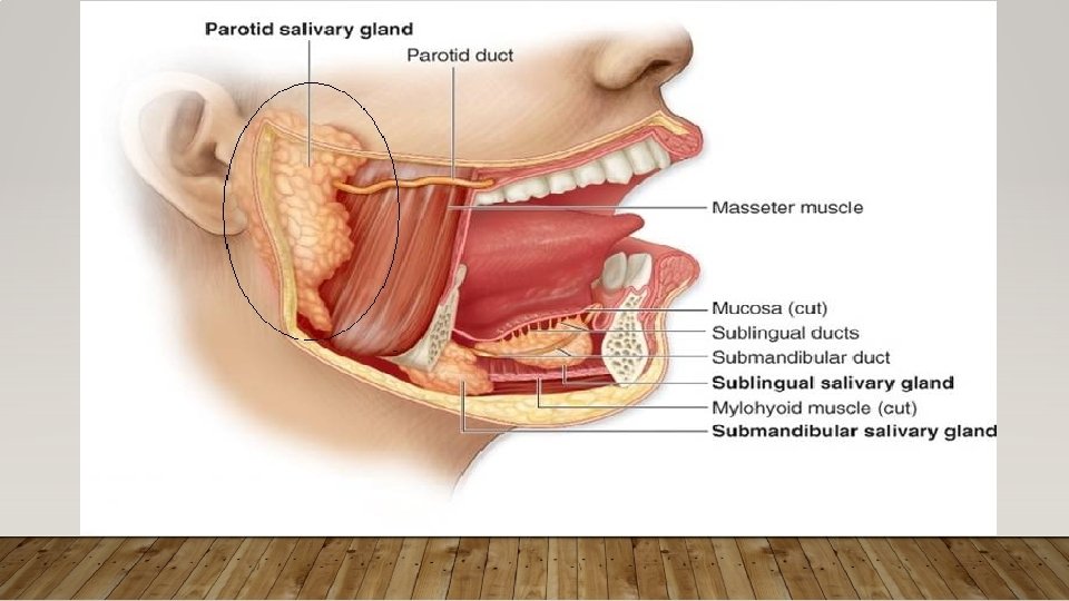
THE PAROTID GLAND • Largest salivary gland • It resembles a 3 sided pyramid the apex is directed downwards • The average weight of a parotid gland is 15 g • Below EAM between ramus of mandible and sternocleidomastoid
The gland has 4 surfaces • Superior (base of the pyramid) • Superficial • Antero medial • Postero medial The surfaces are separated by 3 borders anterior , posterior, medial
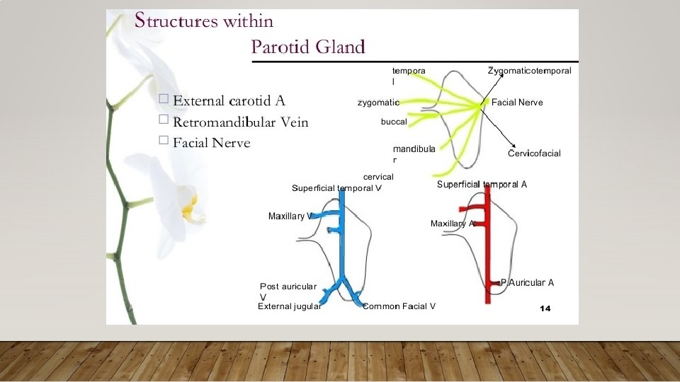
IT LIES IN THE PAROTID COMPARTMENT, A TRIANGULAR SPACE WHICH ALSO 1. Contains CN VII and its branches, sensory and autonomic nerves, 2. The External Carotid artery and its branches, 3. The Retromandibular (Posterior Facial) vein, and 4. Parotid lymphatics.
• 80% of the gland overlies the Masseter and mandible. The remaining 20% of the gland (the retromandibular portion) extends medially through the stylomandibular tunnel
• The tail of the parotid overlies the upper ¼th of the Sternocleidomastoid muscle and extends toward the mastoid process. • Patients with parotitis frequently have pain with mastication • An accessory Parotid gland duct are noted in 20% of people
FASCIA The Parotid fascia consists of 1) Superficial layer – thick adherent to parotid and attached to Zygoma above 2) Deep layer – thin attached to styloid process tympanic plate and angle of mandible and it forms the Stylo mandibular ligament separating the Parotid and Submandibular glands.
COMPARTMENTS The most superficial portion of the Parotid makes up the neural compartment, containing (a) CN VII, (b) the Auriculotemporal nerve, and (C) the Great Auricular nerve.
1. FACIAL NERVE • Cranial Nerve VII is intimately associated with the parotid gland, dividing it into 2 surgical zones (the superficial and deep lobes). • After exiting the stylomastoid foramen, CN VII turns laterally to enter the Parotid gland at its posterior margin. The nerve then branches at the Pes Anserinus (goose’s foot) approximately 1. 3 cm from the stylomastoid foramen. • Followed by 5 terminal branches: 1) Temporal 2) Zygomatic 3) Buccal 4) Marginal Mandibular 5) Cervical
2. GREATER AURICULAR NERVE • The Great Auricular nerve passes from deep to superficial around the posterior border of the SCM • It is the first nerve encountered in surgery, lateral to the Parotid fascia and deep to the Platysma. The nerve is routinely divided during parotidectomy
3. AURICULOTEMPORAL NERVE • The Auriculotemporal nerve, a branch of V-3, runs anterior to the EAM • when this nerve is injured intraoperatively, aberrant parasympathetic innervation to the skin results in Frey’s Syndrome (i. e. , gustatory sweating). This nerve may be resected intentionally to avoid Frey’s Syndrome
VENOUS COMPARTMENT Sits in the middle of the Parotid, deep to CN VII. Venous drainage is provided by the Retromandibular vein, which lies deep to the Facial nerve. This vein emerges at the inferior pole of the Parotid gland.
ARTERIAL COMPARTMENT sits in the deep portion of the gland ( a) External Carotid, ( b) Internal Maxillary ( c) Superficial Temporal arteries. Arterial supply - Transverse Facial artery from the Superficial Temporal artery It providing blood to the Parotid gland, Stensen’s duct, and the Masseter muscle. This artery runs between the zygomatic arch and Stensen’s duct
PATEY’S FACIO-VENOUS PLANE Within the gland the facial nerve and its branches and retro-mandibular vein lie in one plane. In this plane the gland can be split sagittally into two parts, superficial and deep parts without injuring the nerve.
LYMPHATIC DRAINAGE AND AUTONOMIC NERVE SUPPLY Lymphatic drainage is unique in the Parotid with Paraparotid and Intraparotid nodes. The Parotid lymphatics drain into the superficial and deep cervical lymph nodes Parasympathetic nerve supply : Sympathetic: superior cervical ganglion
STENSEN’S DUCT Stensen’s duct is 5 cm long. Arises from the anterior part of the gland runs over the masseter and Pierces Buccinator at 2 nd Molar Clinical examination of the parotid gland should include examination of the duct orifice opposite the upper 2 nd molar for signs of inflammation, and palpated for stone
SUBMANDIBULAR SALIVARY GLAND • This gland lies in the submandibular triangle • The gland forms a ‘C’ around the anterior margin of the Mylohyoid muscle, which divides the Submandibular gland into a superficial and deep lobe. • Submandibular gland is invested in its own capsule, which is continuous with the superficial layer of deep cervical fascia.
SUPERFICIAL AND DEEP RELATIONS
BLOOD AND LYMPHATIC SUPPLY OF SUBMANDIBULAR GLAND • Arterial supply to the Submandibular gland comes from the Submental branch of the Facial artery • The Facial vein courses over the lateral surface of the Submandibular gland. • The Facial artery and vein are the first blood vessels encountered when resecting the Submandibular glands as they cross superficially over the inferior border of the mandible. • Venous drainage is provided by the Anterior Facial vein, which lies deep to the Marginal Mandibular branch of CN VII. • Lymphatic drainage goes to the deep cervical and jugular chains of nodes.
NERVE SUPPLY Innervation to the Submandibular gland derives from 2 important sources: 1) Sympathetic innervation from the Superior Cervical ganglion via plexus on the walls of the facial & the Lingual artery. 2) Parasympathetic supply
WHARTON’S DUCT (5 CM) • The Submandibular duct (Wharton’s duct) exits the medial surface of the deep gland runs between the Mylohyoid (lateral) and Hyoglossus muscles and on to the Genioglossus muscle. • Wharton’s duct empties into the intraoral cavity lateral to the lingual frenulum on the anterior floor of mouth.
• . The Lingual nerve wraps around Wharton’s duct • CN XII parallels the Submandibular duct, running just inferior to it • The identification of CN XII, the Lingual nerve, and Wharton’s duct is absolutely essential prior to resection of the gland.
A stone in the submandibular duct can be felt bimanually in the floor of the mouth and can be seen if large The presence of LN adherent to the gland makes removal of the gland part of block neck dissection Submandibular LN are adherent to the gland partly between it and the mandible
SUBLINGUAL SALIVARY GLAND This is the smallest of the major salivary glands The almond shaped gland lies just deep to the floor of mouth mucosa between the mandible and genioglossus muscle. It is bounded inferiorly by the mylohyoid muscle. No true fascial capsule. Lacks a single dominant duct. 10 small ducts (the ducts of rivinus) Open along the sublingual fold on the floor of mouth. ,
1. Sympathetic innervation from the cervical chain ganglia via the Facial artery 2. Parasympathetic • Arterial supply to this gland is two-fold: 1) The Sublingual branch of the Lingual artery, and 2) The Submental branch of the Facial artery Venous drainage reflects the arterial supply. Lymphatic drainage goes to the Submandibular nodes.
MINOR SALIVARY GLANDS • Unlike the major salivary glands, the minor salivary glands lack a branching network of draining ducts. Instead, each salivary unit has its own simple duct. The minor salivary glands are concentrated in • Buccal, Labial, Palatal and Lingual regions. Most of the minor glands receive parasympathetic innervation from the Lingual nerve, except for the minor glands of the palate, which receive their parasympathetic fibers from the Palatine nerves, fed by the Sphenopalatine ganglion
MICRO ANATOMY OF THE SECRETORY UNIT • Acinus (serous, mucous, mixed) • Myoepithelial cells • Intercalated duct • Striated duct • Excretory duct
All salivary acinar cells contain secretory granules; in serous glands, these granules contain amylase, and in mucous glands, these granules contain mucin. Acini, responsible for producing the primary secretion, are divided into 3 types: 1) Serous (protein-secreting) 2) Mucous (mucin-secreting) 3) Mixed
Myoepithelial cells send numerous processes around the acini and proximal ductal system (intercalated duct), moving secretions toward the excretory duct. The lumen of the acinus is continuous with the ductal system, made up of (from proximal to distal) the intercalated duct the striated duct the excretory duct
• The Parotid gland is a purely serous salivary gland. • The Submandibular gland is mixed, but predominantly serous. Approximately 10% of its acini are mucinous. • The Sublingual gland is mixed, but predominantly mucous. • Of the major salivary glands, the Sublingual gland utilizes a simple system of transport, whereas the Parotid and Submandibular glands involve elaborate networks.
AUTONOMIC INNERVATION Parasympathetic • Abundant, watery saliva • Amylase down Sympathetic • Scant, viscous saliva • Amylase up
MECHANISM OF SALIVA SECRETION • Afferent stimulatory pathway : psychic, taste, smell and inter organ stimuli • Afferent neural pathway Central control of salivation • Efferent neural pathway
PRODUCTION OF SALIVA The production of saliva is an active process occurring in 2 phases: 1) Primary secretion – occurs in the acinar cells. This results in a product similar in composition and osmolality to plasma. 2) Ductal secretion – results in a hypotonic salivary fluid. It also results in decreased sodium and increased potassium in the end product.
Saliva is composed of 99. 5% water in addition to proteins, glycoproteins, and electrolytes. • Saliva is high in potassium (7 x plasma), bicarbonate (3 x plasma), calcium, phosphorous, chloride, thiocyanate, and urea. • Saliva is low in sodium (1/10 x plasma). The normal p. H of saliva is 5. 6 -7
SALIVARY FLOW • The average volume of saliva secreted in a 24 hour period is 11. 5 liters most of which is secreted during meals. • Salivary flow rate from the minor salivary glands is independent of stimulation, constituting 7 -8% of total salivary output.
• In the UNSTIMULATED state the relative contribution of the major salivary glands is as follows: 1) Submandibular gland=69% 2) Parotid gland=26% 3) Sublingual gland=5% Though the Sublingual glands and minor salivary glands contribute only about 10% of all saliva, together they produce the majority of mucous and are critical in maintaining the mucin layer over the oral mucosa.
FUNCTION OF SALIVA v Moistens oral mucosa v Moistens & cools food v v Digestion (Amylase, Lipase) v Antibacterial (Lysozyme, Ig. A, Medium for Peroxidase) dissolved food v Mineralization. v Buffer (HCO 3
SUMMARY • Exocrine glands • 3 major and minor salivary glands • Parotid is the largest • Serous gland produce saliva rich in amylase mucous glands produces mucin • Mediated by parasympathetic stimulation
Reference. . 1. Disease of Ear, Nose, Throat &Head and Neck surgery – PL Dhingra, Shruthi Dhingra Acknowledgement Images from google images and text book(Disease of Ear, Nose, Throat &Head and Neck surgery – PL Dhingra, Shruthi Dhingra)