Cardiovascular Block Physiology Capillary Circulation Intended learning outcomes

Cardiovascular Block Physiology Capillary Circulation
 After reviewing the Power. Point presentation and the associated learning")
Intended learning outcomes (ILOs) After reviewing the Power. Point presentation and the associated learning recourses, the student should be able to: Outline the parts of the microcirculation, and list types of blood capillaries and differentiate between them. Explain regulation of flow in the capillary beds. Compare and contrast diffusion and filtration. State Starling forces acting on the capillary wall. Define edema, state its causes and discuss its mechanisms. Describe the role of the microcirculation in temperature regulation.

Learning Resources Guyton and Hall, Textbook of Medical Physiology; 13 th Edition; Unit IV-Chapter 16. Linda Costanzo, Physiology, 5 th Edition; Chapter 4. Ganong’s Review of Medical Physiology; 25 th Edition; Section V; Chapter 31.

The microcirculation q The microcirculation refers to the microscopic divisions of the vascular system that function to bring exchange of materials (nutrients and oxygen) between the blood and various body tissues. q it also functions to drain waste products from tissues to blood. q It also plays a role in temperature regulation. q The capillary bed is composed of a series of blood vessels through which adequate exchange of gases, nutrients and wastes take place. q How much is the blood flow velocity in the capillaries?


Parts of the microcirculation q The capillary bed is composed of a series of blood vessels through which adequate exchange of gases, nutrients and waste products take place.

Capillary bed: Arterioles q Arterioles are resistance blood vessels which may restrict flow to capillaries or allow greater flow. q Arteriolar diameter is generally 5 – 100 micrometers. q They have a thick smooth muscle layer and endothelial lining. q They have precapillary sphincters which may shut off flow to capillary q When the sphincters are open → nutritional flow. q When the sphincter are closed → non-nutritional flow or shunt flow.

Capillary bed: Metarterioles and A-V shunt q Metarterioles are intermediate resistance blood vessels found in some capillary beds q They arise from arterioles and give rise to capillaries. q They have a diameter of 10 -20 micrometer. q They can connect with venules and/ or supply capillaries. q They have pre-capillary sphincters, which can shut off flow. q An A-V shunt is a small vessel with direct connection between an arteriole and a venule. q An A-V shunt allows bypass of capillaries if precapillary sphincters are closed. q In this case, the flow is 100% nonnutritional.

q Capillaries are blood vessels where exchange of gases, nutrients and wastes occur. q Structure: They are small blood vessels ≈ 0. 5 -1 mm long, and ≈ 0. 01 mm (10 micrometers) diameter. There are no smooth muscle cells in the capillary walls. The walls of the capillaries are only a single endothelial cell thick. This permits rapid diffusion of water, solutes and gases. Capillary density in organs or tissues is indicative of the metabolic activity • High density in skeletal muscle, heart, brain, liver and kidney. • Low density in skin and cartilage. q Function: Sites of exchange between blood and tissues. The arterial system delivers blood to > l billion capillaries throughout the body. Total capillary surface area=1000 m 2. Capillary bed: Capillaries

Capillary bed: Capillaries q Capillaries diameters are not uniform. q Some capillaries have diameter less than that of a red blood cell → temporary deformation of blood cell as they pass through these capillaries. q It is important that sufficient blood moves through capillaries for oxygen demand in tissue q Failure to do this results in circulatory failure or shock.

Types of body capillaries Three structural types: § Continuous capillaries § Fenestrated capillaries § Sinusoids

Types of body capillaries

Continuous Capillaries These capillaries are present in most body tissues, e. g. , muscle, lung, and adipose tissue. q They have continuous endothelial lining and adjacent endothelial cells are closely joined together by tight junctions. q There are thin intercellular slits (clefts) ≈ 2 -10 nm in width in-between the endothelial cells that allow diffusion and bulk flow of water and water soluble small ions (e. g. , Na+, Cl-, and glucose). q Intercellular slits (clefts) are gaps in tight junctions. q Tight junctions are continuous in brain and do not allow passage of water soluble molecules. They form part of the bloodbrain barrier. q Continuous type of capillary found in skeletal muscle. Blood brain barrier

Continuous Capillaries

Fenestrated Capillaries n These are found in the kidney glomeruli, small intestine, and endocrine glands. n Some endothelial cells have wide pores (fenestrations). n They are very permeable : they allow even large substances to pass but not plasma proteins. Fenestrated type of capillary

Fenestrated Capillaries

Sinusoids; discontinuous capillaries n In these capillaries the endothelial cell are widely spaced. n These have large irregular lumens slows blood flow. n Few tight junctions allow large molecules (e. g. , proteins) to pass through. n Located in liver, spleen, bone marrow, lymphoid tissue, some endocrine glands.

Regulation of flow in capillary beds q The arterioles and the precapillary sphincters function as control valves in the tissue they feed: q During the “fight or flight” response, flow to non-essential organs (kidney, skin, etc) is clamped off → increased flow to skeletal muscle. q Metabolic waste products act as vasodilators to relax precapillary sphincters. q Vasomotion: intermittent flow through capillary, in response to altering metabolic needs.

Mechanisms of capillary exchange Transport of substances across the capillary wall occurs by 3 major mechanisms: q Diffusion (according to concentration gradient). q Filtration (according to pressure gradient). q Transcytosis (vesicular transport).

Mechanisms of capillary exchange q Capillary permeability is not the same in all tissues. q It is specialized for different tissues: q Liver sinusoids have discontinuous endothelium → higher permeability → allows exchange of solutes and proteins. q Blood brain barrier capillaries: very low permeability due to tight junctions. q Kidney and intestinal capillaries contain fenestrations (pores) → increased permeability. q Clefts or pores only cover 0. 02% of capillary surface area.

Diffusion q This is the process of movement of particles between blood in the capillaries and the interstitium across the capillary wall according to their concentration (chemical) gradients. q It is the major process by which most nutritional substances and waste products cross the capillary membrane.
 q")
q Capillary Diffusion using modified Fick’s Law: J = P (CB – CT) q P: permeability coefficient; CB: conc. of solute in the blood; CT : conc. of solute in the tissue. P is affected by lipid solubility and molecular diameter. q Lipid solubility influences speed of diffusion: q O 2 and CO 2 are highly lipid soluble and permeable. q Molecular diameter impacts solute diffusion: q It must be small enough for the clefts and pores in most tissues. q Glucose and Na. Cl have relatively high permeability, but albumin does not). q In liver, proteins (such as albumin) can move through the discontinuous endothelium easily. q Concentration gradient impacts permeability. substances are exchanged from a high concentration to a low concentration. Diffusion
 q This is an active process by which large molecules can")
Transcytosis (vesicular transport) q This is an active process by which large molecules can be transported across the capillary membrane. q It includes the formation of vesicles from the endothelial membrane to surround the required particle (endocytosis). The vesicle separates and migrates across the cell to release its contents to the other side (exocytosis). e. g. Peptide hormones are moved this way

Filtration q Filtration is the process by which plasma and its dissolved crystalloids (electrolytes and glucose) can filter across the capillary according to pressure gradient. q Filtration is determined by Starling’s forces. q It is called bulk flow as movement of water drags along with it dissolved substance to which the membrane is permeable. q Although the amount of materials exchanged by filtration are small compared to diffusion (rate of diffusion is 4000 times as the rate of filtration-reabsorption), however, filtration aids diffusion by keeping the fluid across the capillary membrane in a state of continuous motion.

Transcapillary fluid dynamics Filtration forces Four pressures are involved in fluid exchange in the capillary bed: Starling forces: Capillary hydrostatic pressure tends to force fluid out. Plasma colloid osmotic pressure: sucks fluid back in. Interstitial fluid pressure. Interstitial fluid colloid osmotic pressure: sucks fluid out.
 is needed to push substances through slits and")
Hydrostatic pressure q Hydrostatic Pressure (Pc) is needed to push substances through slits and pores. q Pc is equivalent to Plat q ↑resistance in arterioles through the action of norepiniphrine and epinephrine on precapillary sphincter → ↓Pc and ↑ the pressure before the arterioles. q This is not true for capillary beds which have predominately beta-adrenergic receptors in the capillary sphincters: these would be dilated or have reduced resistance. q ↓resistance in arterioles → ↑Pc. q Pc varies from tissue to tissue. In the renal glomeruli, it is about 60 mm Hg (filtering capillaries). In contrast, it is only about 8 -10 mm Hg in those of the intestine and the pulmonary circulation absorptive capillaries). q Pc varies as blood moves along the capillary. Pc is highest at the arteriolar end and lowest at venular end of capillary. q ↑Pc → ↑filtration; can lead to edema. q The interstitial fluid generally has very low hydrostatic pressure (P if), usually given a value near zero q Things which increase Pif, such as support hose, can reduce edema in legs.
 q Oncotic or osmotic pressure in the capillaries is")
Oncotic or Osmotic Pressure (π) q Oncotic or osmotic pressure in the capillaries is a pressure which is mainly related to the presence of plasma proteins which can’t cross the capillary wall. q Albumin contributes to 65% of the osmotic pressure, whereas globulin contributes to only 15%. q However, an ↑solute concentration → an↑osmotic pressure. q Proteins in the surrounding interstitial fluid contribute to the tissue osmotic pressure (πif). q The πc q Leads to reabsorption of water near the venular end of the capillaries, and thus recovers blood volume. q During the process of filtration and reabsorption, there is generally a lose of about 15% of the blood volume. q This is recovered by the lymphatic system.
")
Transcapillary fluid dynamics Arterial end of capillary Venous end of capillary Blood pressure (+40) Osmotic pressure of plasma (- 28) Osmotic pressure of interstitial fluid (+3) Blood pressure (+15) Osmotic pressure of plasma (- 28) Osmotic pressure of interstitial fluid (+3) (40 + 3) - 28 = +15 (15 + 3) - 28 = - 10 Net filtration Net absorption

Transcapillary fluid dynamics

Starling’s equation for net fluid flow across capillaries q Fluid movement = K (filtration forces – reabsorption forces). q Fluid movement = K [(Pc + πIF) – (πc + PIF)]. q K is determined by: q Blood viscosity. q Wall thickness. q Surface area. q Permeability of capillary wall to water. q K is high in liver and kidneys, while it is low in the blood-brain barrier.

At arteriolar end At venular end ↓ ↓ Net reabsorption Net filtration Via lymphatics Filtration Reabsorption Normally the amount filtered slightly exceeds the amount reabsorbed and is eventually returned to the circulation via the lymphatics Using Starling equation, we can calculate fluid movement at the arteriolar and venular ends of the capillary: Filtration: (arterial end) 20 ml fluid/min reabsorption: (venous end) 18 ml fluid/min Hence: there is net filtration of about 2 ml/min for entire body: This is removed by the lymphatics (prevent oedema)

Clinical significance of capillary filtration q In blood loss, vasoconstriction of arterioles decrease capillary hydrostatic pressure. Hence osmotic pressure of plasma proteins favours absorption of interstitial fluid blood volume. q In congestive heart failure, venous pressure rises build-up of blood in capillaries capillary hydrostatic pressure filtration oedema. q In hypoproteinemia (e. g. , starvation, liver disease) plasma protein colloid osmotic pressure loss of fluid from capillaries oedema. q In inflammation: the gaps between the endothelial cells increase because of the inflammatory mediators movement of proteins into the interstitium oedema.

Circulation of ECF Lymph

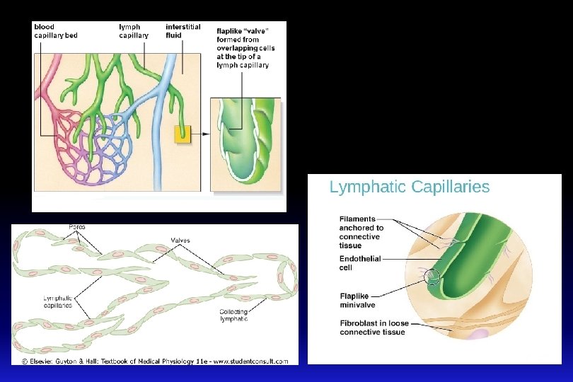
q Theses are blind sacs that collect excess tissue fluid. q They consist of simple squamous epithelium (endothelium). q Cells overlap to form oneway valves within lumen. q The gaps between the endothelial cells are very large. They lack tight junctions. q They possess fine filaments which anchor them to the connective tissue q With contractions, these filaments can pull on the endothelial cells, and thus allowing protein, large particles or cells to enter lymph system. Lymphatic vessels: lymphatic capillaries

§ Lymphatics: These originate as lymph capillaries that unite to form larger vessels - They resemble veins in structure but with thinner walls, less muscle, less connective tissue, and more valves. - They connect to lymph nodes at various intervals. § Lymphatic trunks: These are formed by the union of lymphatics. They carry lymph to lymphatic ducts. § Lymphatic ducts: these are formed by the union of lymphatic trunks. They empty into large veins (right and left subclavian veins) just before they join the superior vena cava. Lymphatic vessels: continued

Lymph Circulation n Lymph moves along pressure gradient; about 2 -4 liters/day. Valves in the lymph vessels keep flow moving in one direction. The mechanisms that may contribute to pressure gradient are: – “milking” by skeletal muscle (contraction of skeletal muscle puts pressure on lymphatic to move fluid forward). – pressure changes during breathing (inspiration lowers pressure in thoracic cavity and increases pressure in abdominal cavity). – Pulsating of neighboring elastic arteries. – contraction of smooth muscle in walls of larger lymphatic vessels and ducts.
 n neutral")
Lymph n Protein: < plasma n Lipids: n cholesterol and phospholipid (lipoproteins) n neutral fat (chylomicrons) n Electrolytes: » similar to plasma n Cells: » lymphocytes of all sizes and maturity » rare monocytes / macrophages » granulocytes following infection
 is abnormal increase in the interstitial fluid volume.")
Edema (swelling in Greek) is abnormal increase in the interstitial fluid volume.

Cause of edema 1 - Increased capillary hydrostatic pressure: q Excess retention of salt and water by kidney: q Renal failure q Excess aldosterone. q Heart failure. q Increased venous pressure: q Heart failure. q Venous obstruction. e. g. thrombus, pregnancy, tumor, local venous occlusion, compression etc. . q Failure of venous pump e. g. varicose veins. q Arteriolar dilatation and decreased arteriolar resistance: q Vasodilator drugs. q Excess body heat.

Cause of edema 2 - Decreased capillary colloid osmotic pressure: - Decreased protein intake (starvation or nutritional edema). - Liver cirrhosis. - Nephrotic syndrome. - Loss from skin (burns). 3 - Increased capillary permeability: - Destruction of the endothelium (burns). - Allergic release of histamine. - Infections (bacterial toxins). - Vitamin C defeciency. 4 - Impaired lymphatic drainage and lymphatic obstruction: - Destruction of lymphatics (trauma, irradiation). - Obstruction of lymph flow (e. g. , filariasis, cancer).
 • Parasitic Worms block the lymphatic system • Transmitted by mosquitoes")
Lymphatic Filariasis (Elephantiasis) • Parasitic Worms block the lymphatic system • Transmitted by mosquitoes

Role of the microcirculation in temperature regulation q Capillaries of the skin are arranged perpendicular to the surface. q The cutaneous circulation is characterized by low density of capillaries. q There are other unique features: q Venous plexus: responsible for heat transfer to the skin. q Arteriovenous anastomosis: opens or closes in response to changes in temperature.
")
q Hypothalamic temperature centers respond to temperature sensors in skin: q If core (blood) or surface temp is low → increased sympathetic outflow to skin → closure of the arteriovenous anastomoses → reduced blood flow in venous plexus and capillaries. q If temperature falls below 50 degrees F, then smooth muscle in cutaneous circulation can’t maintain contraction and an overall dilation is seen. This is responsible for red appearance of exposed skin on a cold winter day. q If core or surface temp high → decreased sympathetic outflow to skin → opening of the arteriovenous anastomoses → increased flow in venous plexus. This allows for a lot of blood in venous plexus, as well as the capillaries. q Blood in venous plexus is used to radiate heat to the skin. Role of the microcirculation in temperature regulation
 After reviewing the Power. Point presentation and the associated learning")
Intended learning outcomes (ILOs) After reviewing the Power. Point presentation and the associated learning recourses, the student should be able to: Outline the parts of the microcirculation, and list types of blood capillaries and differentiate between them. Explain regulation of flow in the capillary beds. Compare and contrast diffusion and filtration. State Starling forces acting on the capillary wall. Define edema, state its causes and discuss its mechanisms. Describe the role of the microcirculation in temperature regulation.

Thank You
- Slides: 46