
Diabetic retinopathy screening NSFbased training Diabetes Tunde Peto
Diabetic retinopathy screening NSF-based training Diabetes Tunde Peto Head of Reading Centre
Diabetes mellitus - pathogenesis, types, management, complications Key issues for discussion • Definition and types of diabetes mellitus • Current medical and surgical management of diabetes mellitus • Acute complications of diabetes mellitus: hypo and hyperglycaemia • Chronic complication of diabetes mellitus: micro- and macrovascular Learning outcomes • Identify types of diabetes and appropriate management strategies • Identify and able to deal with acute complications especially hypoglycaemia • Able to discuss micro- and macrovascular complications competently
Pathogenesis of diabetic eye disease Key issues for discussion • Discuss details of pathogenesis of diabetic eye disease • Discuss the importance of good diabetes and blood pressure control Learning outcome • Able to discuss factors associated with the development and progression of diabetic eye disease
Classification of diabetic eye disease Key issues for discussion • Discuss issues related to diabetes and diabetic retinopathy • Discuss key features of diabetic retinopathy • Discuss the grading systems for diabetic retinopathy and develop an understanding of the severity scale of the diseases Learning outcome • Develop knowledge related to classification of diabetic retinopathy • Use critical appraisal while discussing the features of grading systems in use • Apply the grading system knowledge to teaching slides on diabetic retinopathy
Introduction • • • Definition, symptoms and diagnosis of DM Epidemiology of DM Main aims of management of DM Costs and prevention of DM Right and responsibilities in DM Hypoglycaemia
Definition of diabetes mellitus (DM) • “disease involving a disturbance of metabolism, the underlying cause of which is the defective production or action of the hormone insulin” (WHO Expert Committee, 1985) • Classification of diabetes mellitus is based on this definition
Symptoms and diagnosis of DM • Symptoms – Thirst – Frequent urination especially at night – Tiredness – Sudden change in weight: weight loss – Genital itching and frequent thrush – Blurred vision • Diagnosis – Blood sugar level – Oral glucose tolerance test
Epidemiology of diabetes • The DM epidemic is partly the results of the major public health achievements! • DM 2000: about 151 million people worldwide • DM 2010: about 221 million: 46% increase • The vast majority of the increase will be in the developing countries • Rapid increase in type 2 DM, even in children!! • Type 1 diabetes is the most common chronic disease in children
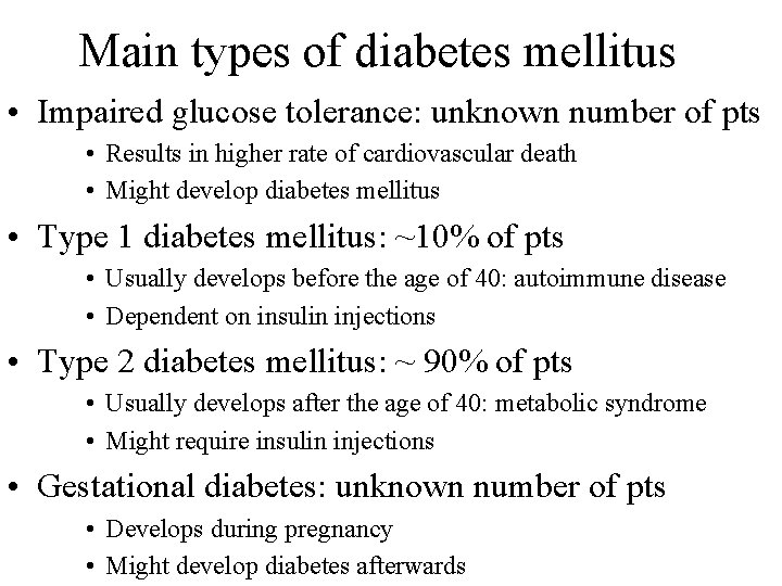
Main types of diabetes mellitus • Impaired glucose tolerance: unknown number of pts • Results in higher rate of cardiovascular death • Might develop diabetes mellitus • Type 1 diabetes mellitus: ~10% of pts • Usually develops before the age of 40: autoimmune disease • Dependent on insulin injections • Type 2 diabetes mellitus: ~ 90% of pts • Usually develops after the age of 40: metabolic syndrome • Might require insulin injections • Gestational diabetes: unknown number of pts • Develops during pregnancy • Might develop diabetes afterwards
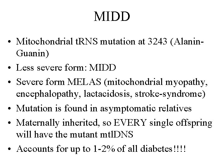
MIDD • Mitochondrial t. RNS mutation at 3243 (Alanin. Guanin) • Less severe form: MIDD • Severe form MELAS (mitochondrial myopathy, encephalopathy, lactacidosis, stroke-syndrome) • Mutation is found in asymptomatic relatives • Maternally inherited, so EVERY single offspring will have the mutant mtl. DNS • Accounts for up to 1 -2% of all diabetes!!!!
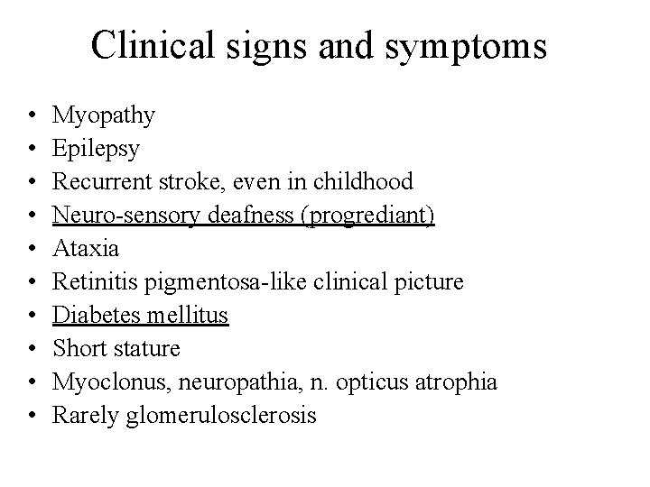
Clinical signs and symptoms • • • Myopathy Epilepsy Recurrent stroke, even in childhood Neuro-sensory deafness (progrediant) Ataxia Retinitis pigmentosa-like clinical picture Diabetes mellitus Short stature Myoclonus, neuropathia, n. opticus atrophia Rarely glomerulosclerosis
Main aims of management of DM • Improve diabetes control • Prevent the development of diabetes-related complications • Provide better quality of life
Improve diabetes control • Diet and exercise • Type 1 diabetes: life depends on insulin injections • Type 2 diabetes: add tablets to diet and exercise regime – If necessary add insulin injection • Manage BP and blood lipids appropriately • Complications depend on blood sugar level control and control of BP!
Acute and chronic complications • Acute complications • Hypoglycaemia and Hyperglycaemia • Chronic complications – Microvascular • Diabetic retinopathy: eye disease • Diabetic nephropathy: kidney disease, can lead to dyalisis • Diabetic neuropathy: small nerve disease, “pins and needles”, numbness, can contribute to amputations • Autonomic neuropathy: orthostatic hypotension, gastroparesis, impotence, bladder dysfunction, gustatory sweating – Macrovascular • Coronary artery disease, including silent AMI • Cerebrovascular disease: strokes • Peripheral vascular disease: amputations
Who is at risk of complications? • All patients with diabetes are at risk, but: • People with poor diabetes and blood pressure control are at even higher risk • The longer the duration of diabetes the higher the risk, so age at diagnosis is important factor as well • There seems to be a genetic component to risk • Certain ethnic groups, especially migrants are at high risk • Smoking can make complications worse • High risk of retinopathy during pregnancy
Provide better quality of life • These complications affect the patients’ life • Also affect ability to cope with the screening episode, such as unable to come, unable to transfer to chair, (mobility); unable to understand commands, etc • Patients with DM tend to be more depressed • Employment problems • Fear of complications • Ignorance
Costs of DM • Patients are hospitalised more and for longer then pts who do not have the disease • Patients utilise more expensive medical intervention (eg. dyalisis) and for longer period • Need more extensive rehabilitation (stroke, AMI, amputations, blindness)
Prevention of DM • Major lifestyle modifications, especially in pts with impaired glucose tolerance, gestational diabetes and family history • Dealing with auto-immune factors in patients with type 1 diabetes • Needs more education and intervention especially in developing countries and migrant populations
Rights of people with diabetes: UK • International Diabetes Federation 1994: – Never use diabetic as an adjunctive – Exemption: diabetic retinopathy / nephropathy – But: patient with diabetes and NEVER DIABETIC • • • Healthcare free of charge Choice of GP Right to see the medical records/ info in NHS Right to refuse treatment Make reasonable complaint against NHS Full or part refund of the cost of treatment
Rights and responsibilities • Patients should know the members of their health team and the reasons they are included • Patients should receive appropriate education on their disease • Patients should take an active part in the management of their diabetes
Hypoglycaemia • Medical term for blood sugar levels (BSL) below 4 mmol/L • Patients normally call it “ a hypo” • It can happen to anyone! • Symptoms vary greatly • It takes time to develop, so usually you have enough time to react • Except in hypoglycaemia unawareness
Mechanism of hypoglycaemia • Insulin works for lowering BSL by making it available for tissues and storage • Glucagon works for increasing BSL by making the liver release some • Eating food that contains carbohydrates leads to a rise in BSL • This stimulates insulin secretion, and BSL is lowered • If BSL is low, glucagon stimulates release of sugar from stores such as liver
Mechanism of hypoglycaemia 2 • In DM this system does not work well • So, another back-up system comes into play • ADRENALIN is released and it is responsible for the warning signs of a hypo • End-result of a hypo: brain does not get enough sugar, so it cannot function, the patient can die
Causes of hypoglycaemia • • • Too much insulin / too many tablets Delayed or missed meals or snacks Not enough food especially carbohydrate Unplanned or strenuous exercise Drinking alcohol without food No obvious cause
Type 2 diabetes: not on insulin • • Occasionally hypo can happen More frequent in the elderly More frequent in active people More frequent in people taking insulin and sulphonylureas • More frequent in people taking Glipizide, Gliclazide, Chlorpopamide, Gliquidone, Glimerpiride, Tolbutamide and Repaglinide
Type 2 diabetes: not on insulin: 2 • Some tablets are unlikely to cause a hypo: Metformin (Glucobay) • People on Acarbose should always be given sg like Lucosade as the absorption of sugar is very slow! • Some other medications such as antibiotics, antidepressants modify the action of the tablets for diabetes
Type 1 diabetes • Hypo is very common in patients with type 1 DM • The better the diabetes control the more common the hypo is • Requires careful balance between insulin – food exercise • Hypoglycaemia unawareness is more common
Mild hypo: treat yourself stage • Signs and symptoms – Hungry – Trembling and sweating – Irritable, aggressive, unreasonable – Pale, fast pulse, tingling of the limps – Blurring vision • Treatment – Immediate: quick carbohydrate: fruit juice, coke, glucose tablets, sugar, chocolate – Long acting: sandwich, biscuits, cereal, fruit
Severe hypo: cannot treat yourself • Signs and symptoms – – Difficulty in concentrating Confused and vague Irritable and irrational Loss of consciousness, fits and coma • Treatment – If conscious and can swallow: Hypostop (sugar gel), honey, treacle or jam inside the mouth and massage gently, once the pt is better, follow up as per mild hypo – If unconscious: if you are trained, use Hypostop etc; if not, place the pt in the recovery position and GET HELP! – Glucagon or iv glucose should only be given by trained personnel – When you get help, always specify that the patient has diabetes!
Other hypos • • • Hypos at night Hypos during or following exercise Hypos following drinking Hypos while driving Hypos while pregnant
Screening and hypos • Patients might be very anxious about screening and after initial rise in BSL, they can have a drop • They might miss a meal while waiting • They might have had to alter their daily routine for getting there • They might bring less food than normal
When to suspect a hypo • If patient becomes irrational, loud, agitated for no apparent reason especially if it is mealtime (morning and afternoon tea, lunch) • If patient complains about sudden blurring of the vision and other signs of hypo • Always check what medication the patient is on • Always ask if it is mealtime or not or if they skipped meal to keep their appointment • Keep sugar/honey or Lucosade at Screening van • KNOW how to get help!!!!
Diabetes education • Availability and type of diabetes education varies a great deal between areas • Always ask if the patient had diabetes education or not • Always ask where the patient received their diabetes care, opportunities for education will differ between hospital clinic patients, GP patients • Always consider practise nurse, a lot of them are trained in basic diabetes education
Where to direct the patient to? • Always have a list of available education opportunities for your patients • Consider local guidelines: practice nurses, GPs, diabetes centre, mobile training units • Diabetes UK is an excellent source of information for all patients and they have excellent leaflets and sessions • If the patient is registered blind or visually impaired, ask if they attend the RNIB or local agencies
Summary • Two types of diabetes both diagnosed by blood tests • Vast majority will have type 2 diabetes and will have multiple disabilities • The aim of the screening is to help improve diabetes control, see complication status and help with giving quality of life • DM is a costly disease for which there is no prevention and is difficult to cure • Both patients and health professionals have right and responsibilities in DM • Hypoglycaemia should be prevented and treated appropriately
Summary • Screening: 80 -90% of pts have type 2 DM • They will have multiple disabilities, including difficulty of learning or responding to commands • They might not see and/or hear you well! • Do not try to hurry them, they are trying to help you and please you if you are nice to them. • Remember, you are only one of many they need to see because of their diabetes! • After all, have fun screening, you are doing a service that gives them better quality of life.
• Diabetic Retinopathy – 200, 000 Type 1, > 1 million Type 2 persons with diabetes (PWDs), may double by 2012 – Commonest complication of diabetes mellitus – Commonest cause of blindness in UK between 20 and 55 years of age
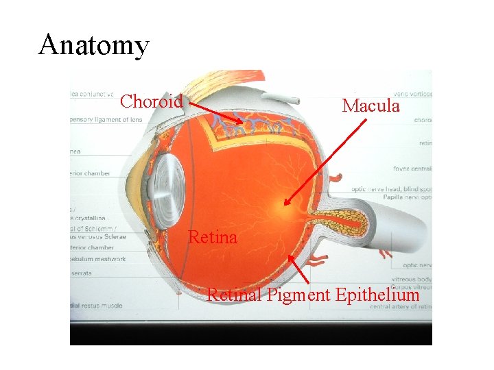
Anatomy Choroid Macula Retinal Pigment Epithelium
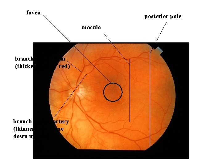
fovea posterior pole macula branch retinal vein (thicker, darker red) branch retinal artery (thinner, white line down middle)
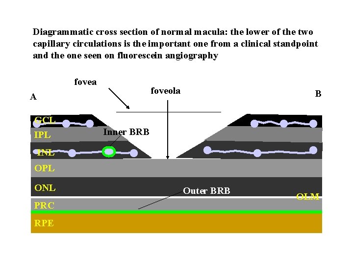
Diagrammatic cross section of normal macula: the lower of the two capillary circulations is the important one from a clinical standpoint and the one seen on fluorescein angiography fovea foveola A B GCL IPL Inner BRB INL OPL ONL PRC RPE Outer BRB OLM
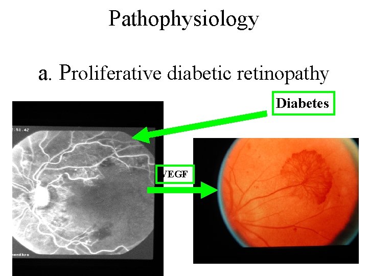
Pathophysiology a. Proliferative diabetic retinopathy Diabetes VEGF
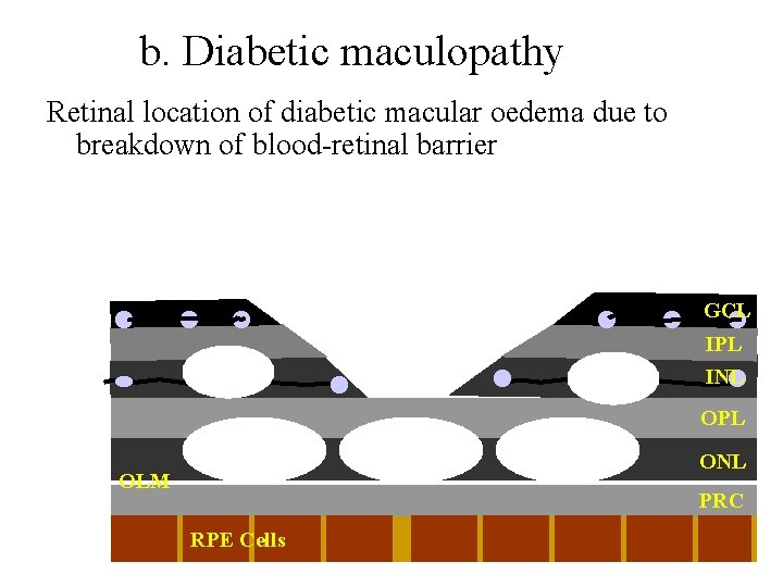
b. Diabetic maculopathy Retinal location of diabetic macular oedema due to breakdown of blood-retinal barrier GCL IPL INL OPL ONL OLM PRC RPE Cells RPE
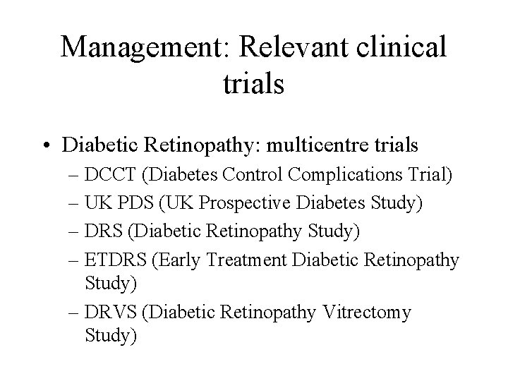
Management: Relevant clinical trials • Diabetic Retinopathy: multicentre trials – DCCT (Diabetes Control Complications Trial) – UK PDS (UK Prospective Diabetes Study) – DRS (Diabetic Retinopathy Study) – ETDRS (Early Treatment Diabetic Retinopathy Study) – DRVS (Diabetic Retinopathy Vitrectomy Study)
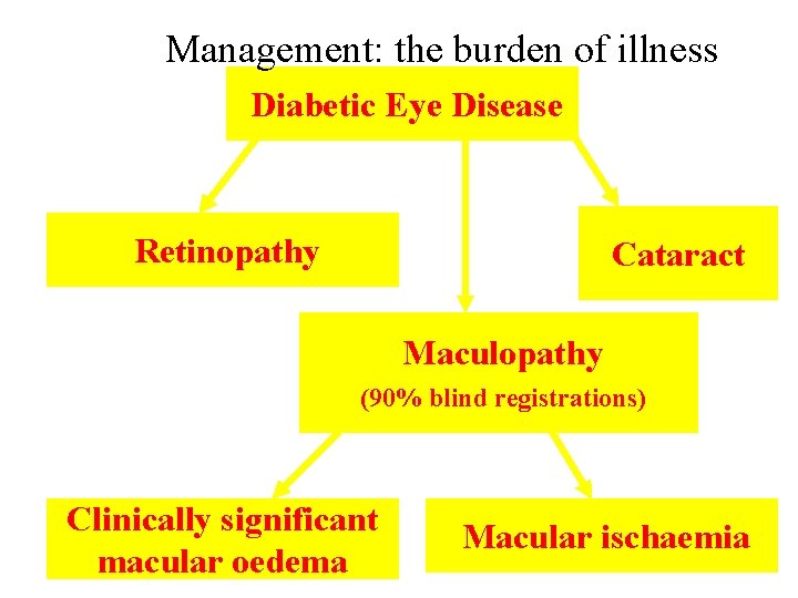
Management: the burden of illness Diabetic Eye Disease Retinopathy Cataract Maculopathy (90% blind registrations) Clinically significant macular oedema Macular ischaemia
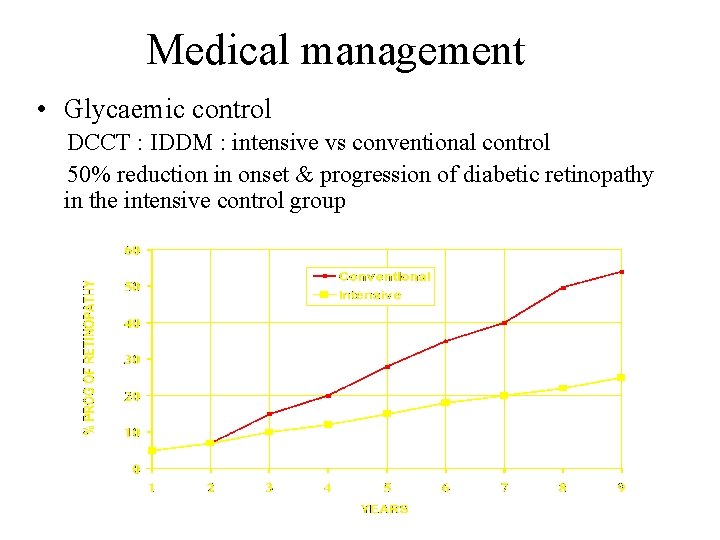
Medical management • Glycaemic control DCCT : IDDM : intensive vs conventional control 50% reduction in onset & progression of diabetic retinopathy in the intensive control group
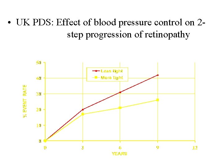
• UK PDS: Effect of blood pressure control on 2 step progression of retinopathy
Management: Laser therapy Proliferative diabetic retinopathy – panretinal photocoagulation (PRP) – 2 sessions, 1 -2 weeks apart, 2000 shots – may need adjunctive sessions – vitreous haemorrhage and tractional retinal detachment can still occur – extensive treatment may affect ability to drive
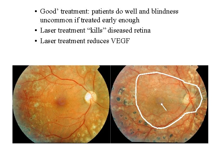
• Good’ treatment: patients do well and blindness uncommon if treated early enough • Laser treatment “kills” diseased retina • Laser treatment reduces VEGF 6 weeks post treatment 3 months post treatment
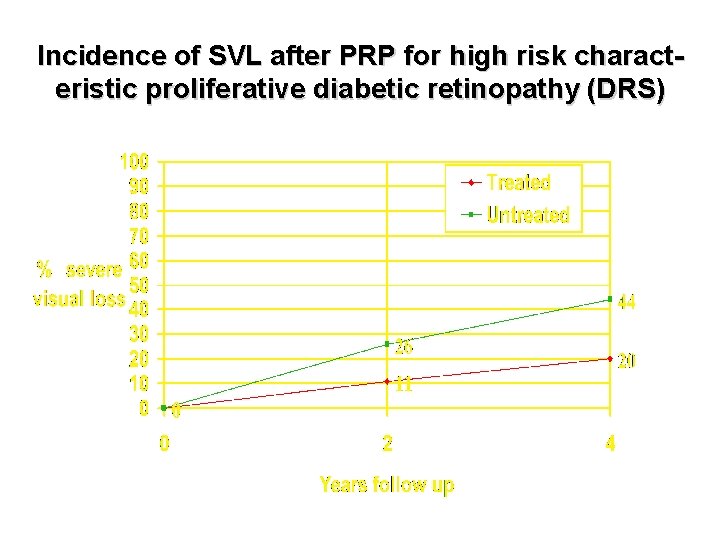
Incidence of SVL after PRP for high risk characteristic proliferative diabetic retinopathy (DRS)
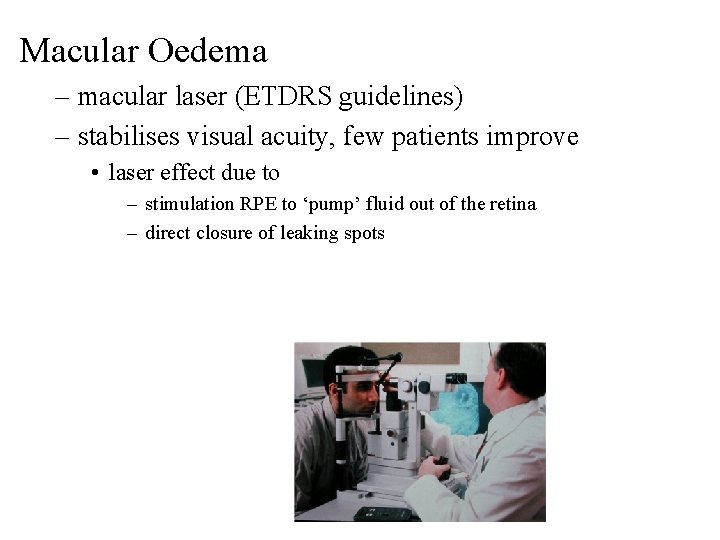
Macular Oedema – macular laser (ETDRS guidelines) – stabilises visual acuity, few patients improve • laser effect due to – stimulation RPE to ‘pump’ fluid out of the retina – direct closure of leaking spots
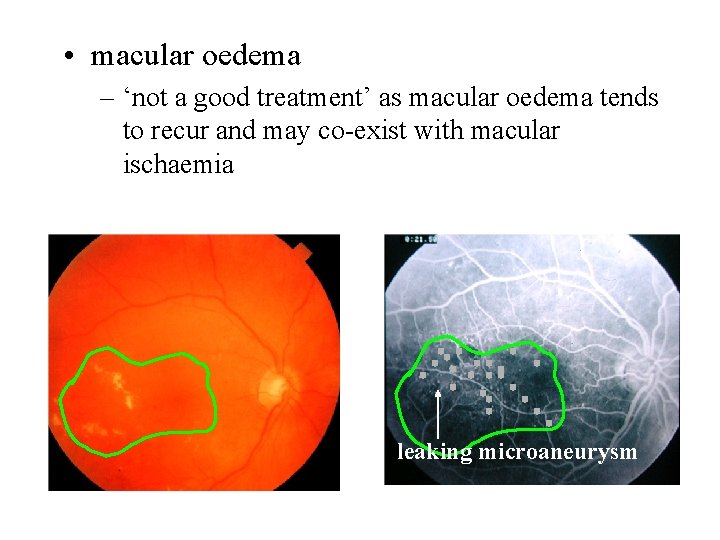
• macular oedema – ‘not a good treatment’ as macular oedema tends to recur and may co-exist with macular ischaemia leaking microaneurysm
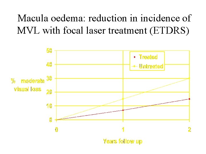
Macula oedema: reduction in incidence of MVL with focal laser treatment (ETDRS)
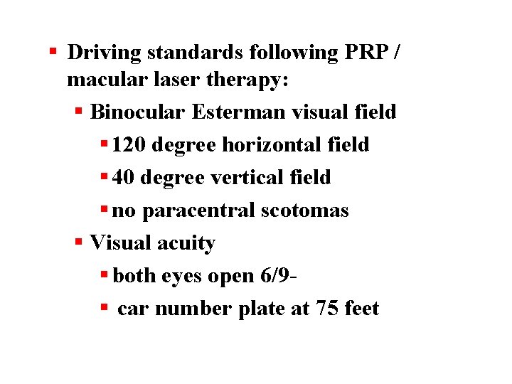
§ Driving standards following PRP / macular laser therapy: § Binocular Esterman visual field § 120 degree horizontal field § 40 degree vertical field § no paracentral scotomas § Visual acuity § both eyes open 6/9§ car number plate at 75 feet
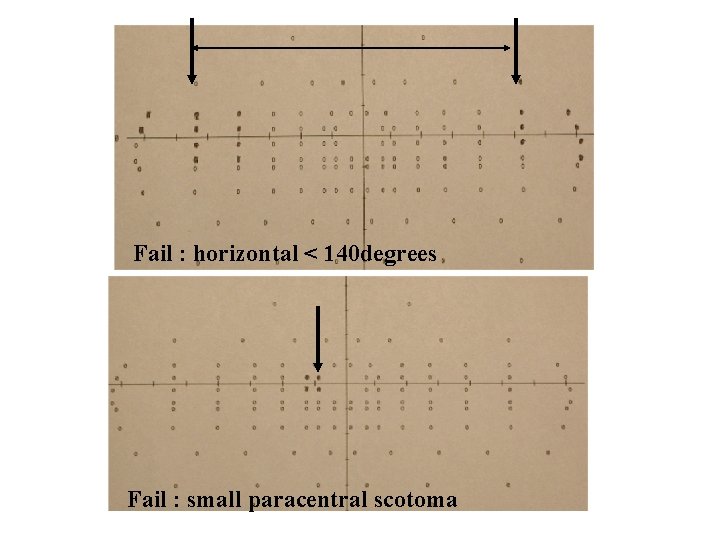
Fail : horizontal < 140 degrees Fail : small paracentral scotoma
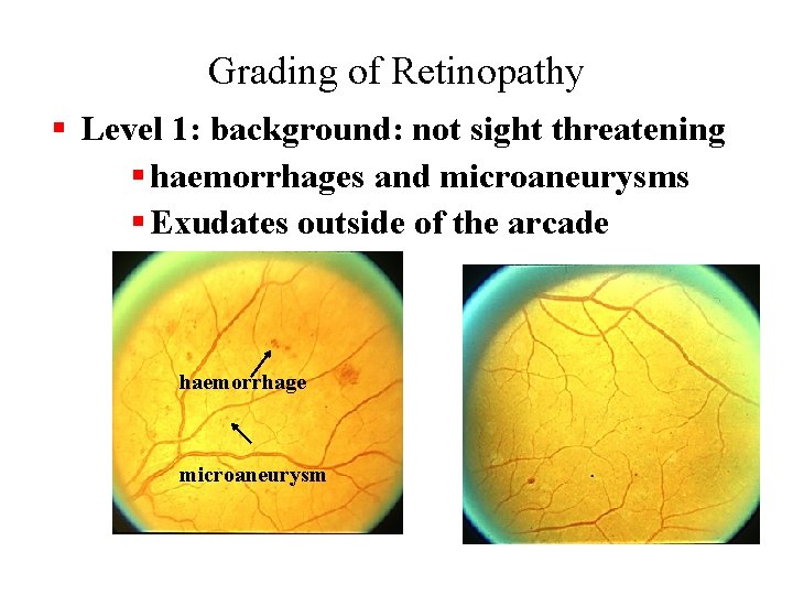
Grading of Retinopathy § Level 1: background: not sight threatening § haemorrhages and microaneurysms § Exudates outside of the arcade haemorrhage microaneurysm
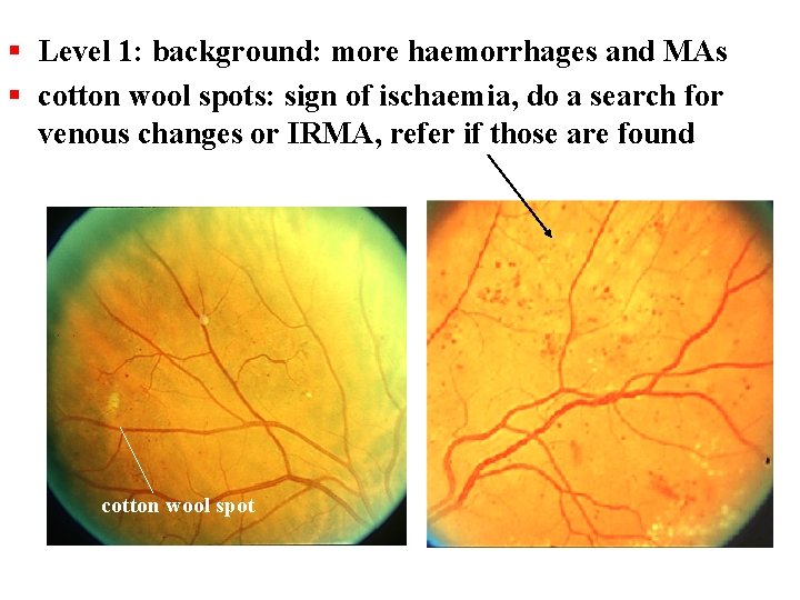
§ Level 1: background: more haemorrhages and MAs § cotton wool spots: sign of ischaemia, do a search for venous changes or IRMA, refer if those are found cotton wool spot
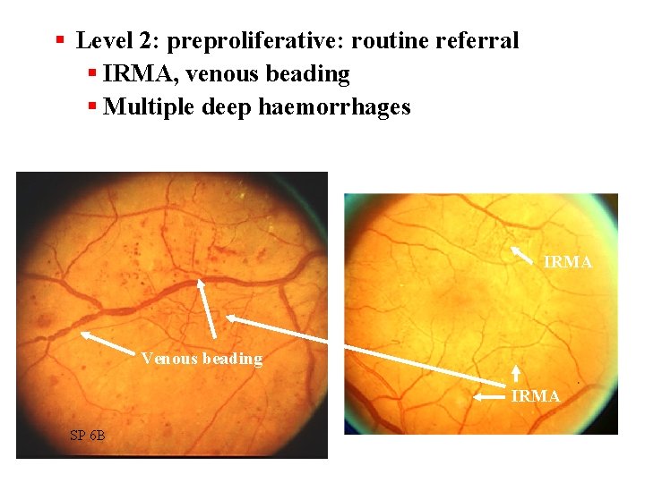
§ Level 2: preproliferative: routine referral § IRMA, venous beading § Multiple deep haemorrhages IRMA Venous beading IRMA SP 6 B
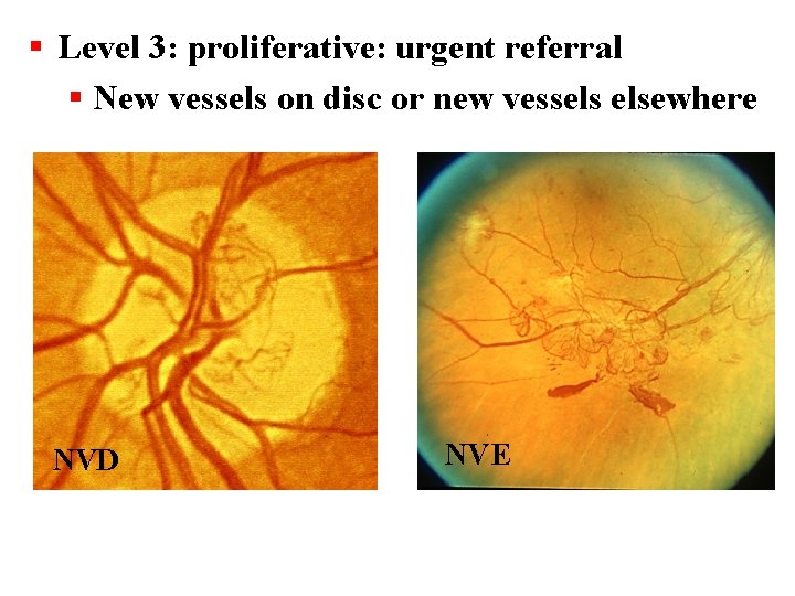
§ Level 3: proliferative: urgent referral § New vessels on disc or new vessels elsewhere NVD NVE
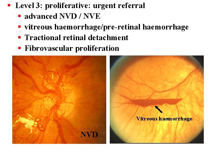
§ Level 3: proliferative: urgent referral § advanced NVD / NVE § vitreous haemorrhage/pre-retinal haemorrhage § Tractional retinal detachment § Fibrovascular proliferation Vitreous haemorrhage NVD
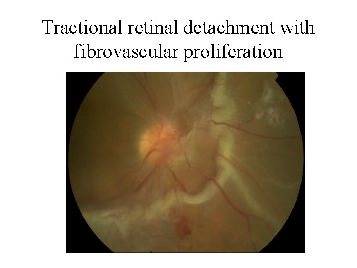
Tractional retinal detachment with fibrovascular proliferation
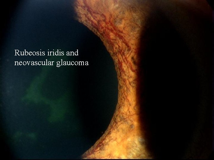
Rubeosis iridis and neovascular glaucoma
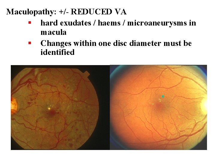
Maculopathy: +/- REDUCED VA § hard exudates / haems / microaneurysms in macula § Changes within one disc diameter must be identified
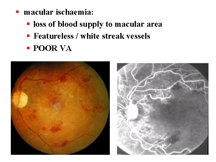
§ macular ischaemia: § loss of blood supply to macular area § Featureless / white streak vessels § POOR VA
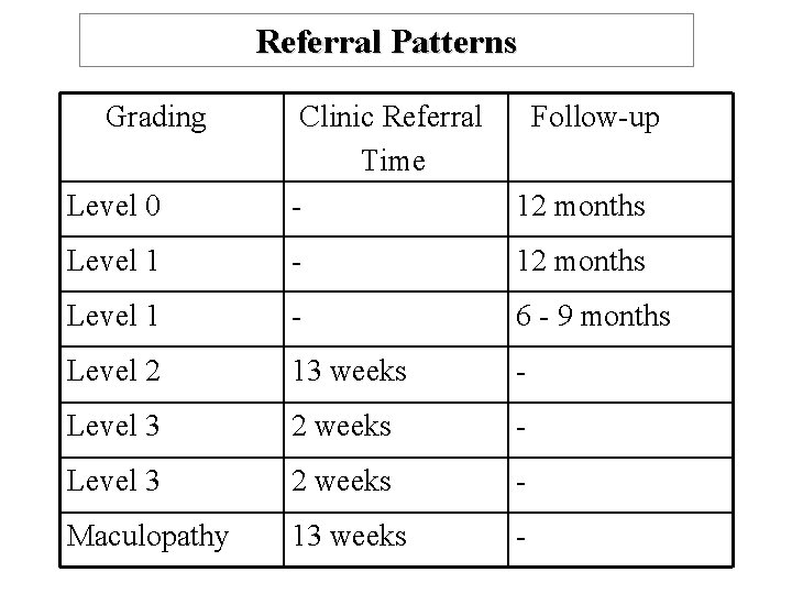
Referral Patterns Grading Level 0 Clinic Referral Time - Follow-up 12 months Level 1 - 6 - 9 months Level 2 13 weeks - Level 3 2 weeks - Maculopathy 13 weeks -
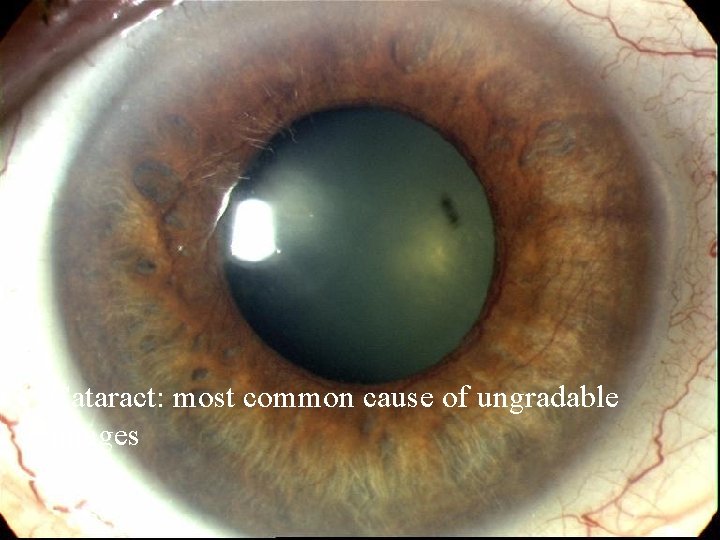
Cataract: most common cause of ungradable images
Cataract in patients with diabetes • Changes in blood sugar level can affect vision as lens swells when blood sugar is high (myopic change) and releases water and shrinks when BSL is low (hypermetropic change) • Lens returns to normal when BSL normal, so wait for stabilisation in newly diagnosed and unstable patients before sending them to optometrist • Patients with diabetes are 2 -4 times more likely to develop cataract (15 - 20 times more if <40)
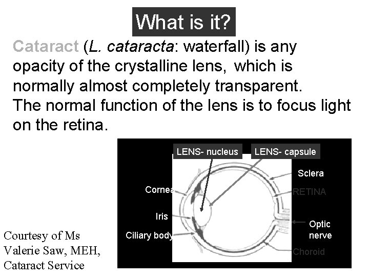
What is it? Cataract (L. cataracta: waterfall) is any opacity of the crystalline lens, which is normally almost completely transparent. The normal function of the lens is to focus light on the retina. LENS- nucleus LENS- capsule Sclera Cornea Iris Courtesy of Ms Valerie Saw, MEH, Cataract Service Ciliary body RETINA Optic nerve Choroid
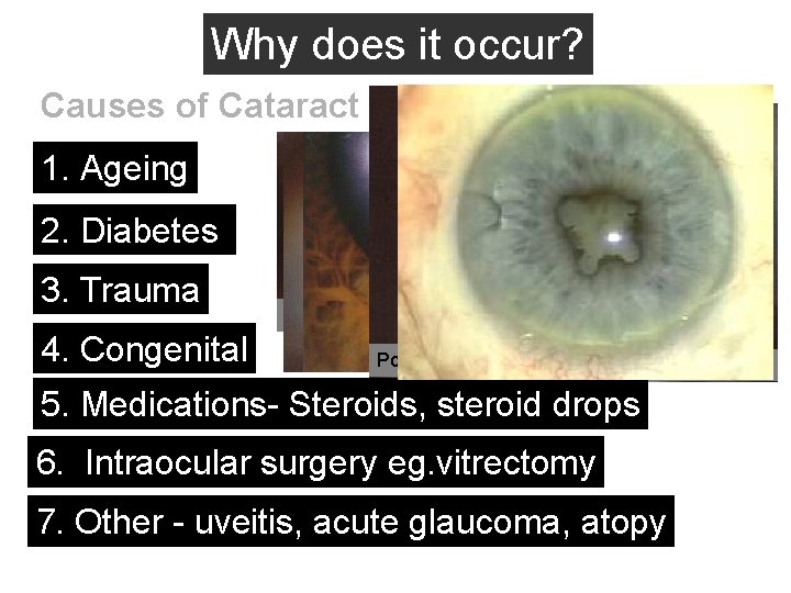
Why does it occur? Causes of Cataract 1. Ageing 2. Diabetes 3. Trauma 4. Congenital Nuclear cataract Cortical cataract Posterior subcapsular cataract Blue dot cataract 5. Medications- Steroids, steroid drops 6. Intraocular surgery eg. vitrectomy 7. Other - uveitis, acute glaucoma, atopy
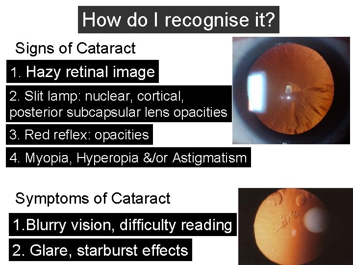
How do I recognise it? Signs of Cataract 1. Hazy retinal image 2. Slit lamp: nuclear, cortical, posterior subcapsular lens opacities 3. Red reflex: opacities 4. Myopia, Hyperopia &/or Astigmatism Symptoms of Cataract 1. Blurry vision, difficulty reading 2. Glare, starburst effects
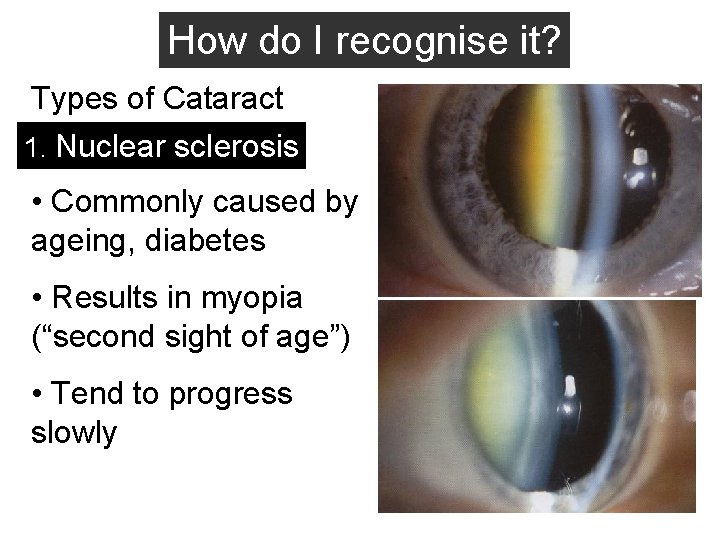
How do I recognise it? Types of Cataract 1. Nuclear sclerosis • Commonly caused by ageing, diabetes • Results in myopia (“second sight of age”) • Tend to progress slowly
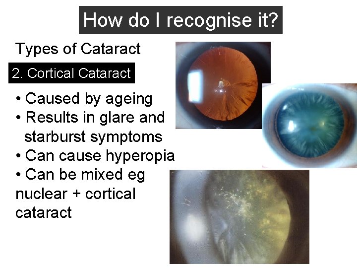
How do I recognise it? Types of Cataract 2. Cortical Cataract • Caused by ageing • Results in glare and starburst symptoms • Can cause hyperopia • Can be mixed eg nuclear + cortical cataract
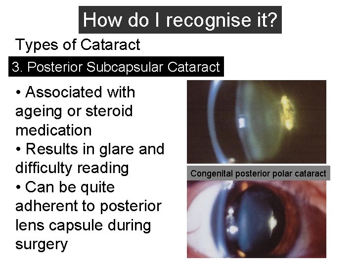
How do I recognise it? Types of Cataract 3. Posterior Subcapsular Cataract • Associated with ageing or steroid medication • Results in glare and difficulty reading • Can be quite adherent to posterior lens capsule during surgery Congenital posterior polar cataract
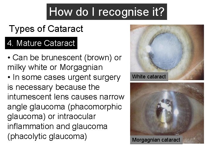
How do I recognise it? Types of Cataract 4. Mature Cataract • Can be brunescent (brown) or milky white or Morgagnian • In some cases urgent surgery is necessary because the intumescent lens causes narrow angle glaucoma (phacomorphic glaucoma) or intraocular inflammation and glaucoma (phacolytic glaucoma) White cataract Morgagnian cataract
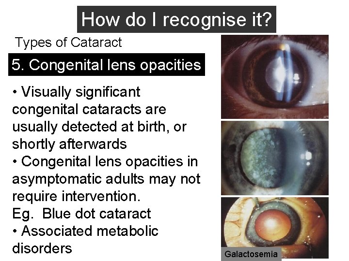
How do I recognise it? Types of Cataract 5. Congenital lens opacities • Visually significant congenital cataracts are usually detected at birth, or shortly afterwards • Congenital lens opacities in asymptomatic adults may not require intervention. Eg. Blue dot cataract • Associated metabolic disorders Galactosemia
How to deal with cataract in screening? • Understand local and national guidelines • If ungradable: <6 weeks referral • If gradable: refer first to optometrist to correct with glasses if possible, surgery might not be needed for years • Refer for surgery if cataract influences daily activities or vision is <6/12; or images are ungradable; when benefits outweigh risks • Remember: corneal opacity, vitreous haze!
What are the risks of cataract surgery? • 95% of patients achieve better vision then before surgery, BUT • 1% achieves no improvement or deteriorates after surgery • 1: 1000 chance of retinal detachment • 1: 1000 chance of infection • 1: 1000 chance of blindness • Retinopathy and maculopathy must be treated before cataract surgery if fundus is visible!
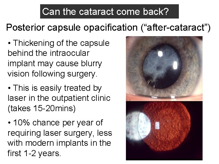
Can the cataract come back? Posterior capsule opacification (“after-cataract”) • Thickening of the capsule behind the intraocular implant may cause blurry vision following surgery. • This is easily treated by laser in the outpatient clinic (takes 15 -20 mins) • 10% chance per year of requiring laser surgery, less with modern implants in the first 1 -2 years.
Summary • Diabetic retinopathy is a consequence of a multisystem disease. • Management of DR needs to reflect the chronicity of the disease. • Screening is effective, but setting it up is costly • Grading of retinopathy & maculopathy can be achieved by non-medically trained graders • Patient education might be the key factor to achieve compliance with NSF.
- Slides: 78