Fetal Face Neck HHHOLDORF Normal Anatomy o Face


 o A condition where the jaw is undersized and is sometimes called")


 o The congenital absence of one or both eyes. o True or")
 Secondary (extremely tiny eyes, eyes start to develop then")


 o Similar to epignathus")

- Slides: 21
Fetal Face & Neck HHHOLDORF
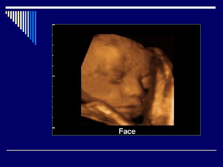
Normal Anatomy o Face: o Evaluation of the face is a vital part of the clinical genetic examination that is performed post-natally. Any time a fetal anomaly is identified, the diagnostic workup should include a detailed examination of the fetal face. Many details of facial anatomy can be identified as early as 11 weeks, particularly by using transvaginal sonography. It is necessary that it is accessible and there is a pocket of amniotic fluid in front of it for three dimensional scanning. o The upper lip and nares may be visualized in an oblique coronal plane and is useful in searching for facial clefts and some types of proboscis.
o The profile shows the forehead, nose and jaw which are important to assess the integrity of these structures.
Eyes o The eyes may be imaged in either a true coronal or a transverse plane. It is important to measure the outer orbital distance because it is valuable in diagnosing hypertolerism or hypotelorism
Neck o Transverse sections allow the measurement of the nuchal fold. Studies have shown an association with Down syndrome when this measurement exceeds 6 mm between 15 and 21 weeks. It is considered nuchal thickening when there is increased soft tissue thickness over the posterior aspect of the neck.
Facial Clefting o Typical facial clefts are cleft lip and Cleft palate anomalies. Cleft palate refers to the defect of the posterior portion of the palate in the presence of a normal upper lip and anterior palate. CL-CP can be unilateral or bilateral, and usually results from a failure of mesenchymal masses of the lateral palatine processes to fuse with each other, with the nasal septum and/ or median palatine process. It can be genetic or non genetic causing a minor developmental defect.
o Sonographic presentation demonstrates a groove extending from one nostril through the lip and possibly the alveolar ridge.
o A cleft lip or palate can be successfully treated with surgery soon after birth. o The edges of the cleft between the lip and nose are cut (A and B). The bottom of the nostril is formed with suture (C). The upper part of the lip tissue is closed (D), and the stitches are extended down to close the opening entirely (E). ( o Illustration by Argosy. )
Median cleft lip A malformation of the upper lip with/without cleft palate. The development of this anomaly is related to the differentiation process of the forebrain and is often associated with other midline defects or the face and brain such as holoprosencephaly. The incidence is about 1: 10, 000 births o It can vary from being a small notch to a complete division of the lip and alveolar part of the maxilla. o When it is unilateral it results from failure of maxillary prominence to fuse with medial nasal prominences. o When it is bilateral it results from failure of maxillary processes to meet and merge with medial prominences. o
Epignathus o A congenital tumor and a rare type of teratoma arising from the oral cavity or pharynx. It is associated with midline abnormalities. It may also arise from the sphenoid bone, hard or soft palate, pharynx, tongue or jaw. o Sonographic Findings: o Solid, complex tumor seen extruding from the fetal mouth, calcifications may be present within mass o Calcifications may be present within mass.
Micrognathism (Micrognathia) o A condition where the jaw is undersized and is sometimes called “Mandibular hypoplasia. ” It is common in infants but is usually self correcting during growth, due to the jaws increasing in sized. It may be a cause of abnormal tooth alignment and in severe cases can hamper feeding. It can present as a birth defect in multiple syndromes (fetal alcohol syndrome, congenital rubella, Trisomy 13 and 18).
Macroglossia o Minor or severe enlargement of the tongue can cause cosmetic and or functional difficulties, such as speaking, eating, swallowing and sleeping.
ORBITAL DEFECTS o Hypotelorism- a medical condition pertaining to abnormally close eyes. o Hypertelorism – an abnormally increased distance between two organs or bodily parts, usually referring to an increased distance between the eyes, seen in a variety of syndromes. o Microphthalmia or micropthlamos means small eye.
Anophthalmia (Anopththalmos) o The congenital absence of one or both eyes. o True or primary anopththalmos is very rare. The diagnosis for this can ONLY be made when there is complete absence of the ocular tissue within the orbit. Extreme micropthlamos is seen more commonly. In this condition, a very small globe is present within the orbital soft tissue, which is not visible on the first examination.
o Primary (No eye tissue) Secondary (extremely tiny eyes, eyes start to develop then stops) o Degenerative (eye started to form and for some reason degenerated
Retinoblastoma o Cancer of the retina. This tumor can begin in one or both eyes and can spread to the brain through the optic nerve. Development of the tumor is initiated by mutations
Holoprosencephaly o A spectrum of defects or malformations of the brain and face. The most severe cases involve serious malformations of the brain, and malformations so severe that they often cause miscarriage or stillbirth. There can also be facial defects – which may affect the eyes, nose, and upper lip- and normal or nearnormal brain development. Seizures and mental retardation may occur. The etiology is unknown. Suggested risk factors include: maternal diabetes, infections during pregnancy such as herpes, syphilis, rubella etc. , and various drugs taken during pregnancy. Women with previous pregnancy loss and 1 st trimester bleeding are likely to give birth to a child with this defect. o Symptoms: o Range from mild (no facial/organ defects), moderate ( cleft lip or palate) to severe ( synopththalmia proboscis or cyclopia)
Holoprosencephaly o The alobar type, which is the most severe, is characterized by a monoventricular cavity and fusion of the thalami. In the semilobar type there is partial segmentation of the ventricles and cerebral hemispheres posteriorly with incomplete fusion of the thalami. In lobar holoprosencephaly there is normal separation of the ventricles and thalami but absence of the septum pellucidum.
Anomalies of the Neck Teratoma of the Neck (Cervical teratoma) o Similar to epignathus except the tumor arises from the neck. Sonographic appearance includes complex, cystic/solid tumor seen near the fetal neck. o It is vital to be able to identify the origin of the mass in order to distinguish it from epignathus. o
Cystic Hygroma o Benign developmental anomaly of lymphatic origin characterized by sing or multiple cystic areas within soft tissues surrounding the neck. Sonographically it can appear to be a thin walled, multi-septated cyst usually located posterior to fetal head/neck but also may be anterior.