Neck Region PA 544 Clinical Anatomy Neck Topic


")


")
 Parafollicular cells calcitonin")


 –Ab may stimulate thyroid without")


 Arytenoid cartilage Thyroid cartilage Cricoid cartilage (posterior)")


 External carotid a. Internal carotid a. Common carotid a.")

 Superficial Temporal Post. Auricular Maxillary Occipital Facial Lingual Asc. Pharyngeal Sup.")


")


 tube Tubal tonsils (small holes) Pharyngeal recess")


")

")
")

- Slides: 73
Neck Region PA 544 Clinical Anatomy
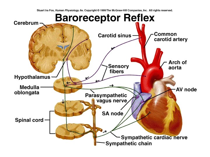
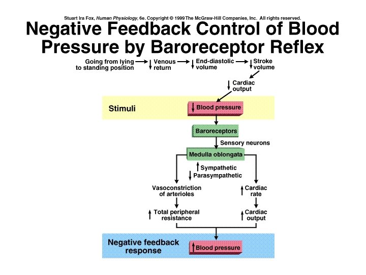
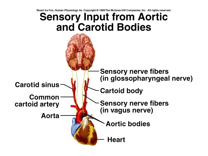
Neck: Topic Objectives • Be able to describe and identify the major muscles, bones and surface anatomy of the neck. • Be able to identify the architecture of the spinal cord anatomically and functionally • Be able to list the components of ascending and descending pathways of the cord and the cervical and brachial plexi • Be able to describe the fascia of the neck and apply its significance to clinically important conditions • Be able to describe the borders of triangles of the neck and list their components • Be able to identify thyroid structures including anatomical variations • Be able to predict level of thyroid malfunction physiologically • Be able to describe hypo and hyper thyroid causes and effects • Be able to describe the action of PTH • Be able to identify the cartilage and soft components of the larynx • Be able to predict the response of the baroreceptors to changing BP • Be able to describe the sequence of the swallowing reflex
Anterior Surface
Triangles at Surface
Hyoid 1. C 3 level 2. Suprahyoid muscles 3. Infrahyoid muscles
Vertebral Column
Intervertebral disc
Vertebra Parts
C 1 & C 2
Cervical Vertebra
Typical Cervical Vertebra
Spinal Cord • Receives and generates signals to body through the spinal nerves
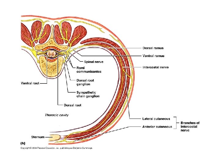
Cord in Spinal Canal Posterior Median Sulcus Posterior Root Denticulate Ligament Dorsal Root Ganglion Anterior Root Spinal Nerve
Spinal Cord (X. S. )
Spinal Nerve Anatomy
Functional Arrangement of SC Ascending and descending pathways
Ascending Pathways
Descending Pathways
Cervical Plexus Serves neck and diaphragm
Brachial Plexus and pectoral nerves
Brachial Plexus Innervates most of the arm and some of the body wall.
Spinal Cord Segments • 4 segments: Cervical, Thoracic, Lumbar, and Sacral (only 1 coccygeal nerve) • 31 pairs of spinal nerves
Cervical Segment
Neck Fascia Superficial fascia Prevertebral fascia Alar fascia Pretracheal fascia Investing fascia
Platysma
SCM and Trapezius SCM Trapezius
Cervical Triangles Posterior Triangle Anterior Triangle
Posterior Triangle Occipital triangle Supraclavicular triangle
Superficial and Deep Posterior Triangle Ext. jugular v. SCM Splenius Levator scapulae Accessory n. Nerve Point of Neck Phrenic n. Prevertebral fascia Platysma Scalenes Brachial plexus
Supraclavicular Triangle Brachial Plexus SCM Subclavian vessels
Anterior Triangles Submandibular Carotid Submental Muscular
Muscular Triangle
Muscles of Triangle Thyrohyoid Sternothyroid Thyroid Sternohyoid
Thyroid Location
Accessory Thyroid gland Accessory thyroid along thyroglossal duct
Pyramidal lobe (50% of people have this lobe structure)
Thyroid Follicle (follicular cells thyroxine) Parafollicular cells calcitonin
T 3 & T 4
T 3 & T 4 Formation and Secretion
Control of Thyroxine Secretion Short loop Long loop
Thyroid Malfunction • Hypothyroidism • Endemic goiters –due to iodine deffeicency • Cretinism –i thyroxine in child results in igrowth (dwarf) and severe mental retardation • Myxedema –i thyroxine in adult, leads to swelling of tissues plus other symptoms
Cretinism
Thyroid Malfunction • Hyperthyroidism • Toxic goiters (Graves disease) –Ab may stimulate thyroid without negative feedback control • Exophthalmos –symptom present in many hyperthyroid patients
Parathyroid Location
PTH Actions • Stimulates resorption of bone h. Ca+ and PO 4 - in blood • Stimulates Ca+ absorption in intestine (active Vit. D 3 necessary for Ca+ absorption) • Stimulates Ca+ reabsorption and PO 4 - excretion in kidney • Stimulates Vit. D 3 formation (skin) and activation (kidney) • Vital for life
Laryngeal Cartilages Hyoid Epiglottis (anterior) Arytenoid cartilage Thyroid cartilage Cricoid cartilage (posterior)
Voice Box Vestibular fold Ventricle Vocal fold
CN X: supply to laryngeal muscles Inf. Vagal ganglion Internal Laryngeal n. (sensory & autonomic) External Laryngeal n. (motor to inf. Pharnygeal constrictor and the cricohyoid) Recurrent Laryngeal n. (motor to all other laryngeal muscles)
Digastric Mylohyoid Submental Triangle -contains submental lymph nodes
Carotid Triangle Digastric (posterior belly) External carotid a. Internal carotid a. Common carotid a. Ansa cervicalis SCM IJV Omohyoid
Ext. Carotid Branches Hypoglossal n. Occipital Post. Auricular Int. carotid Superficial Temporal Maxillary Ext. carotid Facial Lingual Sup. Thyroid IJV Common Carotid Ansa cervicalis Asc. Pharyngeal (not show)
External carotid (isolated) Superficial Temporal Post. Auricular Maxillary Occipital Facial Lingual Asc. Pharyngeal Sup. Thyroid Note: Internal carotid has NO neck branches.
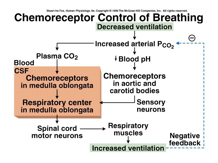
Carotid bodies
IJV Sup. Bulb of IJV Facial Sup. Thyroid Inf. Bulb of IJV
Lymph nodes of neck Deep Cervical Lymph nodes (most located in carotid sheath)
Submandibular Triangle Submandibular glands Digastrics
Submandibular Lymph nodes
Pharynx Function: deglutition Mucosa: Str. Squamous Muscularis: Skeletal
Oro- and nasopharynx Pharyngeal tonsil Pharyngotympanic (Eustachian) tube Tubal tonsils (small holes) Pharyngeal recess Palatine bone Salpingopharyngeal fold Soft palate Uvula Palatoglossal arch Tonsillar cleft Palatopharyngeal arch Lingual Tonsil
Longitudinal pharyngeal muscles Palatopharyngeus
Tonsils Pharyngeal Tonsil
Pharyngeal Constrictors Superior Middle Inferior
Peristalsis
Deglutition (Swallowing)
Fluid Drinking
Deglutition (cont’d)
Deglutition (cont’d)
Laryngopharynx Epiglottis Piriform recess