Topographical anatomy of the lower limb gait mechanism

Topographical anatomy of the lower limb, gait mechanism http: //anatomymasterclass. com/wp-content/uploads/2015/03/12 -1 -Lower-Limb-Anatomy-1. jpg Mark Kozsurek, M. D. , Ph. D. mark@kozsurek. hu EM II. , 05/02/2018

palpable: ant. sup. iliac spine * Subinguinal region Note first the epifascial structures! Saphenus hiatus: an opening of the fascia lata through which the great saphenous vein arrives into the femoral vein. Is considered as the external opening of the femoral canal. Superficial circumflex iliac, superficial epigastric and external pudendal vessels also pass through this opening. Superficial inguinal lymph nodes are tipically the ONLY palpable lymph nodes in healthy people! Cutaneous nerves (from lateral to medial): iliohypogastric, lateral femoral cutaneous, anterior femoral cutaneous, femoral branch of genitofemoral and ilioinguinal nerves.

https: //www. researchgate. net/Illustration-ofthe-inguinal-nerves-and-their-dermatomes. The-GFN-forms-from-L 1 -and-L 2_267875395 Lateral and anterior femoral cutaneous nerves

Inguinal ligament and the hip bone encloses the subinguinal hiatus which consists of the lacuna musculonervosa, lacuna vasorum and lacuna lymphatica. Note the iliopectineal arch and the lacunar ligament! A schematic drawing might be required on the final exam!

Iliopectineal fossa is a downward extension of the subinguinal hiatus. This triangular region is not identical to the femoral triangle! Fascia lata isolates superficial and deep inguinal lymph nodes. That big one found in the lacuna lymphatica is called Rosenmüller’s node. In the iliopectineal fossa the femoral artery gives off the deep femoral artery which somewhat later gives the lateral and medial circumflex femoral arteries and the perforating branches. Note femoral and obturator nerves! Femoral canal is a virtual canal normally filled by some connective tissue and lymph nodes. Femoral canal extends from the lacuna lymphatica to the saphenous hiatus and the walls are: the femoral vein laterally, the Pectineus muscle medially and the fascia lata anteriorly.

Gluteal region Cutaneous innervation is provided by the dorsal branches of lumbar and sacral spinal nerves and the posterior femoral cutaneous nerve (superior, middle and inferior cluneal nerves). Please note the greater and lesser sciatic foramina and sacrotuberous, sacrospinous ligaments! Pirifomis and Obturator internus muscles exit through te greater and lesser sciatic foramina, respectively.

After the removal of the Gluteus maximus muscle it is clearly seen that Gluteus medius, Piriformis, Gemelus superior, Obturator intenus, Gemellus inferior and Quadratus femoris muscles project toward the greater trochanter. palpable: ischial tuberosity and greater trochanter Suprapiriformis hiatus: - superior gluteal a. , v. and n. * * Infrapiriformis hiatus: - inferior gluteal a. , v. and n. - internal pudendal a. and v. , but (!) pudendal nerve - sciatic n. and its artery - postrior femoral cutaneous n. Note that the pudendal triad exits through the infrapiriformis hiatus but returns into the pelvis via the lesser sciatic foramen!

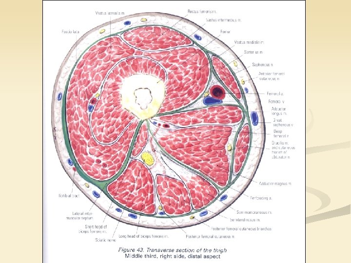
Anterior femoral region Cutaneous innervation is provided by the lateral and anterior femoral cutaneous nerves, medially the cutaneous branch of obturator nerve should be mentioned. All these are found superficial to the fascia lata. The main contents of the region are the extensors of the tigh. Femoral artery and vein together with the saphenous nerve arising from the femoral nerve as its longest sensory branch enter the adductor canal at the inferior corner of the femoral triangle enclosed by the inguinal ligament, the Sartorius and the Adductor longus muscles. Adductor canal is bounded by the vastoadductorial membrane, Vastus medialis and Adductor longus and magnus muscles.

palpable: ischial tuberosity * Posterior femoral region Cutaneous innervation is made by the posterior femoral cutaneos nerve. After the removal of the fascia lata the hamstring muscles (flexors of the tigh) become observable. It is clearly seen that all the hamstrings arise from the ischial tuberosity. The floor of the region is constituted by the Adductor magnus musle which is pierced in three distinct locations by the perforating arteries of the deep femoral artery. Sciatic nerve tipically divides into the tibial and common fibular (peroneal) nerves after being crossed by the long head of the Biceps femoris muscle.

Anterior genicular region Note the pes anserinus, the site of attachment of Sartorius, Gracilis and Semitendinosus muscles. Descending genicular artery and the saphenous nerve pierce the vastoadductorial membrane and join the great saphenous vein.

After cutting the capsule of the knee joint, medial and lateral menisci and the anterior cruciate ligament is seen.

Posterior genicular region Within the posterior genicular region popliteal fossa is a diamond-shaped area (borders are seen on the picture). Subcutaneously the small saphenous vein accampanied by the sural nerve is found. Sural nerve is formed by the fusion of the lateral (from common peroneal) and medial (from tibial) sural cutaneous nerves. Where the small saphenous vein drains into the popliteal vein somy lymph nodes are found. Adductor hiatus enclosed by the muscular and tendinous attachments of the Adductor magnus muscle is the distal opening of the adductor canal. Femoral vessels exit the adductor canal as popliteal ones. The floor of the region is made by the popliteal surface of the femur, the knee joint capsule and the popliteus muscle.

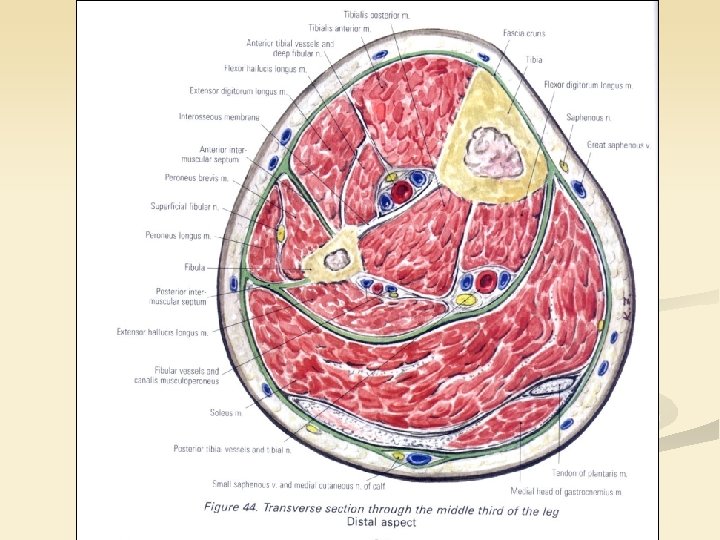
palpable: head of fibula * Anterior crural region Medially, superficial to the crural fascia the saphenous nerve and the great saphenous vein is found. Mainly the extensors of the leg are found in this region but at the lateral margin the peroneus compartment also appears. Common peroneal (fibular) nerve bifurcates at the head of the fibula: deep peroneal nerve descends among the extensors, while the superficial peroneal nerve innervates the Peroneus muscles. Anterior tibial artery and vein accompany the deep peroneal (fibular) nerve.

Posterior crural region Superficially the small saphenous vein ascends along the sural nerve on the lateral aspect of the leg. Between the superficial and deep flexors the posterior tibial artery and vein as well as the tibial nerve is present. The most important is the „abnormal” site of origin of the Flexor digitorum longus muscle: as it arises from the posterior surface of the tibia, first has to cross the tendon of Tibialis posterior (above the medial ankle – crural chiasm) then Flexor hallucis longus tendon already on the sole (plantar chiasm).

Lateral malleolar region Behind and below the lateral ancle sural nerve and small saphenos nerve descend. Superior and inferior peroneal retinacula attach the synovial sheaths of Peroneus muscles to the tarsal bones. Lateral margin of the foot is made by the Abductor digiti minimi muscle.

Medial malleolar region Great saphenous vein and saphenous nerve is seen around the medial ancle. Flexor retinaculum consists of a superficial and a deep layer. Vessels and nerves are found between the two layers, while flexor tendons and their synovial sheaths are found under the deep lamina. Note the order of structures: (1) Tibialis posterior, (2) Flexor digitorum longus, (3) tibial nerve and posterior tibial vessels superficially and the tendon of Flexor hallucis longus in the deep.

Dorsum of the foot Right under the skin the dorsal veinous plexus of the foot is seen from which the small and great saphenous nerves arise. Note the intresting pattern of the cutaneous innervation at the junction of the first and second toes. Also note that the toe tips always get sensory branches from the plantar surface!

Under the extensor retinaculum tendons of extensors arrive to the toes. A tendon of the Extensor digitorum longus inserts at the lateral side of the foot together with the tendon of Peroneus brevis muscle. This is ofter reffered to as the „third” peroneus or „Peroneus tertius”. Under the tendons of long extensors their short pairs can be identified: Extensor digitorum brevis and Extensor hallucis brevis. Dorsalis pedis artery (dorsal artery of the foot) is palpable along the tendon of Extensor hallucis longus and provides information about the peripheral circulation of the lower limb.

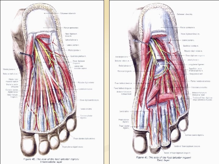
Plantar region – basic concept medial plantar eminence median plantar eminence lateral plantar eminence Muscles of the foot can be divided into three compartments (analogous to thenar, mesothenar and hypothenar). Especially in the central comparment a clearlayered arrangement can be sen.

https: //www. orthobullets. com/foot-and-ankle/7003/layers-of-the-plantar-foot 1 st 2 nd 3 rd 4 th

Cutaneous innervation of the sole

https: //www. clinicalkey. com/#!/content /book/3 -s 2. 0 -B 9780702052514500134 Great saphenous vein is formed around the medial ankle and ascends subcutaneously along the medial surface of the lower limb until it passes throuht the saphenous hiatus of the fascia lata and drains into the femoral vein. Small saphenous vein starts at the lateral ankle and as it ascends along the leg moves toward the midline and terminates within the popliteal fossa. Lymph vessels of the lower limb follow these two veins. Some lymph nodes occure in the popliteal fossa but much more are found around the inguinal ligament (superficial and deep inguinal lymph nodes. )

https: //www. clinicalkey. com/#!/content/book/3 -s 2. 0 -B 9780702051319000067

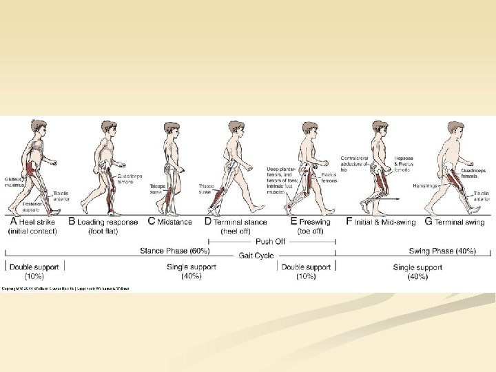
Gait mechanism

Thank you for your attention!
- Slides: 29