Tumors of Respiratory System Do Dr In Doan











- Slides: 107
Tumors of Respiratory System Doç. Dr. Işın Doğan Ekici School of Medicine Department of Pathology
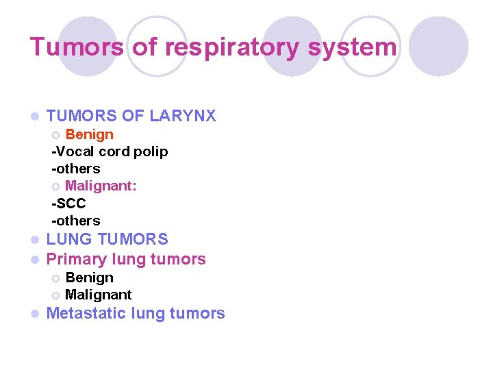
Tumors of respiratory system l TUMORS OF LARYNX Benign -Vocal cord polip -others ¡ Malignant: -SCC -others ¡ l l LUNG TUMORS Primary lung tumors ¡ ¡ l Benign Malignant Metastatic lung tumors
Lung Tumors l Primary lung tumors ¡ Benign ¡ Malignant l Metastatic lung tumors
Primary Lung Tumors
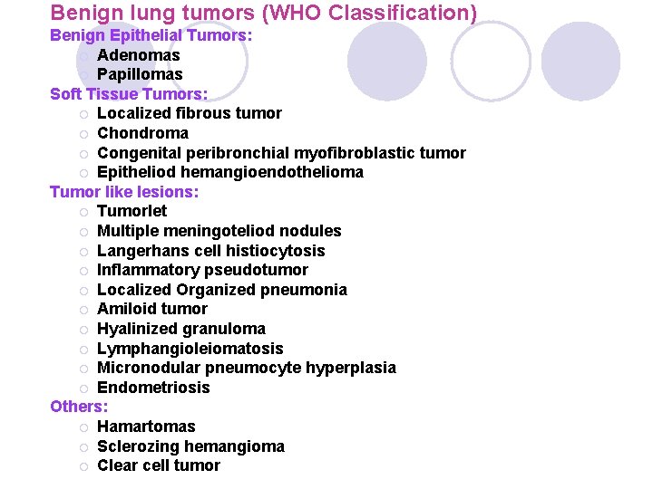
Benign lung tumors (WHO Classification) Benign Epithelial Tumors: ¡ Adenomas ¡ Papillomas Soft Tissue Tumors: ¡ Localized fibrous tumor ¡ Chondroma ¡ Congenital peribronchial myofibroblastic tumor ¡ Epitheliod hemangioendothelioma Tumor like lesions: ¡ Tumorlet ¡ Multiple meningoteliod nodules ¡ Langerhans cell histiocytosis ¡ Inflammatory pseudotumor ¡ Localized Organized pneumonia ¡ Amiloid tumor ¡ Hyalinized granuloma ¡ Lymphangioleiomatosis ¡ Micronodular pneumocyte hyperplasia ¡ Endometriosis Others: ¡ Hamartomas ¡ Sclerozing hemangioma ¡ Clear cell tumor
Bronchial adenomas l Bronchial adenomas make up 50% of all benign pulmonary tumors. l The use of the term bronchial adenoma should be discouraged because it encompasses several benign and malignant tumors.
Mucous gland adenomas l Mucous gland adenomas are true benign bronchial adenomas. l Mucous gland adenomas are also called bronchial cystadenomas, and they arise in the main or local bronchi. l Histologically, they consist of columnar cell– lined cystic spaces with a papillary appearance.
l Localized fibrous tumor Most frequent benign soft tissue neoplasm of lung. It was historically called as “benign mesothelioma” or “fibroma”. It is generally related to the visceral pleura. Mitotic figures must be <4/10 hpf (high power field)
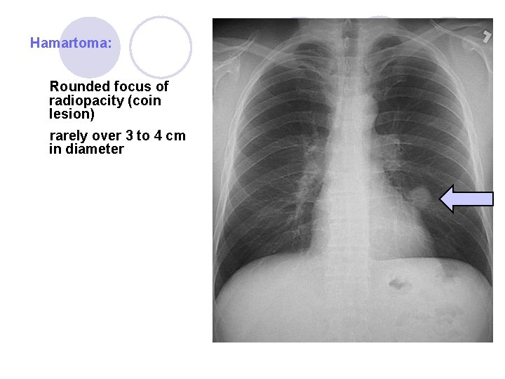
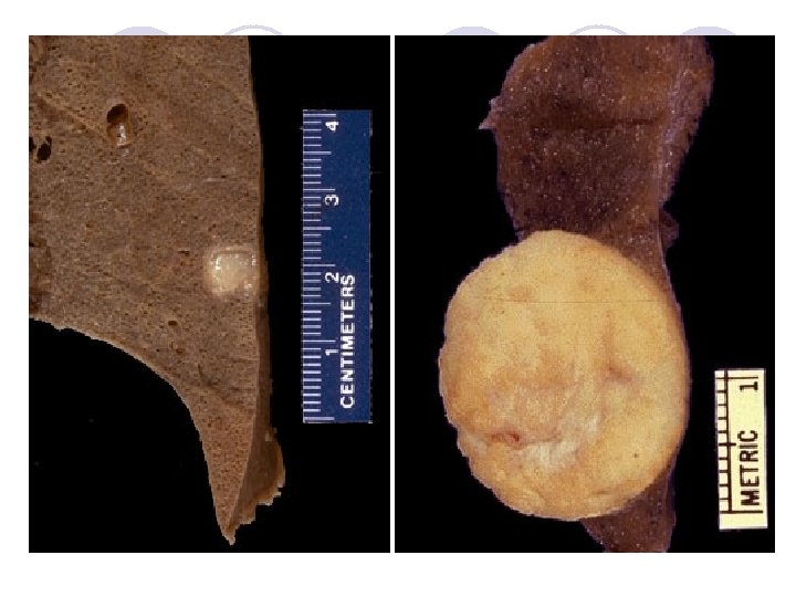
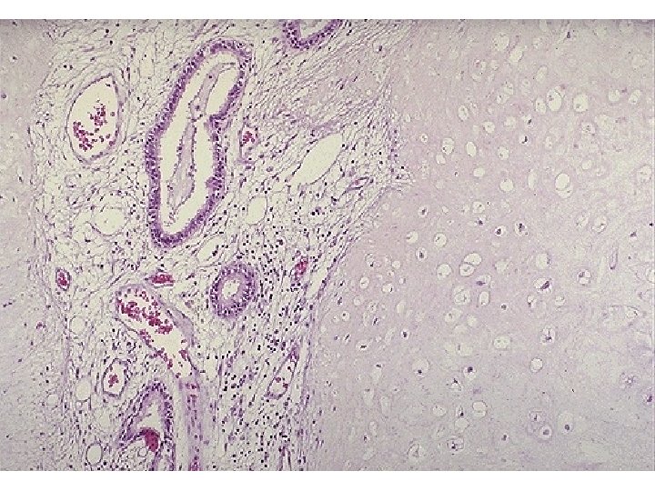
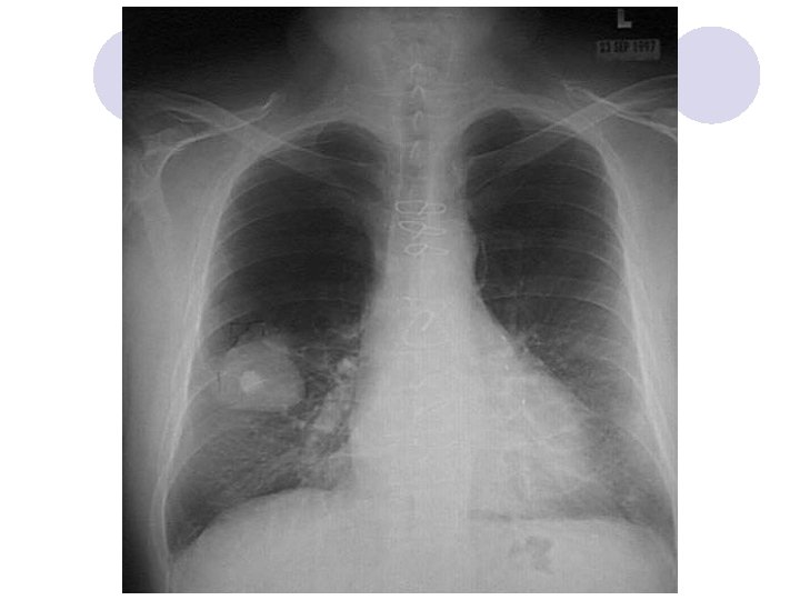
Hamartomas: a focal growth that resembles a neoplasm but results from faulty development in an organ. Hamartomas are the most common type of benign lung tumor. l They mainly occur in adults but, on occasion, occur in children. l Hamartomas are peripherally located. l Rounded focus of radiopacity (coin lesion). l Grossly, they have a firm marble like consistency. Rarely over 3 to 4 cm in diameter. l Histologically, hamartomas generally consist of the cells normally found in lung such as: epithelial tissue and other tissues such as fat and cartilage. l
Hamartoma: Rounded focus of radiopacity (coin lesion) rarely over 3 to 4 cm in diameter
NOTICE: Carney’s triade: ¡ 1. Gastric epitheloid leiomyosarcoma ¡ 2. Functioning extra-adrenal paraganglioma ¡ 3. Pulmonary chondroma (hamartoma).
Primary Malignant Lung Tumors
Lung Cancers l Most common visceral malignancy. l 3 rd of all cancer deaths are due to lung cancer. l Significant increase in incidence. l Dramatic increase among females. l 90% of lung cancers are related to smoking (passive smoking in 5%).
Lung Cancer & Smoking Depends on (1)duration, (2)amount of daily smoking, (3)deep inhaling: ¡ 10 fold greater risk than non smokers. ¡ 20 fold risk if >40 cigarettes per day. l Atypical cells in 96. 7% of smokers compared to 0. 9% in non smokers. l Significant proportion of over 1200 substances in smoke are carcinogenic (not nicotine). ¡ Initiaters – Benzo[a]pyrenes ¡ Promoters – Phenol derivatives, nitrous compounds ¡ Radioactive substances – Polonium, Carbon 14, K 40 l
Etiology of Lung Cancer l Tobacco ¡ ¡ the predominant cause of SCLC, as of NSCLC, is tobacco smoking. Of all histologic types of lung cancer, in fact, SCLC and squamous cell carcinoma have the strongest correlation to tobacco. l Uranium ¡ smoking: miners: All types of lung cancers occur with increased frequency in uranium miners, but SCLC is most common. The incidence is increased further in smokers. l Radon: ¡ Exposure to radon, which is an inert gas developing from the decay of uranium.
Kiss a non-smoker Enjoy the difference !
Molecular Genetics in Lung Cancer l Oncogene dominancy and/or loss or inactivation of tumor suppressor genes plays role in lung cancer; Most common dominant oncogenes in lung cancer are: *C-myc in small cell carcinoma *K-ras in adenocarcinoma Most common deleted or inactive recessive genes in lung cancer are; *p 53, retinoblastoma gene and some genes in the short arm of chromosome 3
TNM classification of lung tumors l The primary tumor (T) is classified according to its size and local invasion. ¡ Tis : Carcinoma insitu ¡ T 1: A tumor 3 cm or less in its greatest dimension, surrounded by lung or visceral pleura, without involvement of the main bronchus, ¡ T 2 : A tumor with any of the following features: • Larger than 3 cm in largest dimension • Involvement of the mainstem bronchus more than 2 cm from the carina • Invades the visceral pleura • Associated with atelectasis or post obstructive pneumonitis extending to the hilar region but not involving the entire lung.
¡ T 3 - A tumor of any size with any of the following features: l Tumor directly invading the chest wall, diaphragm, mediastinal pleura, or parietal pericardium l Tumor associated with atelectasis or obstructive pneumonitis of the entire lung. ¡ T 4 - A tumor of any size with any of the following features: l Tumor invading the mediastinum, heart, great vessels, trachea, esophagus, vertebral body, or carina l Any tumor with a malignant pleural or pericardial effusion l Any tumor with satellite tumor nodules within the ipsilateral primary tumor lobe of the lung.
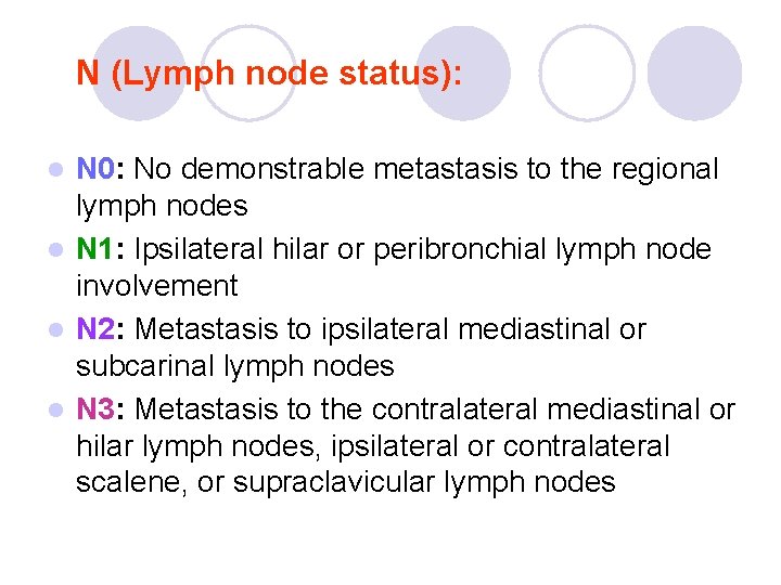
N (Lymph node status): N 0: No demonstrable metastasis to the regional lymph nodes l N 1: Ipsilateral hilar or peribronchial lymph node involvement l N 2: Metastasis to ipsilateral mediastinal or subcarinal lymph nodes l N 3: Metastasis to the contralateral mediastinal or hilar lymph nodes, ipsilateral or contralateral scalene, or supraclavicular lymph nodes l
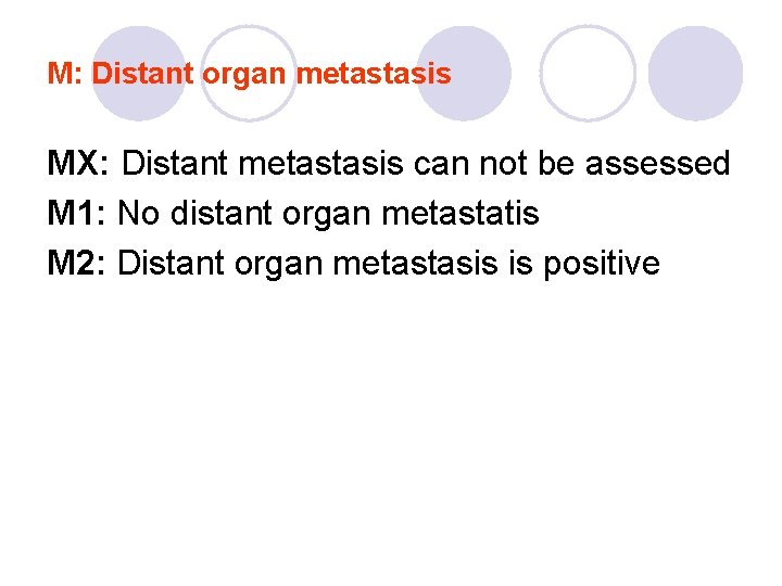
M: Distant organ metastasis MX: Distant metastasis can not be assessed M 1: No distant organ metastatis M 2: Distant organ metastasis is positive
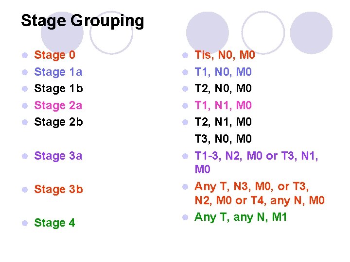
Stage Grouping l l Stage 0 Stage 1 a Stage 1 b Stage 2 a Stage 2 b l Stage 3 a l l Stage 3 b l l Stage 4 l l l l l Tis, N 0, M 0 T 1, N 0, M 0 T 2, N 0, M 0 T 1, N 1, M 0 T 2, N 1, M 0 T 3, N 0, M 0 T 1 -3, N 2, M 0 or T 3, N 1, M 0 Any T, N 3, M 0, or T 3, N 2, M 0 or T 4, any N, M 0 Any T, any N, M 1
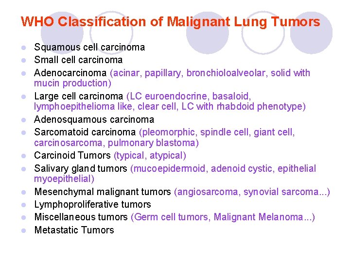
WHO Classification of Malignant Lung Tumors l l l Squamous cell carcinoma Small cell carcinoma Adenocarcinoma (acinar, papillary, bronchioloalveolar, solid with mucin production) Large cell carcinoma (LC euroendocrine, basaloid, lymphoepithelioma like, clear cell, LC with rhabdoid phenotype) Adenosquamous carcinoma Sarcomatoid carcinoma (pleomorphic, spindle cell, giant cell, carcinosarcoma, pulmonary blastoma) Carcinoid Tumors (typical, atypical) Salivary gland tumors (mucoepidermoid, adenoid cystic, epithelial myoepithelial) Mesenchymal malignant tumors (angiosarcoma, synovial sarcoma. . . ) Lymphoproliferative tumors Miscellaneous tumors (Germ cell tumors, Malignant Melanoma. . . ) Metastatic Tumors
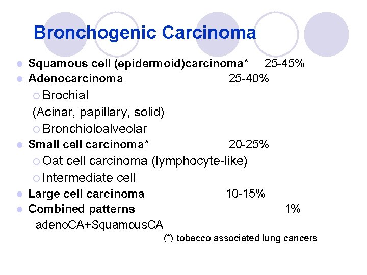
Bronchogenic Carcinoma Squamous cell (epidermoid)carcinoma* 25 -45% l Adenocarcinoma 25 -40% l ¡ Brochial (Acinar, papillary, solid) ¡ Bronchioloalveolar l Small cell carcinoma* 20 -25% ¡ Oat cell carcinoma (lymphocyte-like) ¡ Intermediate cell Large cell carcinoma l Combined patterns adeno. CA+Squamous. CA l 10 -15% 1% (*) tobacco associated lung cancers
In Clinical Oncology Small cell lung cancer (SCLC) is considered distinct from the other lung cancers, called non–small-cell lung cancers (NSCLCs), because of their clinical and biologic characteristics.
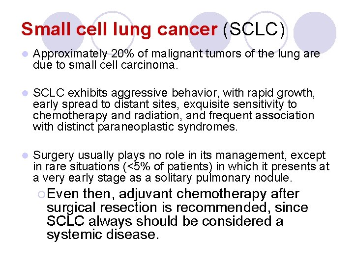
Small cell lung cancer (SCLC) l Approximately 20% of malignant tumors of the lung are due to small cell carcinoma. l SCLC exhibits aggressive behavior, with rapid growth, early spread to distant sites, exquisite sensitivity to chemotherapy and radiation, and frequent association with distinct paraneoplastic syndromes. l Surgery usually plays no role in its management, except in rare situations (<5% of patients) in which it presents at a very early stage as a solitary pulmonary nodule. ¡ Even then, adjuvant chemotherapy after surgical resection is recommended, since SCLC always should be considered a systemic disease.
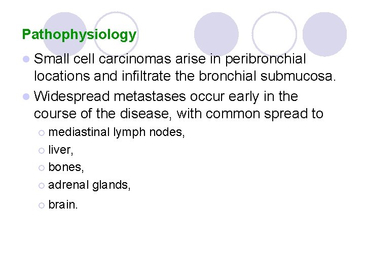
Pathophysiology l Small cell carcinomas arise in peribronchial locations and infiltrate the bronchial submucosa. l Widespread metastases occur early in the course of the disease, with common spread to ¡ mediastinal lymph nodes, ¡ liver, ¡ bones, ¡ adrenal ¡ brain. glands,
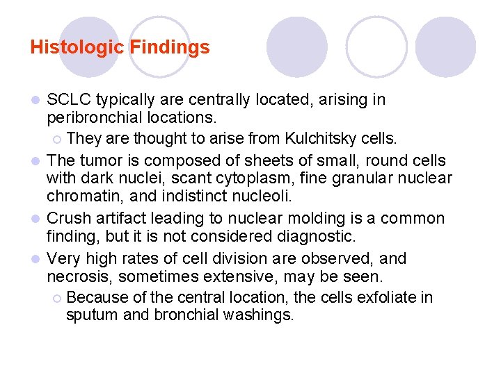
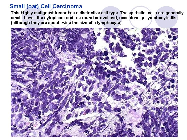
Histologic Findings SCLC typically are centrally located, arising in peribronchial locations. ¡ They are thought to arise from Kulchitsky cells. l The tumor is composed of sheets of small, round cells with dark nuclei, scant cytoplasm, fine granular nuclear chromatin, and indistinct nucleoli. l Crush artifact leading to nuclear molding is a common finding, but it is not considered diagnostic. l Very high rates of cell division are observed, and necrosis, sometimes extensive, may be seen. ¡ Because of the central location, the cells exfoliate in sputum and bronchial washings. l
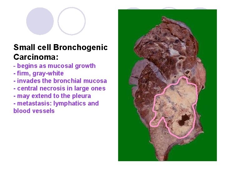
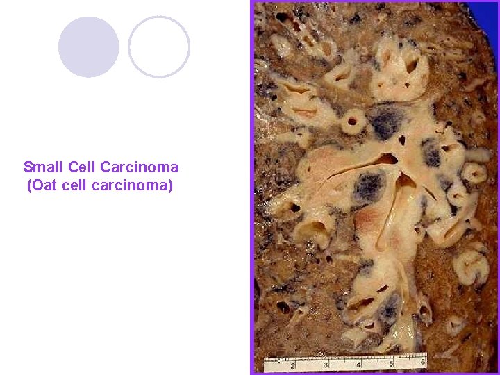
Small cell Bronchogenic Carcinoma: - begins as mucosal growth - firm, gray-white - invades the bronchial mucosa - central necrosis in large ones - may extend to the pleura - metastasis: lymphatics and blood vessels
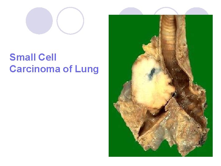
Small Cell Carcinoma of Lung
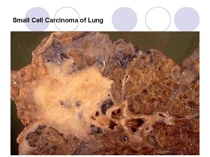
Small Cell Carcinoma of Lung
Small Cell Carcinoma (Oat cell carcinoma)
Small (oat) Cell Carcinoma This highly malignant tumor has a distinctive cell type. The epithelial cells are generally small, have little cytoplasm and are round or oval and, occasionally, lymphocyte-like (although they are about twice the size of a lymphocyte).
l Neurosecretory granules can be identified on electron microscopy, and the neuroendocrine nature of the neoplasm is suggested by its frequent association with paraneoplastic syndromes caused by peptide hormones. l Immunohistochemical stains for chromogranin, neuron-specific enolase, and synaptophysin usually are positive. l Approximately 5% of SCLCs exhibit features of mixed small cell and large cell components and, less frequently, may exhibit mixed small cell and squamous cell components.
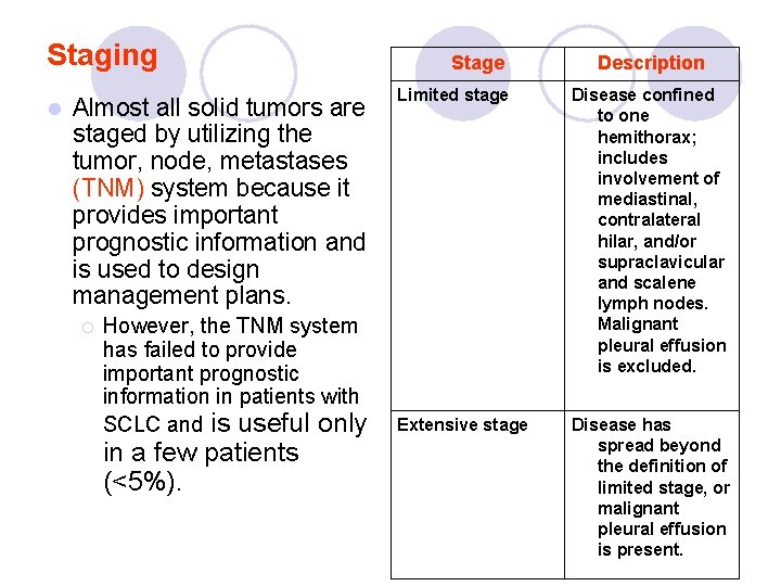
Staging l Almost all solid tumors are staged by utilizing the tumor, node, metastases (TNM) system because it provides important prognostic information and is used to design management plans. ¡ However, the TNM system has failed to provide important prognostic information in patients with SCLC and is useful only in a few patients (<5%). Stage Description Limited stage Disease confined to one hemithorax; includes involvement of mediastinal, contralateral hilar, and/or supraclavicular and scalene lymph nodes. Malignant pleural effusion is excluded. Extensive stage Disease has spread beyond the definition of limited stage, or malignant pleural effusion is present.
Complications l Tumor lysis syndrome: ¡ Tumor lysis can occur rapidly in patients with SCLC on institution of chemotherapy, especially in extensive-stage disease. ¡ The laboratory features of tumor lysis syndrome (TLS) are l hyperuricemia, l hyperphosphatemia, l hypocalcemia, l hyperkalemia.
l Spinal cord compression: ¡ A thorough neurologic examination and radiologic evaluation of the spine is indicated with any suspicion of spinal cord compression (neurological deficit). l Hyponatremia ¡ Results from inappropriate secretion of ADH, which results in the inability of the kidneys to excrete free water. ¡ SIADH is reported in 5 -10% of patients with SCLC.
Paraneoplastic syndromes in Lung Cancers Production of a variety of peptide hormones ¡ The syndrome of inappropriate secretion of antidiuretic hormone (SIADH) ¡ The syndrome of ectopic adrenocorticotropic hormone (ACTH) production. l Hypercalcemia l Eaton-Lambert syndrome (peripheral neuropathy with myasthenia-like symptoms) l Acanthosis nigricans l Hypertrophic osteoarthropathy. l
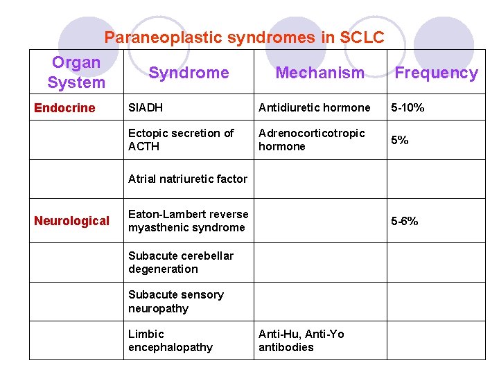
Paraneoplastic syndromes in SCLC Organ System Endocrine Syndrome Mechanism Frequency SIADH Antidiuretic hormone 5 -10% Ectopic secretion of ACTH Adrenocorticotropic hormone 5% Atrial natriuretic factor Neurological Eaton-Lambert reverse myasthenic syndrome 5 -6% Subacute cerebellar degeneration Subacute sensory neuropathy Limbic encephalopathy Anti-Hu, Anti-Yo antibodies
Non–small-cell lung cancers (NSCLCs) l Non-small cell cancer requires meticulous staging, because the treatment and prognosis vary widely depending on the stage. ¡ Surgical resection offers patients the best chance for survival. Surgery may be curative for stage I and stage II disease.
Pathophysiology Bronchogenic carcinoma is the most common cancer and the most common cause of cancer-related death in both men and women. l Risk factors for lung cancer include the following: ¡ Cigarette smoking: Smoking increases the risk of bronchogenic carcinoma by 4 -120 times. ¡ Exposure to asbestos: Asbestos exposure increases the risk 4 - or 5 -fold, or as much as 100 -fold if the exposed individual is also a smoker. l Fibrosis/Scarring in the Lung: ¡ Peripheral lung cancers are sometimes associated with areas of fibrous scar. In most cases of these "scar cancers, " the fibrosis may result from, rather than precede the development of the neoplasia. ¡ As many as 6 -12% of patients with idiopathic pulmonary fibrosis develop bronchogenic carcinoma (adenocarcinoma). l
¡ Exposure to toxic agents: Agents such as arsenic, nickel (squamous cell carcinoma), chromium, and chloromethyl ether (small cell carcinoma) increase the risk. ¡ Exposure to uranium or radon: Exposure to breakdown products of uranium increases the risk of non-small cell carcinoma, too. ¡ Prior lung cancer: Approximately 10 -32% of patients who survive resection for lung cancer may develop a second primary lung tumor. ¡ Lung disease: The presence of concomitant chronic obstructive pulmonary disease is a risk factor for lung cancer. ¡ HIV infection: In patients with HIV infection the risk of non-small cell lung carcinoma is increased by 6. 5 times.
Location The relative frequency of lung cancer is 3: 2 in the right compared with the left lung and in the upper lobe compared with the lower lobe. l Squamous cell carcinomas occur predominantly in a central location. l Adenocarcinoma presents in approximately 50% of patients as a peripheral lesion. l Tumors arising endobronchially are located in segmental or lobar bronchi. ¡ Fewer than 4% of cancers arise in the apex of the upper lobes, and fewer than 1% arise from the trachea. l
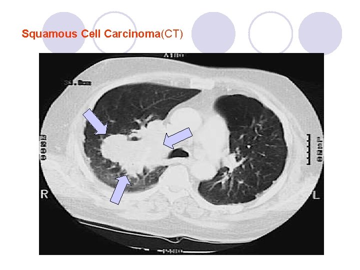
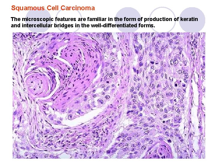
Squamous cell carcinoma (SCC) l l l SCC accounts for 30 -40% of bronchogenic Ca. strong association with smoking. centrally located among all bronchogenic carcinomas, it is most likely to cavitate. SCC grow intraluminally and are least likely to metastasize distantly (<20% of cases at presentation). The mode of spread is direct extension to the local lymph nodes.
l SCC are commonly associated with palmar clubbing and hypertrophic osteoarthropathy. l Hypercalcemia is also commonly observed secondary to a parathormone-like peptide created by the tumor. l Tumors of squamous histology can sometimes elicit a sarcoid reaction in nodes, resulting in nodal enlargement without metastatic spread.
Squamous Cell Carcinoma(CT)
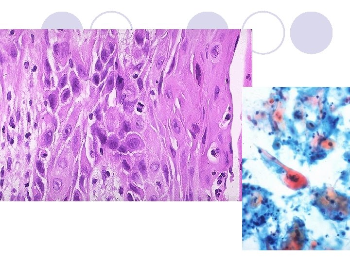
Squamous Cell Carcinoma The microscopic features are familiar in the form of production of keratin and intercellular bridges in the well-differentiated forms.
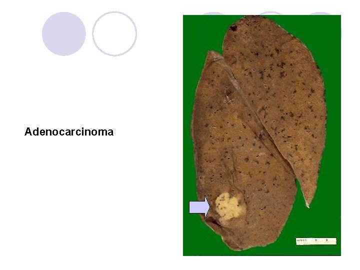
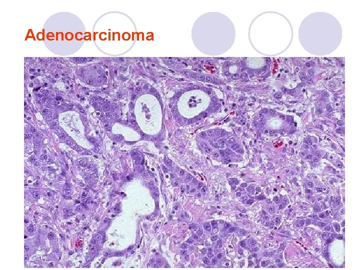
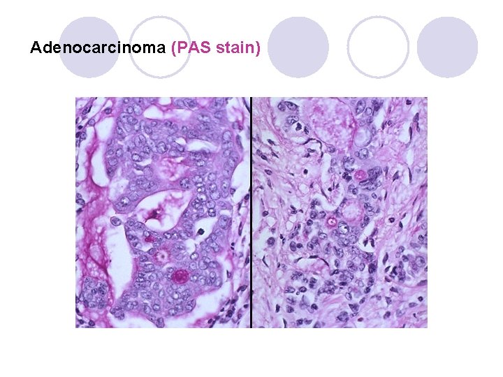
Adenocarcinoma l Adenocarcinoma occurs with a frequency of 3040%, which is higher than the incidence of squamous cell carcinoma. l The lesion is located peripherally in approximately one half of cases. l Adenocarcinoma may arise from a previous scar, it rarely cavitates, and an eccentric pattern of calcification may be evident. l An early propensity is noted of metastases to the lymph nodes, pleura, adrenal glands, central nervous system (CNS), and bone.
Adenocarcinoma
Adenocarcinoma
Adenocarcinoma (PAS stain)
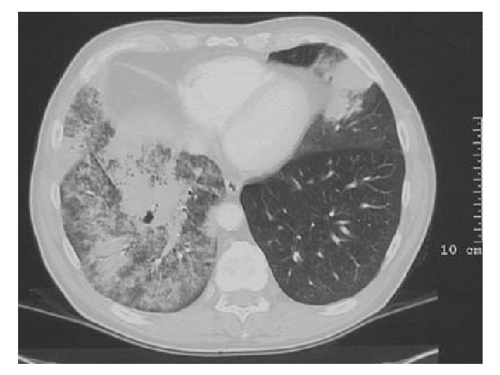
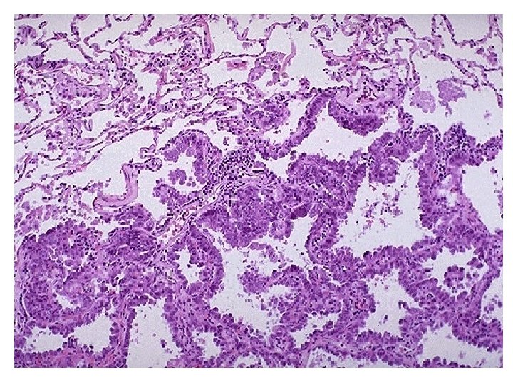
Bronchioloalveolar carcinoma l Bronchioloalveolar carcinoma is a subtype of adenocarcinoma that accounts for as many as 5% of bronchogenic carcinomas and occur in terminal brochioloalveolar regions. l Bronchioloalveolar carcinoma may appear in a variety of ways, including a solitary pulmonary nodule (45%), multiple nodules (25%), and consolidation (30%). l The incidence of bronchioloalveolar carcinoma is increased in patients who have underlying interstitial lung disease, parenchymal scaring, and exogenous lipoid pneumonia.
l Bronchioloalveolar carcinoma is classified as mucinous, nonmucinous and mixed mucinous and nonmucinous on the basis of histopathologic features. The mucinous form is most common (80%) and arises from columnar mucous containing cells. ¡ The mucinous form is likely to be multicentric, it occasionally appears with bronchorrhea, and it has a worse prognosis. ¡ The nonmucinous form arises from type II pneumocytes or Clara cells, it is more likely to be localized, and it has a better prognosis. ¡
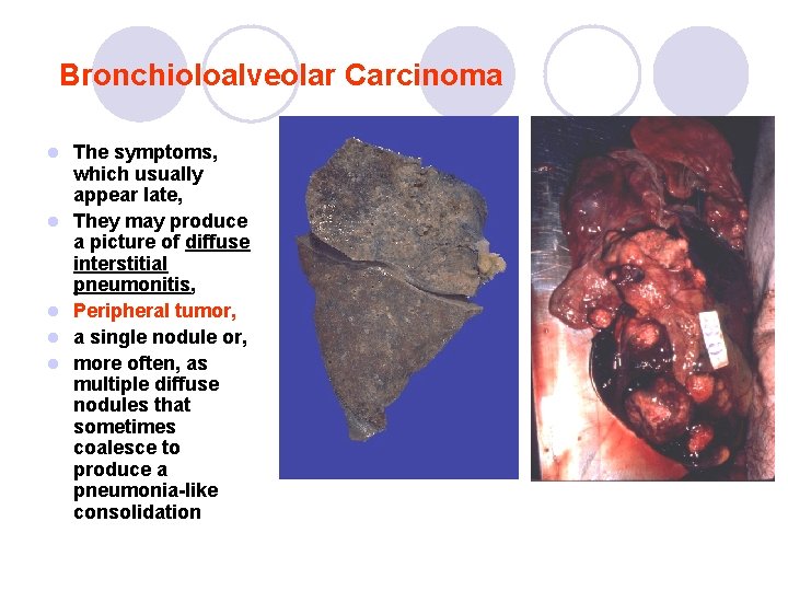
Bronchioloalveolar Carcinoma l l l The symptoms, which usually appear late, They may produce a picture of diffuse interstitial pneumonitis, Peripheral tumor, a single nodule or, more often, as multiple diffuse nodules that sometimes coalesce to produce a pneumonia-like consolidation
Large cell carcinoma l Large cell carcinomas account for only 7 -10% of bronchogenic carcinomas and are strongly associated with cigarette smoking. l The lesion occurs peripherally and grows rapidly, with early metastases and a poor outcome. l A subtype of large cell carcinoma is giant cell carcinoma. ¡ This is highly malignant and associated with a poor prognosis.
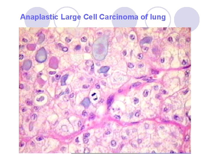
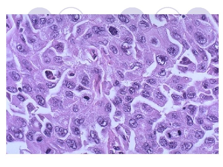
Microscopy l Anaplastic carcinoma ¡ larger, more polygonal cells, vesicular nuclei, l some contain intracellular mucin, l some exhibit multinucleate cells (giant cell carcinoma), l some have cleared cells (clear cell carcinoma), l some have a distinctly spindly histologic appearance (rhabdoid phenotype).
Anaplastic Large Cell Carcinoma of lung
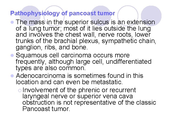
Pancoast tumor is extrathoracic, originating in an extreme peripheral location with a plaquelike extension over the lung apex and principally involving the chest wall structures rather than the underlying lung parenchyma. l Pancoast tumors are a subset of cancers of the lung that invades the apical chest wall. ¡ Because of their location in the pleural apex, they invade adjoining tissue. l The location of the tumor, rather than its pathology or histology of origin, is significant in producing its characteristic clinical pattern. l l LOCATION: Arises in superior sulcus of lung.
Carcinomas in the superior pulmonary sulcus produce the Pancoast syndrome: ¡ pain in the shoulder and along the ulnar distribution of the arm and hand (Brachial plexus involvement) ¡ destruction of adjacent rib or vertebra (1 st rib most often affected), ¡ Horner syndrome (sympathetic chain or sympathetic cervical ganglia invasion Ptosis, Myosis, Anhydrosis). l These apical chest tumors tend to be locally invasive early. l
Pathophysiology of pancoast tumor l The mass in the superior sulcus is an extension of a lung tumor; most of it lies outside the lung and involves the chest wall, nerve roots, lower trunks of the brachial plexus, sympathetic chain, ganglion, ribs, and bone. l Squamous cell carcinoma occurs more frequently, although large cell, undifferentiated types are also common. l Adenocarcinoma is sometimes found in this location and can even be metastatic. ¡ Involvement of the phrenic or recurrent laryngeal nerve or superior vena cava obstruction is not representative of the classic Pancoast tumor.
Neuroendocrine tumors l Benign tumorlets l Carcinoid tumors Benign tumorlets small, hyperplastic neuroendocrine cell populations, l in the areas of scarring or chronic inflammation. l
Carcinoid tumors l Bronchial carcinoid tumors are rare, accounting for as many as 2. 5% of all pulmonary neoplasms and for 12 -15% of carcinoid tumors overall. l They originate from the neurosecretory cells (Kulchitsky cells; argentaffin cells) of bronchial mucosa. l Bronchial carcinoids are now called as low-grade malignant neoplasms because of their potential to cause local invasion, their tendency for local recurrence, and their occasional metastases to extrathoracic sites.
l Findings & complications: ¡ potential to synthesize and secrete peptide hormones and neuroamines, particularly adrenocorticotropic hormone (ACTH), seratonin, somatostatin, and bradykinin ¡ persistent cough, hemoptysis, impairment of drainage or respiratory passages ¡ secondary infections, ¡ symptoms of the obstructive lung disease l bronchiectasis, l emphysema, l atelectasis.
Complications l Metastasis ¡ Regional lymph nodes ¡ Distant organ metastases: l l Bone Liver Adrenal gland Brain Hemorhhage (common) l Pneumothorax (common) l Pleural effusion (due to invasion) l Infections (pneumonia) l Paraneoplastic syndromes (Hormone producing neoplasms): ADH - Hyponatremia ¡ ACTH – Cushings syndrome ¡ Parathyroid hormone – Hypercalcemia ¡ Gonadotrophins – Gynecomastia ¡ Neuromuscular syndromes ¡ Hypertrophic pulmonary osteoarthropaty (clubbing) ¡ Hematologic manifastations (DIC, thromboflebitis, nonbacterial endocarditis) ¡
Complications: Secondary Pathology l Partial Obstruction of airways: Partial: focal emphysema ¡ Total: atelectasis. ¡ Pulmonary abscesses (silent carcinoma) l Superior vena cava syndrome: l ¡ compression or invasion of the superior vena cava can ¡ venous congestion, ¡ dusky head ¡ arm edema, ¡ circulatory compromise l The impaired drainage of the airways: severe suppurative bronchitis ¡ ulcerative bronchitis ¡ bronchiectasis. ¡ Pleural irritation (invasion) l Pericarditis l
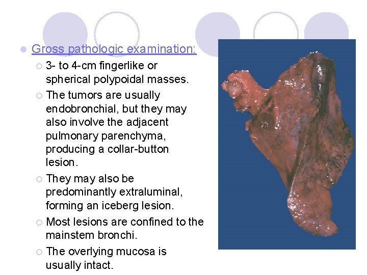
l Gross pathologic examination: 3 - to 4 -cm fingerlike or spherical polypoidal masses. ¡ The tumors are usually endobronchial, but they may also involve the adjacent pulmonary parenchyma, producing a collar-button lesion. ¡ They may also be predominantly extraluminal, forming an iceberg lesion. ¡ Most lesions are confined to the mainstem bronchi. ¡ The overlying mucosa is usually intact. ¡
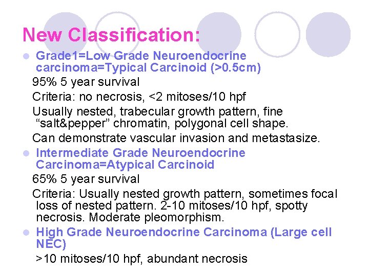
l Histology: Histological classification: Bronchial carcinoids were categorized into 3 groups in the past: l (1) Typical carcinoids, l (2) Atypical carcinoids, l (3) Large-cell neuroendocrine carcinoma (is now classified under “Large cell carcinoma” of lung).
New Classification: Grade 1=Low Grade Neuroendocrine carcinoma=Typical Carcinoid (>0. 5 cm) 95% 5 year survival Criteria: no necrosis, <2 mitoses/10 hpf Usually nested, trabecular growth pattern, fine “salt&pepper” chromatin, polygonal cell shape. Can demonstrate vascular invasion and metastasize. l Intermediate Grade Neuroendocrine Carcinoma=Atypical Carcinoid 65% 5 year survival Criteria: Usually nested growth pattern, sometimes focal loss of nested pattern. 2 -10 mitoses/10 hpf, spotty necrosis. Moderate pleomorphism. l High Grade Neuroendocrine Carcinoma (Large cell NEC) >10 mitoses/10 hpf, abundant necrosis l
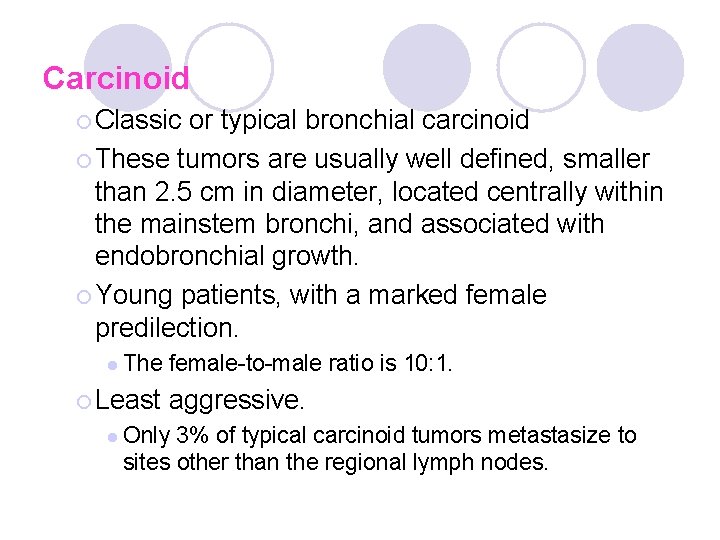
Carcinoid ¡ Classic or typical bronchial carcinoid ¡ These tumors are usually well defined, smaller than 2. 5 cm in diameter, located centrally within the mainstem bronchi, and associated with endobronchial growth. ¡ Young patients, with a marked female predilection. l The ¡ Least female-to-male ratio is 10: 1. aggressive. l Only 3% of typical carcinoid tumors metastasize to sites other than the regional lymph nodes.
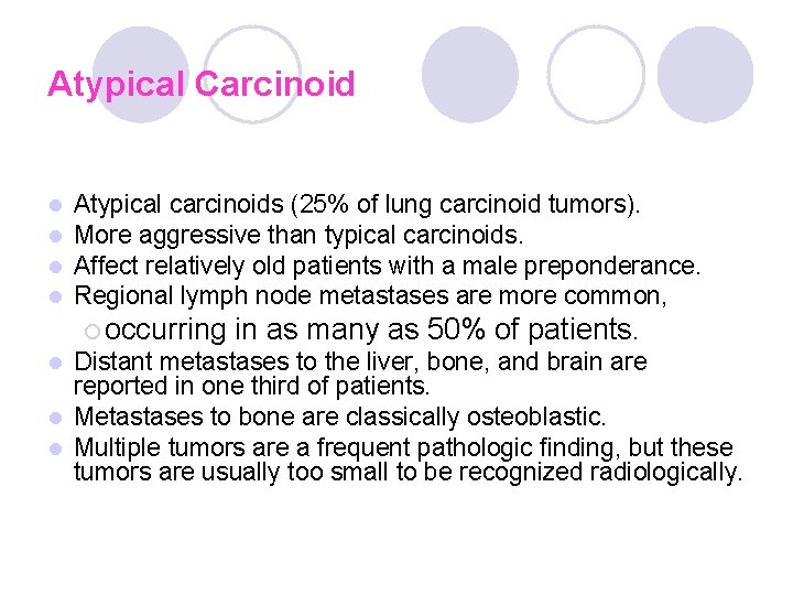
Atypical Carcinoid l l Atypical carcinoids (25% of lung carcinoid tumors). More aggressive than typical carcinoids. Affect relatively old patients with a male preponderance. Regional lymph node metastases are more common, ¡ occurring in as many as 50% of patients. Distant metastases to the liver, bone, and brain are reported in one third of patients. l Metastases to bone are classically osteoblastic. l Multiple tumors are a frequent pathologic finding, but these tumors are usually too small to be recognized radiologically. l
Large-cell neuroendocrine carcinoma l. A newly recognized clinicopathologic entity. l This disease is distinct from small-cell carcinoma and has a poorer prognosis. l The clinical features and optimal treatment of a large-cell carcinoma has not yet been established. l Surgical resection is recommended rather than chemoteraphy.
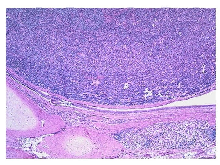
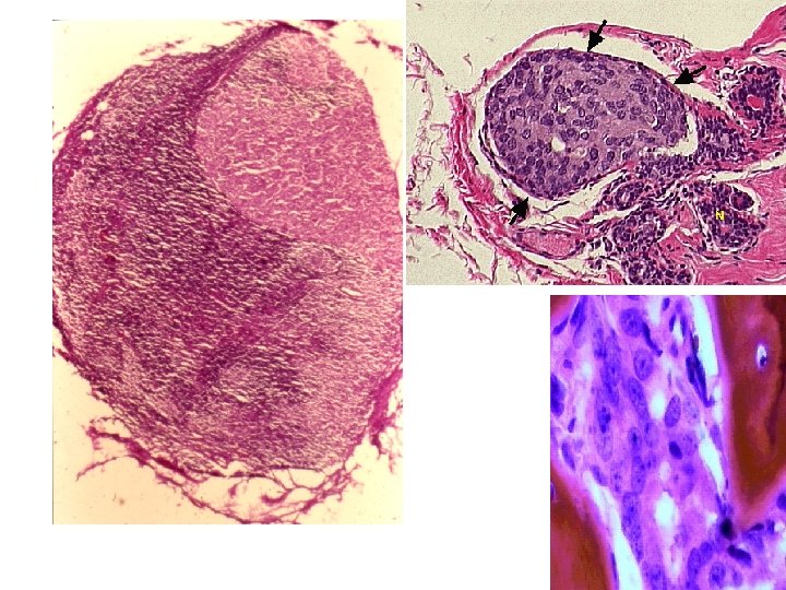
Histologic findings l l l Uniform nests, cords, and masses of small polygonal cells separated by fine fibrovascular stroma. Cells are usually regular, with uniform and round nuclei, Occasional mitoses, Abundant eosinophilic cytoplasm, Pleomorphism with variation in cell or nuclear sizes or shapes is unusual and indicates a more aggressive tumor, as does increased mitotic activity, hyperchromatism, increased cellularity, disorganization and necrosis.
Metastatic Lung Tumors
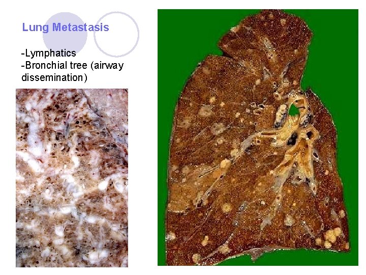
The lung is frequently the site of metastatic neoplasms l Carcinomas and sarcomas arising anywhere l Spread to the lungs via ¡ the blood or ¡ lymphatics or ¡ direct invasion (esophageal carcinomas and mediastinal lymphomas). l
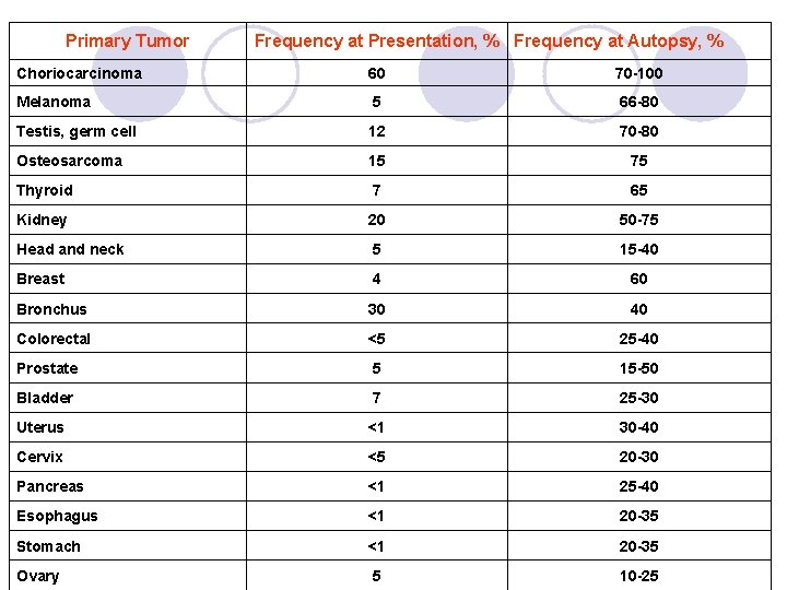
l Pulmonary metastases are common. l They most frequently occur with tumors that have rich systemic venous drainage. ¡ Examples include renal cancers, bone sarcomas, choriocarcinomas, melanomas, testicular teratomas, and thyroid carcinomas. l Most pulmonary metastases arise from common tumors such as breast, colorectal, prostate, bronchial, head and neck, and renal cancers.
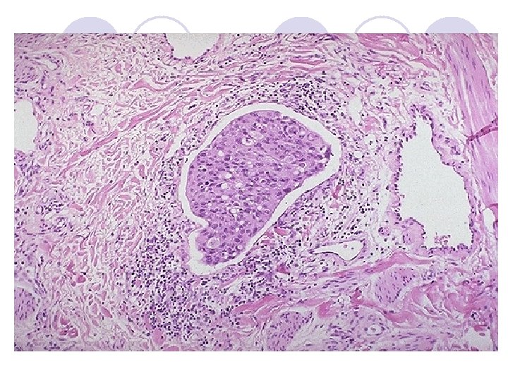
l Intravascular emboli occur most commonly with hepatocellular carcinoma and adenocarcinoma of the breast or stomach. ¡ They may be associated with so called “lymphangitis carcinomatosa”: Lymphatic invasion
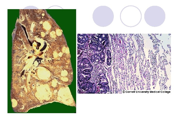
Pathophysiology ¡ Pulmonary nodules are the most common manifestation of secondary neoplastic disease in the lungs. ¡ They are usually derived from tumor emboli arising from invasion of tumor capillaries. ¡ The tumor emboli drain via the systemic veins and pulmonary arteries. ¡ They subsequently lodge in the small pulmonary arteries or arterioles and extend into adjacent lung tissue. ¡ Pulmonary nodules are usually multiple, spherical, and variably sized.
Primary Tumor Frequency at Presentation, % Frequency at Autopsy, % Choriocarcinoma 60 70 -100 Melanoma 5 66 -80 Testis, germ cell 12 70 -80 Osteosarcoma 15 75 Thyroid 7 65 Kidney 20 50 -75 Head and neck 5 15 -40 Breast 4 60 Bronchus 30 40 Colorectal <5 25 -40 Prostate 5 15 -50 Bladder 7 25 -30 Uterus <1 30 -40 Cervix <5 20 -30 Pancreas <1 25 -40 Esophagus <1 20 -35 Stomach <1 20 -35 Ovary 5 10 -25
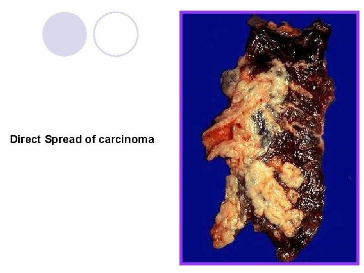
Direct Spread of carcinoma
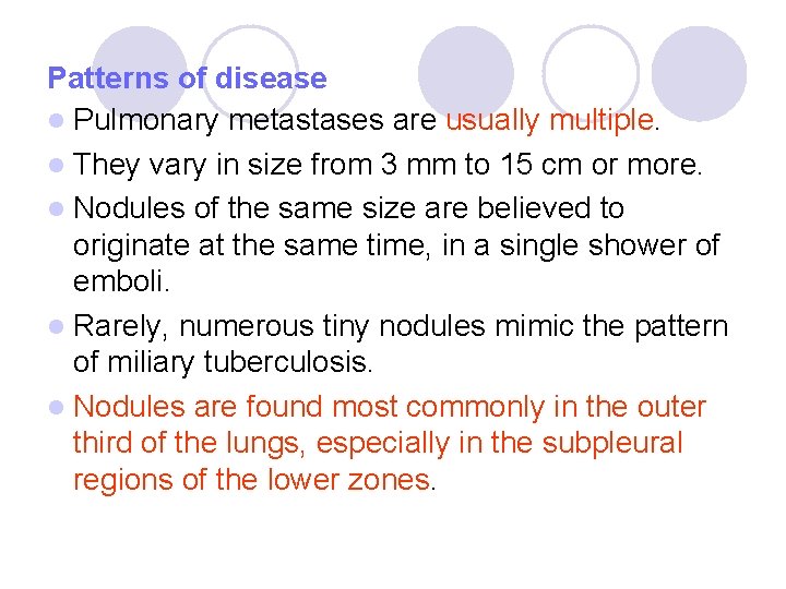
Patterns of disease l Pulmonary metastases are usually multiple. l They vary in size from 3 mm to 15 cm or more. l Nodules of the same size are believed to originate at the same time, in a single shower of emboli. l Rarely, numerous tiny nodules mimic the pattern of miliary tuberculosis. l Nodules are found most commonly in the outer third of the lungs, especially in the subpleural regions of the lower zones.
Lung Metastasis -Lymphatics -Bronchial tree (airway dissemination)
A pneumothorax associated with pulmonary metastases usually indicates that an osteosarcoma is the primary site. l Calcification is seen in metastases from osteogenic sarcoma, synovial sarcoma, or chondrosarcoma. l Hemorrhagic metastases, with a halo of hazy opacity, are most often seen in choriocarcinoma, but also occasionally appear with other vascular tumors such as angiosarcoma or renal cell carcinoma. l Metastases from teratoma of the testis may show complete fibrosis or necrosis after chemotherapy. l
Pathology of Pleura and Mediastinum
l Pleuritis and Pleural effusion l Pneumothorax l Hemothorax and Chylothorax l Neoplastic lesions
Pleural effusion and Pleuritis l Fluid in the pleural space= Effusion l Effusions may be “Transudate” or “exudate” TRANSUDATE l Hydrothrax ¡ Seroangious transudate ¡ Cause: l Congestive heart failure
EXUDATE l Seroangious-fibrinous-suppurative l Causes: ¡ Microbial invasion ¡ Blood-borne infections ¡ Lung cancer invasion ¡ Pulmonary infarction ¡ Viral pleuritis ¡ Systemic connective tissue diseases (SLE, RA) ¡ Uremia ¡ Thoracic surgery
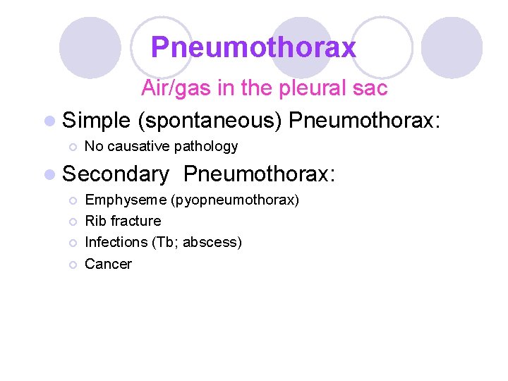
Pneumothorax Air/gas in the pleural sac l Simple (spontaneous) Pneumothorax: ¡ No causative pathology l Secondary ¡ ¡ Pneumothorax: Emphyseme (pyopneumothorax) Rib fracture Infections (Tb; abscess) Cancer
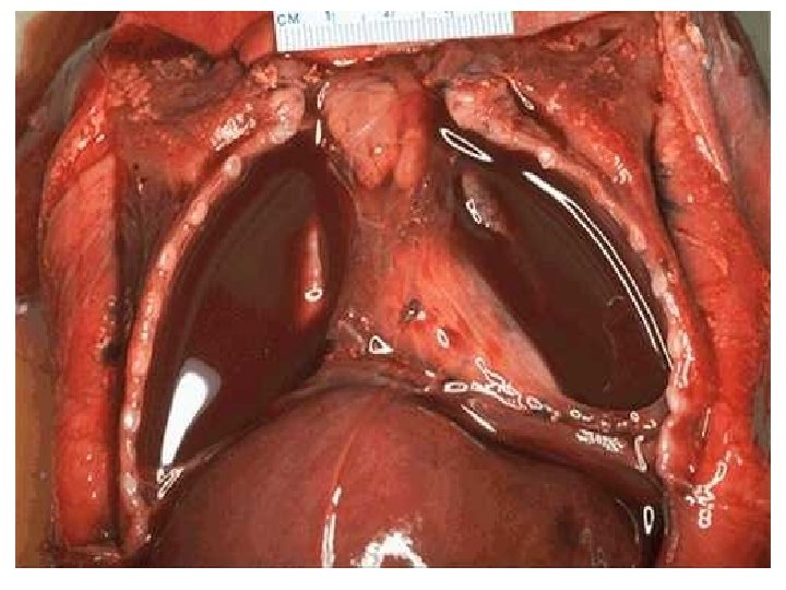
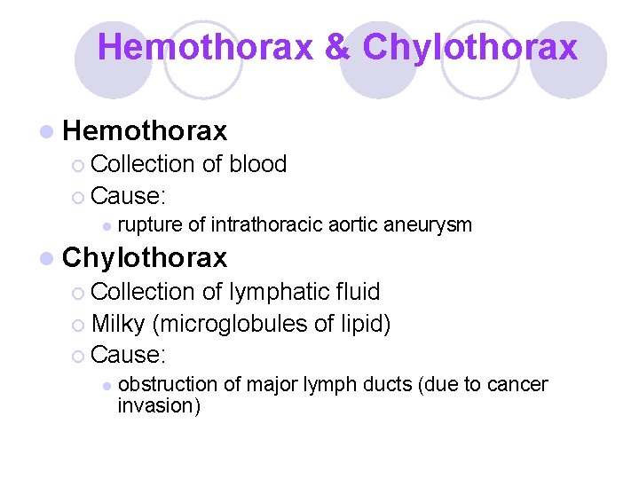
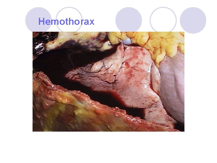
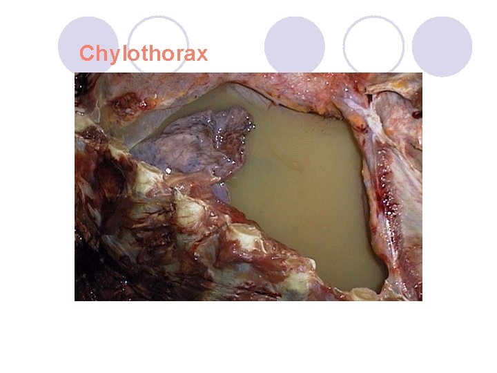
Hemothorax & Chylothorax l Hemothorax ¡ Collection of blood ¡ Cause: l rupture of intrathoracic aortic aneurysm l Chylothorax ¡ Collection of lymphatic fluid ¡ Milky (microglobules of lipid) ¡ Cause: l obstruction of major lymph ducts (due to cancer invasion)
Hemothorax
Chylothorax
Neoplastic lesions of pleura: Malignant mesothelioma l l l Rare. Epitheliod, biphasic and spindle cell variants are present. Pleura (less commonly peritoneum and pericardium) Asbestosis (50% of total cases) Yellow-firm mass obliterating the pleural cavity Distant metastases are rare.
Differential diagnosis: Malignant mesothelioma must be distinguished from adenocarcinoma. Immunhistochemistry and electron microscopy is helpful in the differential diagnosis: Mesothelioma Markers Calretinin Adenocarcinoma Markers TTF-1 WT-1 Carcinoembryonic Antigen Ber. Ep-4 Cytokeratin 5/6