
The Respiratory System Undergraduate Graduate Histology Lecture Series




- Slides: 103
The Respiratory System Undergraduate – Graduate Histology Lecture Series Larry Johnson, Professor Veterinary Integrative Biosciences Texas A&M University College Station, TX 77843
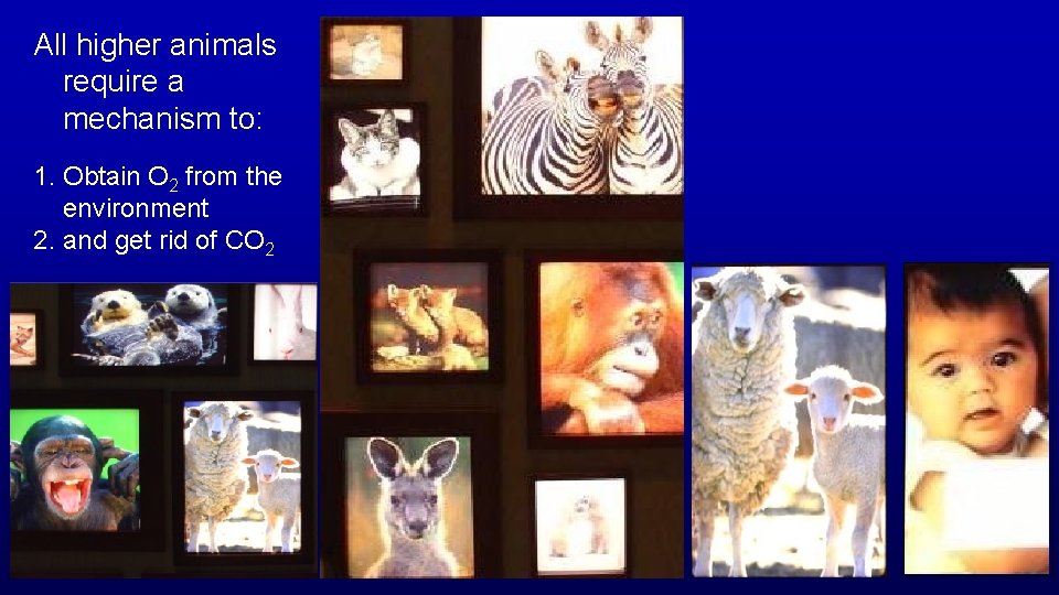
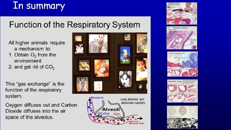
All higher animals require a mechanism to: 1. Obtain O 2 from the environment 2. and get rid of CO 2
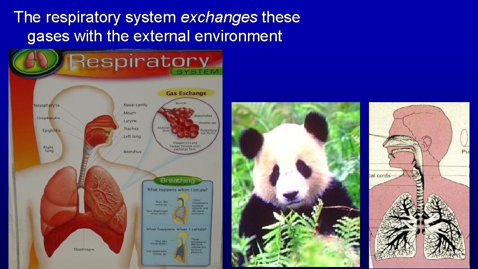
The respiratory system exchanges these gases with the external environment
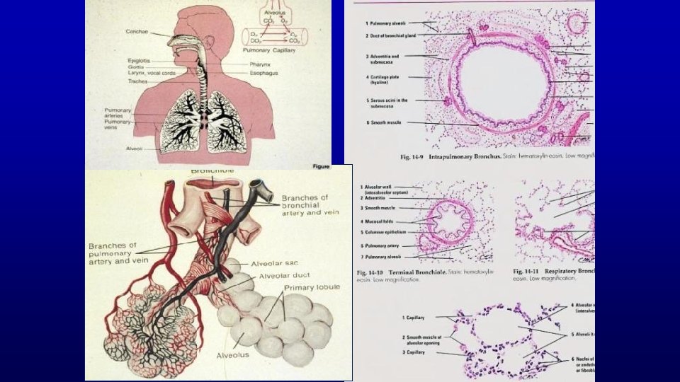
Objectives The histologic characteristics of the components of conducting portion and respiratory portion of the respiratory system How these characteristics allow each component to contribute to the overall function of the respiratory system.
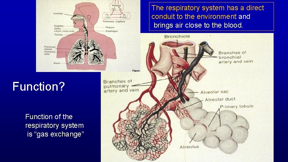
The respiratory system has a direct conduit to the environment and brings air close to the blood. Function? Function of the respiratory system is “gas exchange”
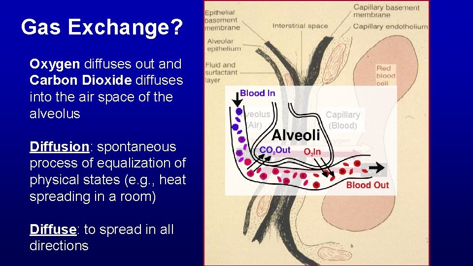
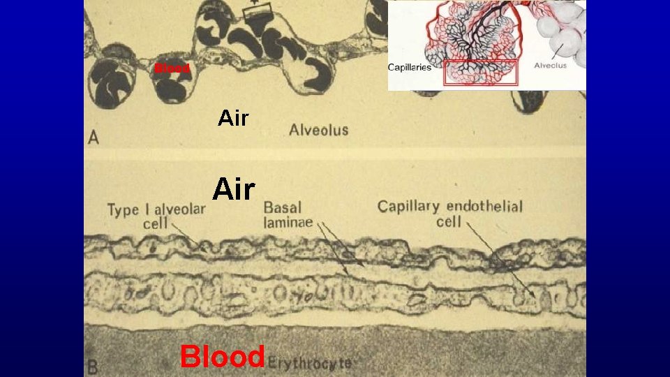
Gas Exchange? Oxygen diffuses out and Carbon Dioxide diffuses into the air space of the alveolus Diffusion: spontaneous process of equalization of physical states (e. g. , heat spreading in a room) Diffuse: to spread in all directions Alveolus (Air) Capillary (Blood) Oxygen CO 2
The respiratory system has a direct conduit to the environment and brings air close to the blood.
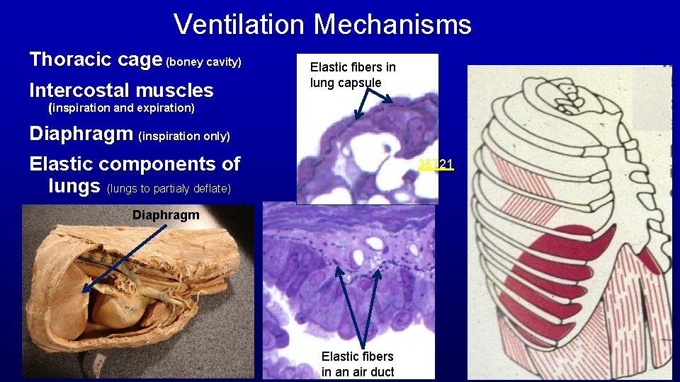
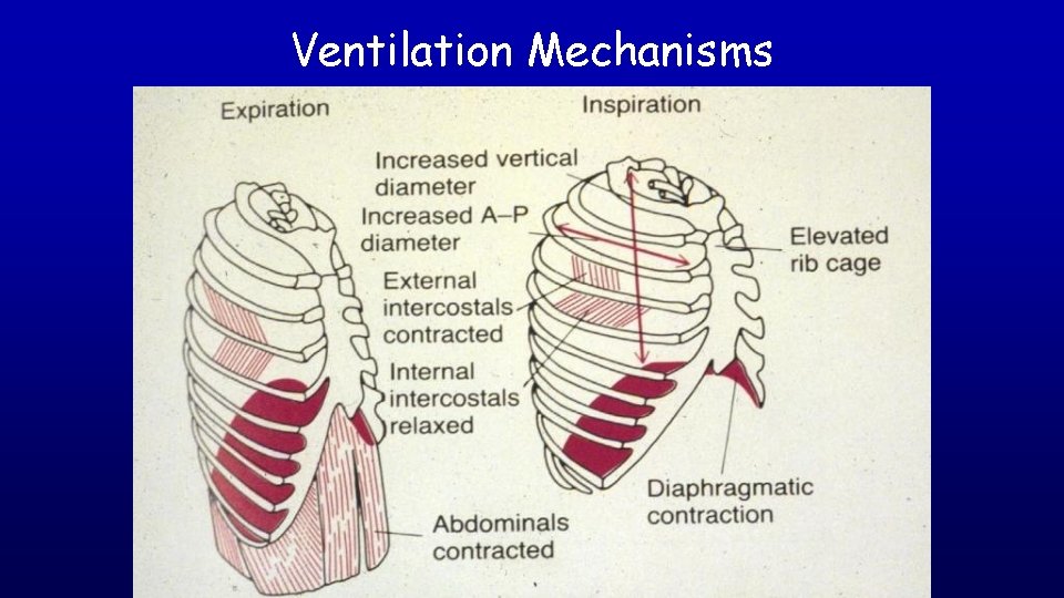
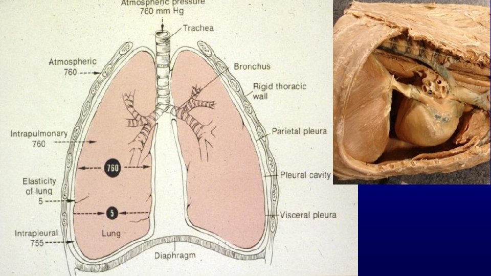
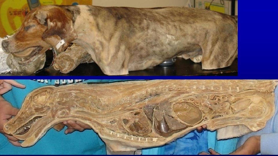
Ventilation Mechanisms Thoracic cage (boney cavity) Intercostal muscles Elastic fibers in lung capsule (inspiration and expiration) Diaphragm (inspiration only) Elastic components of lungs (lungs to partialy deflate) 36721 Diaphragm Elastic fibers in an air duct
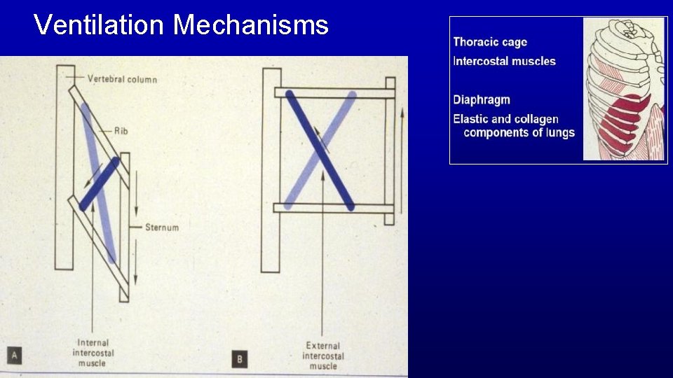
Ventilation Mechanisms
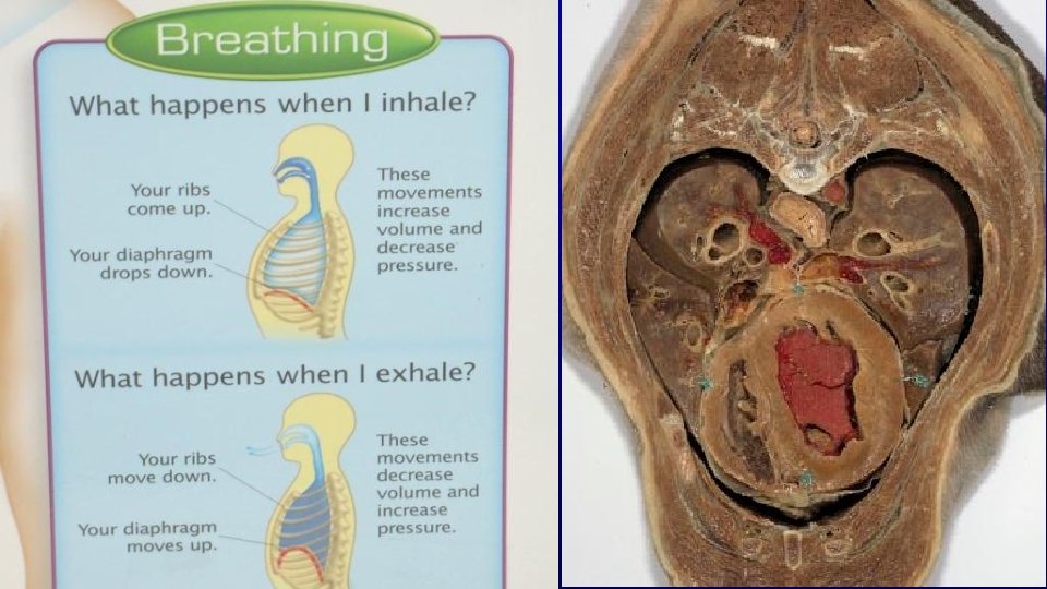
Ventilation Mechanisms
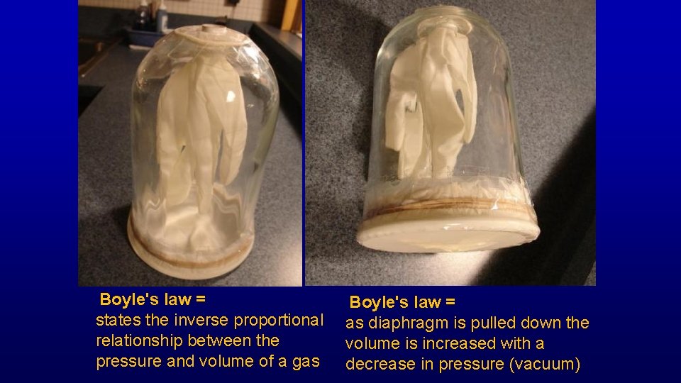
Boyle's law = states the inverse proportional relationship between the pressure and volume of a gas Boyle's law = as diaphragm is pulled down the volume is increased with a decrease in pressure (vacuum)
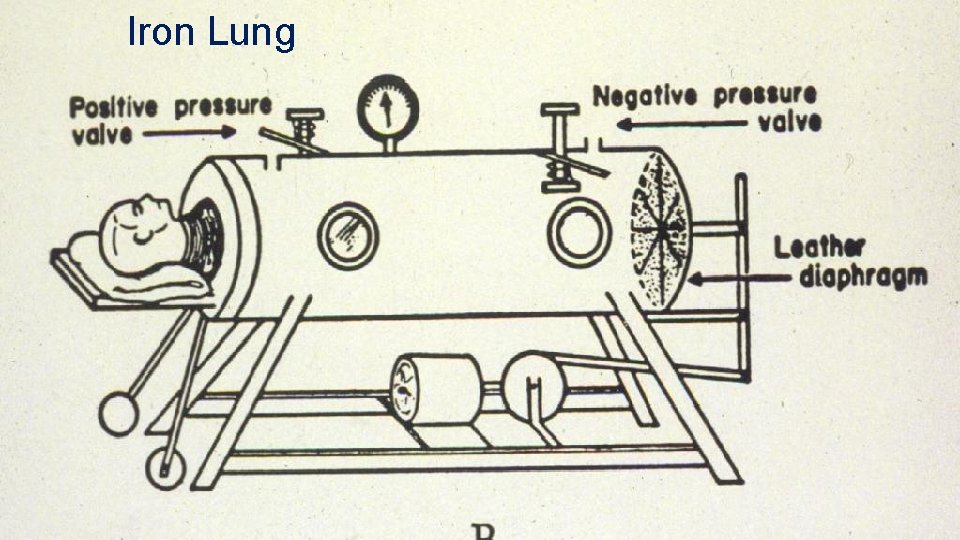
Iron Lung
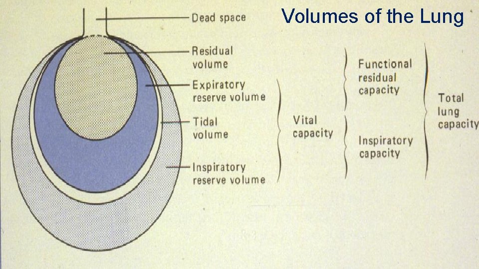
Volumes of the Lung
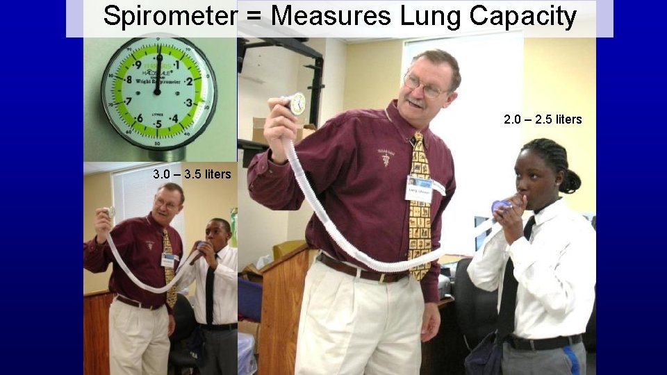
Spirometer = Measures Lung Capacity 2. 0 – 2. 5 liters 3. 0 – 3. 5 liters
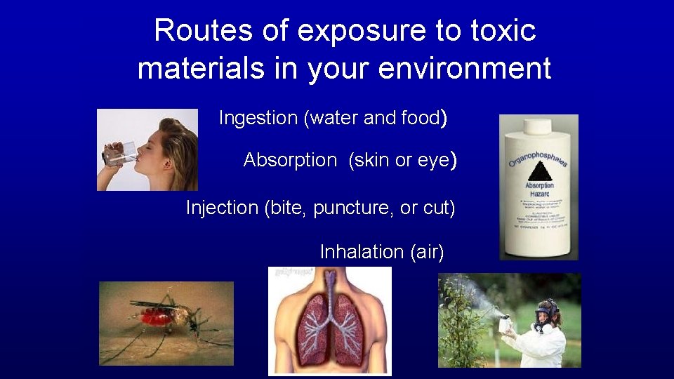
Routes of exposure to toxic materials in your environment Ingestion (water and food) Absorption (skin or eye) Injection (bite, puncture, or cut) Inhalation (air)
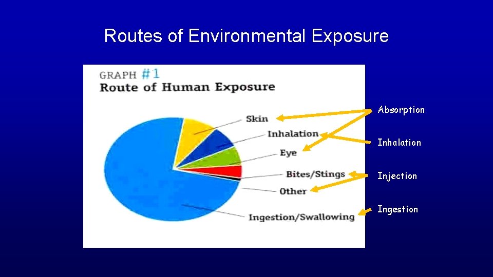
Routes of Environmental Exposure Absorption Inhalation Injection Ingestion
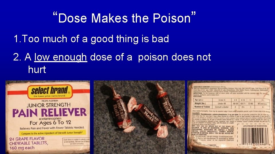
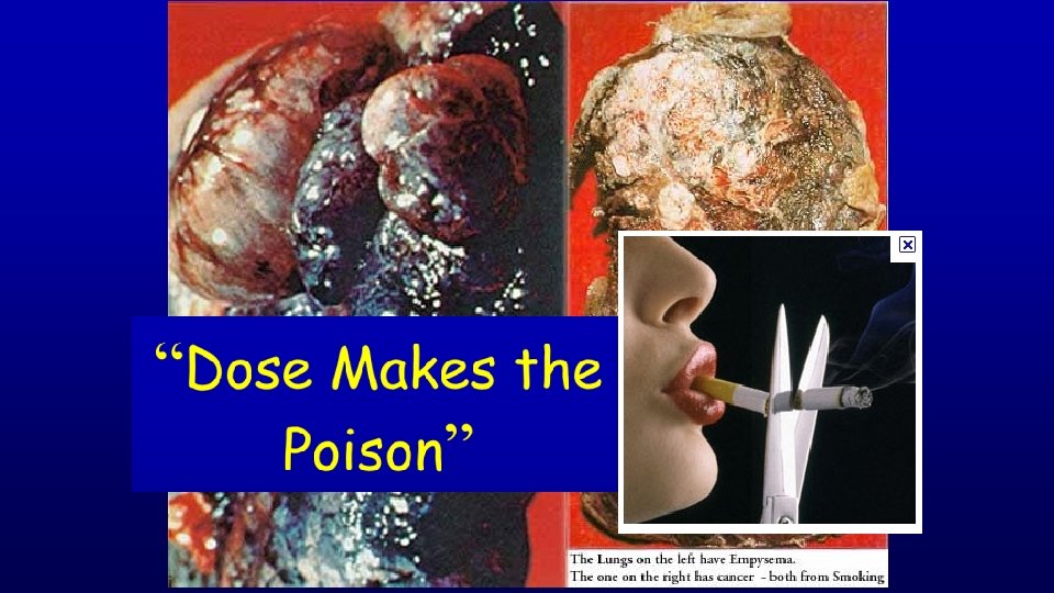
“Dose Makes the Poison” 1. Too much of a good thing is bad 2. A low enough dose of a poison does not hurt
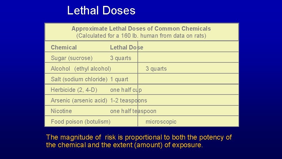
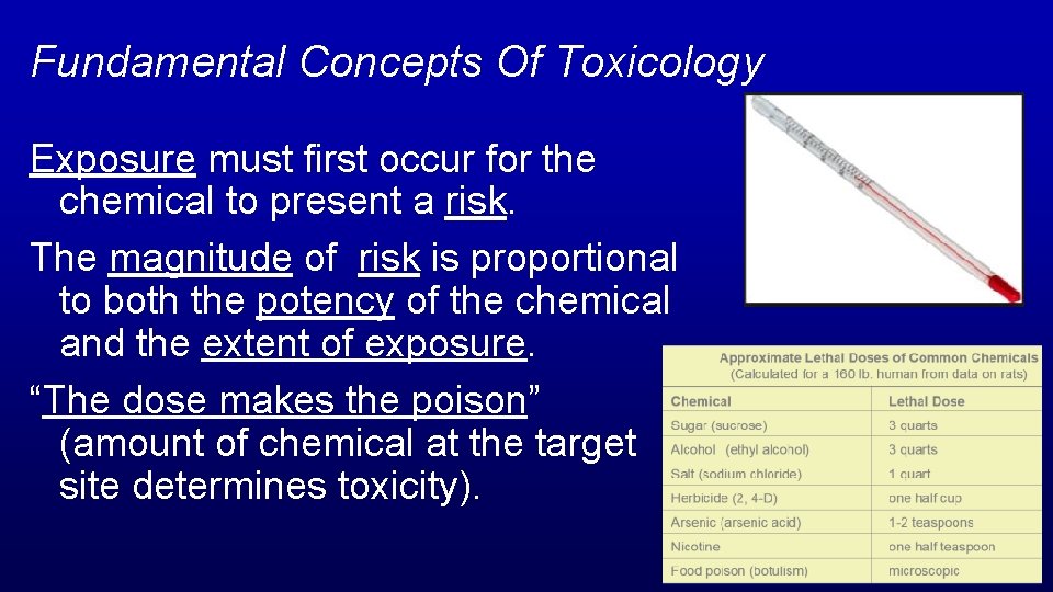
Lethal Doses Approximate Lethal Doses of Common Chemicals (Calculated for a 160 lb. human from data on rats) Chemical Lethal Dose Sugar (sucrose) 3 quarts Alcohol (ethyl alcohol) 3 quarts Salt (sodium chloride) 1 quart Herbicide (2, 4 -D) one half cup Arsenic (arsenic acid) 1 -2 teaspoons Nicotine one half teaspoon Food poison (botulism) microscopic The magnitude of risk is proportional to both the potency of the chemical and the extent (amount) of exposure.
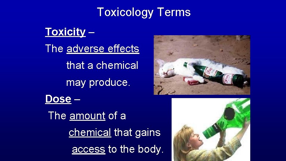
Toxicology Terms Toxicity – The adverse effects that a chemical may produce. Dose – The amount of a chemical that gains access to the body.
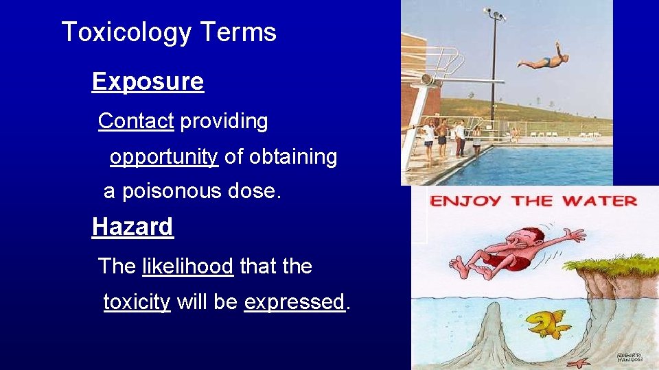
Toxicology Terms Exposure Contact providing opportunity of obtaining a poisonous dose. Hazard The likelihood that the toxicity will be expressed.
Fundamental Concepts Of Toxicology Exposure must first occur for the chemical to present a risk. The magnitude of risk is proportional to both the potency of the chemical and the extent of exposure. “The dose makes the poison” (amount of chemical at the target site determines toxicity).
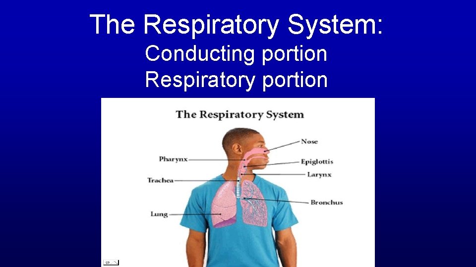
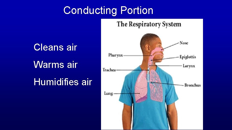
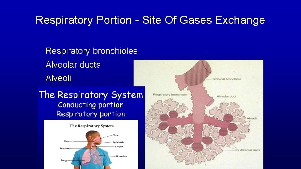
The Respiratory System: Conducting portion Respiratory portion
Conducting Portion Cleans air Warms air Humidifies air
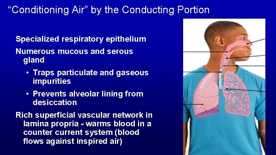
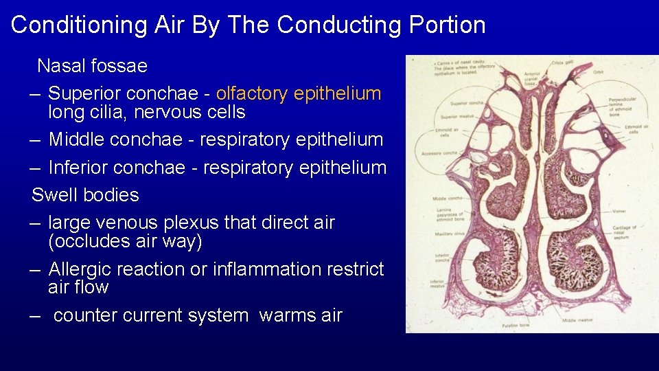
“Conditioning Air” by the Conducting Portion Specialized respiratory epithelium Numerous mucous and serous gland • Traps particulate and gaseous impurities • Prevents alveolar lining from desiccation Rich superficial vascular network in lamina propria - warms blood in a counter current system (blood flows against inspired air)
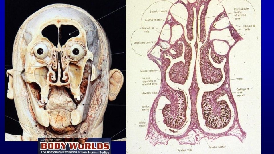
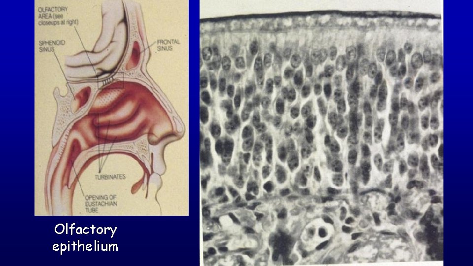
Conditioning Air By The Conducting Portion Nasal fossae – Superior conchae - olfactory epithelium long cilia, nervous cells – Middle conchae - respiratory epithelium – Inferior conchae - respiratory epithelium Swell bodies – large venous plexus that direct air (occludes air way) – Allergic reaction or inflammation restrict air flow – counter current system warms air
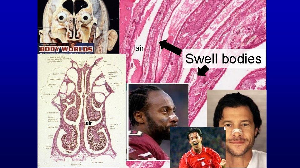
air Swell bodies
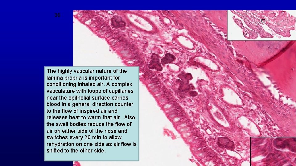
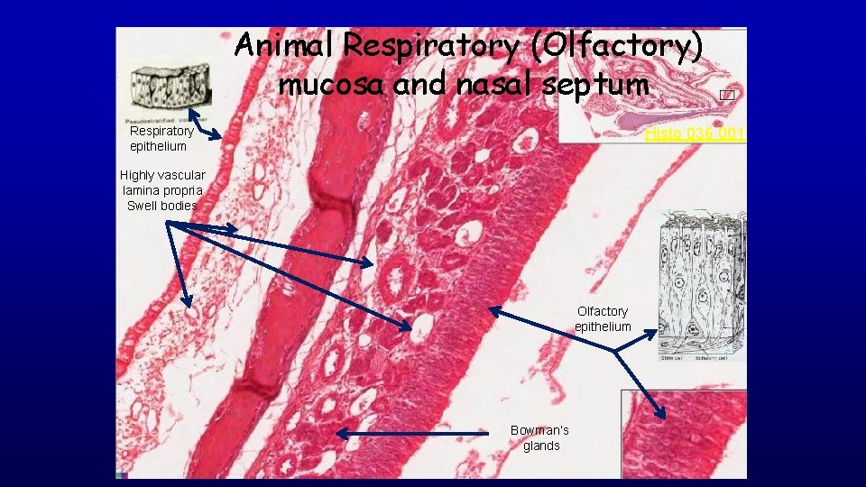
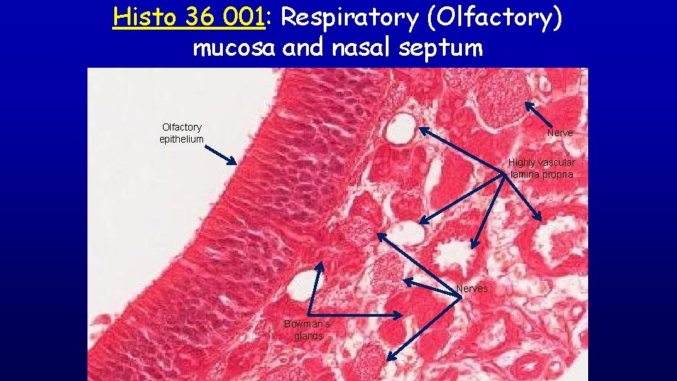
36 The highly vascular nature of the lamina propria is important for conditioning inhaled air. A complex vasculature with loops of capillaries near the epithelial surface carries blood in a general direction counter to the flow of inspired air and releases heat to warm that air. Also, the swell bodies reduce the flow of air on either side of the nose and switches every 30 min to allow rehydration on one side as air flow is shifted to the other side.
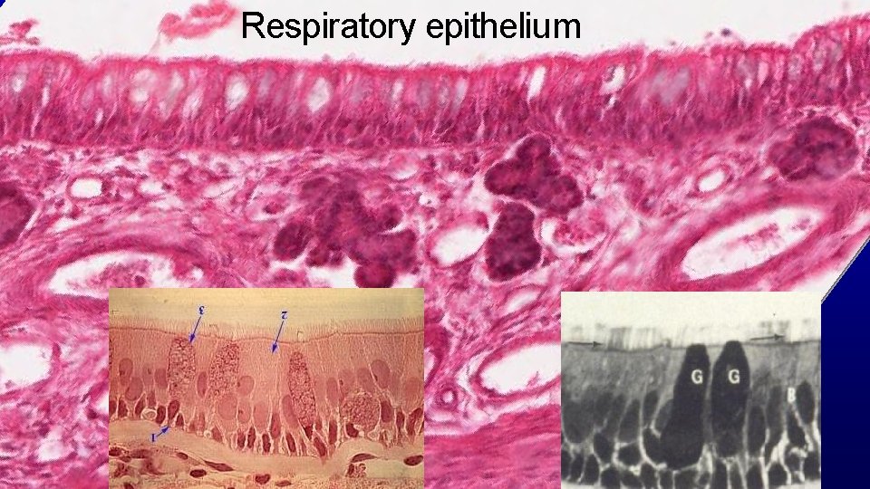
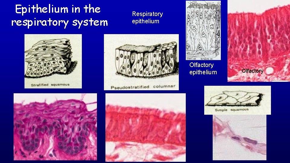
Respiratory epithelium 36
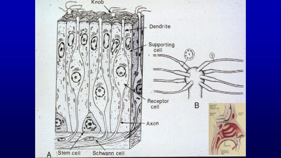
Olfactory epithelium
Animal Respiratory (Olfactory) mucosa and nasal septum Respiratory epithelium Histo 036 001 Highly vascular lamina propria Swell bodies Olfactory epithelium Bowman’s glands
Histo 36 001: Respiratory (Olfactory) mucosa and nasal septum Olfactory epithelium Nerve Highly vascular lamina propria Nerves Bowman’s glands
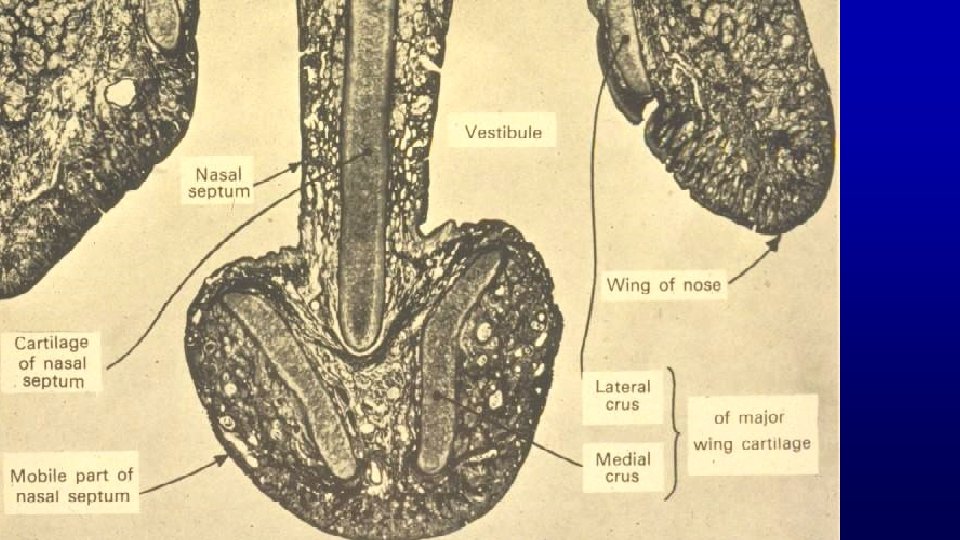
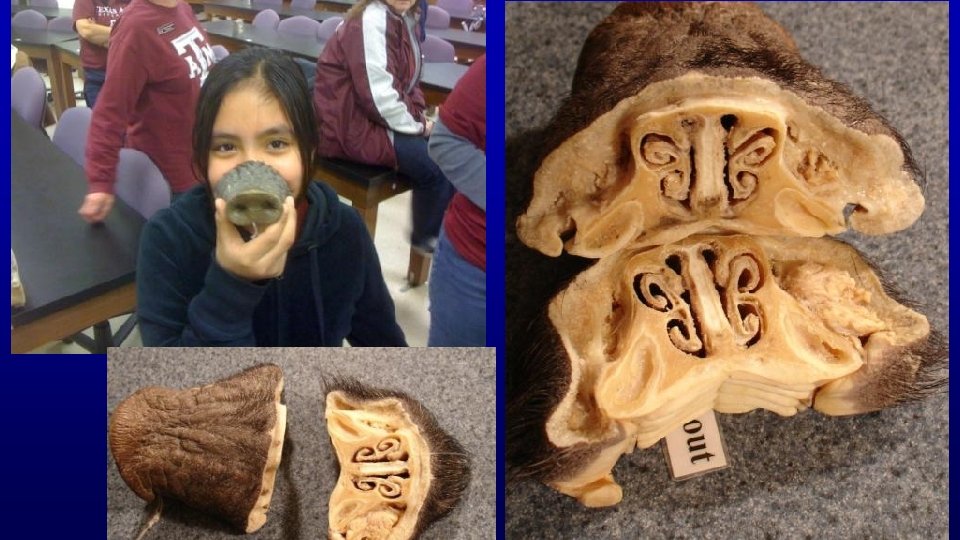
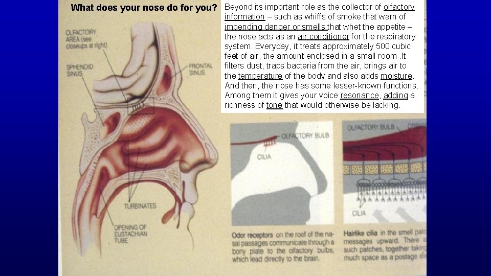
What does your nose do for you? Beyond its important role as the collector of olfactory information – such as whiffs of smoke that warn of impending danger or smells that whet the appetite – the nose acts as an air conditioner for the respiratory system. Everyday, it treats approximately 500 cubic feet of air, the amount enclosed in a small room. It filters dust, traps bacteria from the air, brings air to the temperature of the body and also adds moisture. And then, the nose has some lesser-known functions. Among them it gives your voice resonance, adding a richness of tone that would otherwise be lacking.
Epithelium in the respiratory system Respiratory epithelium Olfactory
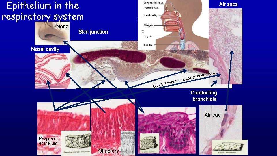
Epithelium in the respiratory system Nose Air sacs Skin junction Nasal cavity mple ted si helium colu epit mnar Cilia Histo 36 Conducting bronchiole Air sac Respiratory epithelium Olfactory
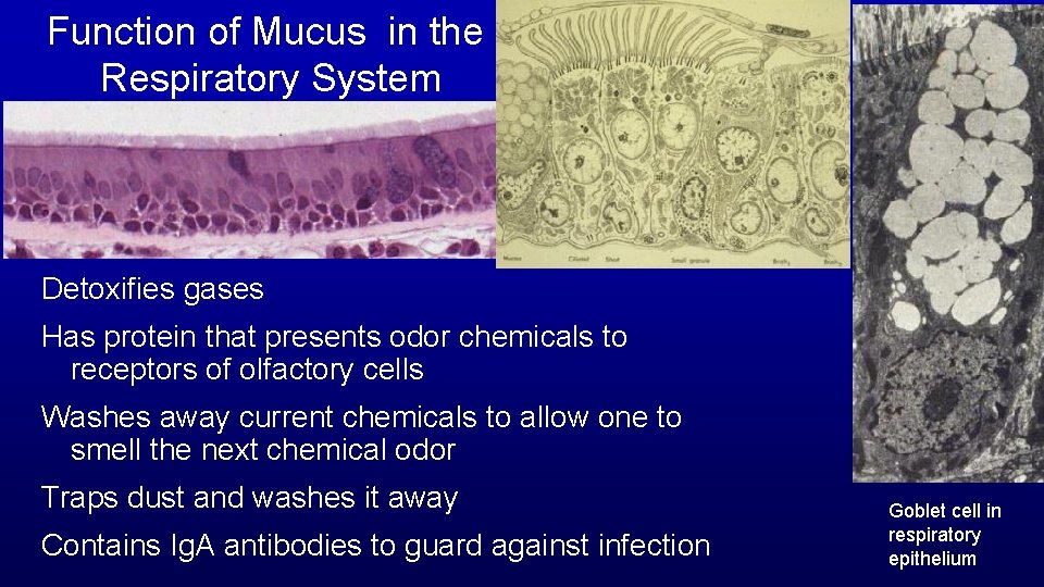
Function of Mucus in the Respiratory System Detoxifies gases Has protein that presents odor chemicals to receptors of olfactory cells Washes away current chemicals to allow one to smell the next chemical odor Traps dust and washes it away Contains Ig. A antibodies to guard against infection Goblet cell in respiratory epithelium
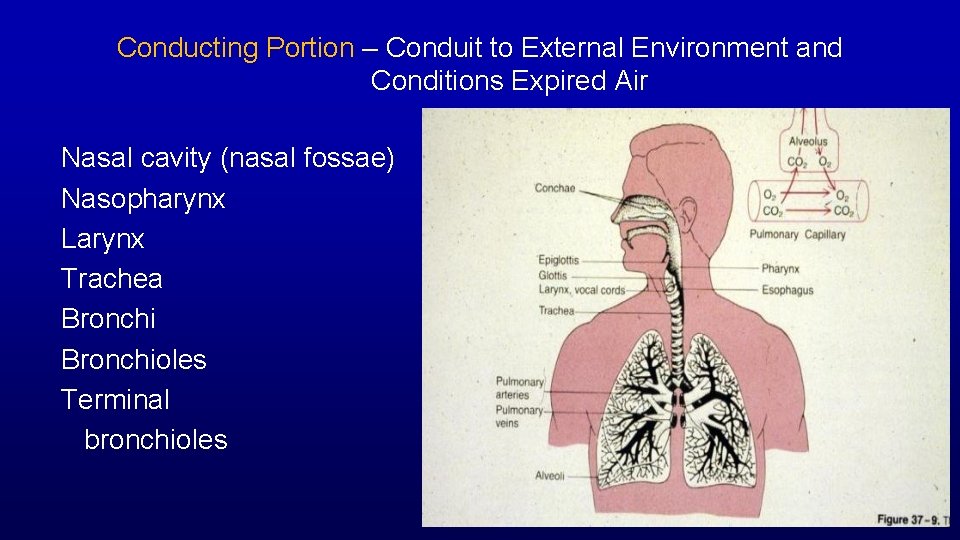
Conducting Portion – Conduit to External Environment and Conditions Expired Air Nasal cavity (nasal fossae) Nasopharynx Larynx Trachea Bronchioles Terminal bronchioles
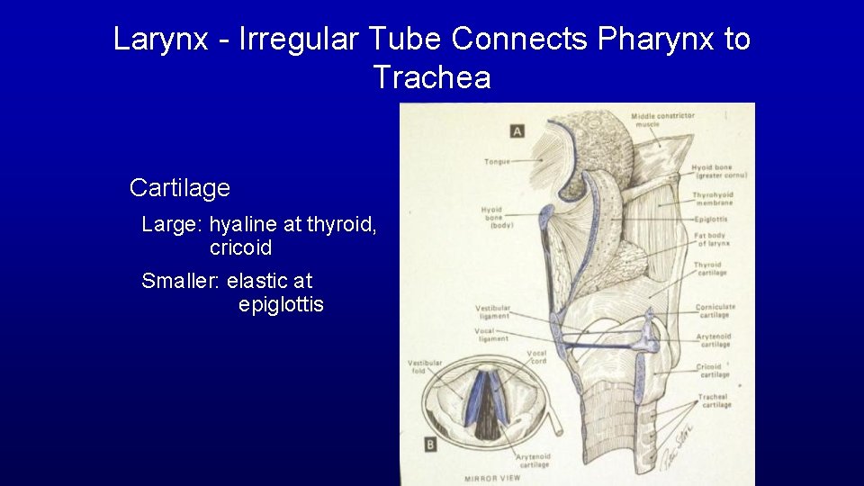
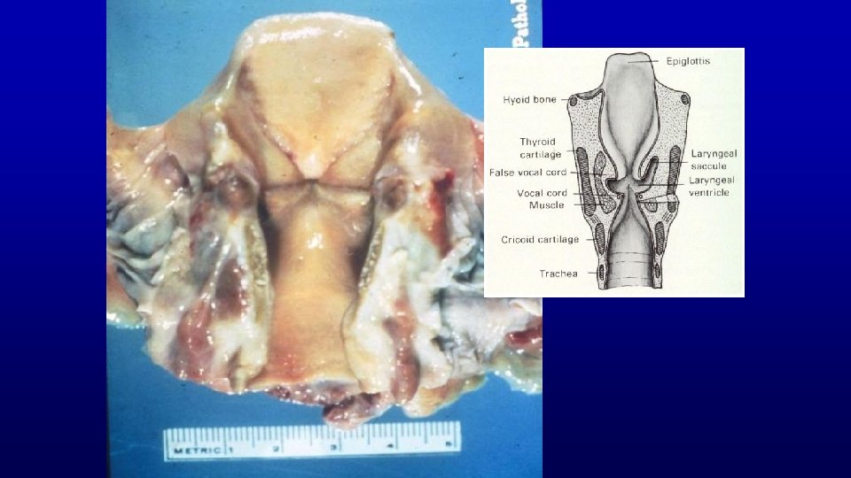
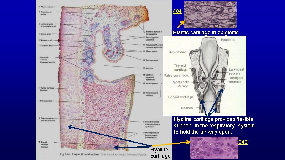
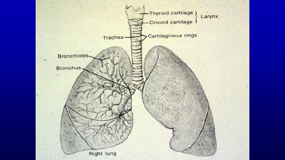
Larynx - Irregular Tube Connects Pharynx to Trachea Cartilage Large: hyaline at thyroid, cricoid Smaller: elastic at epiglottis
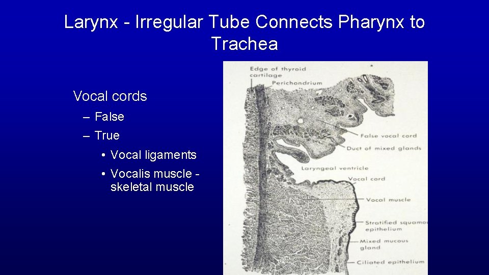
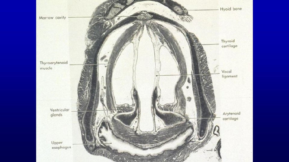
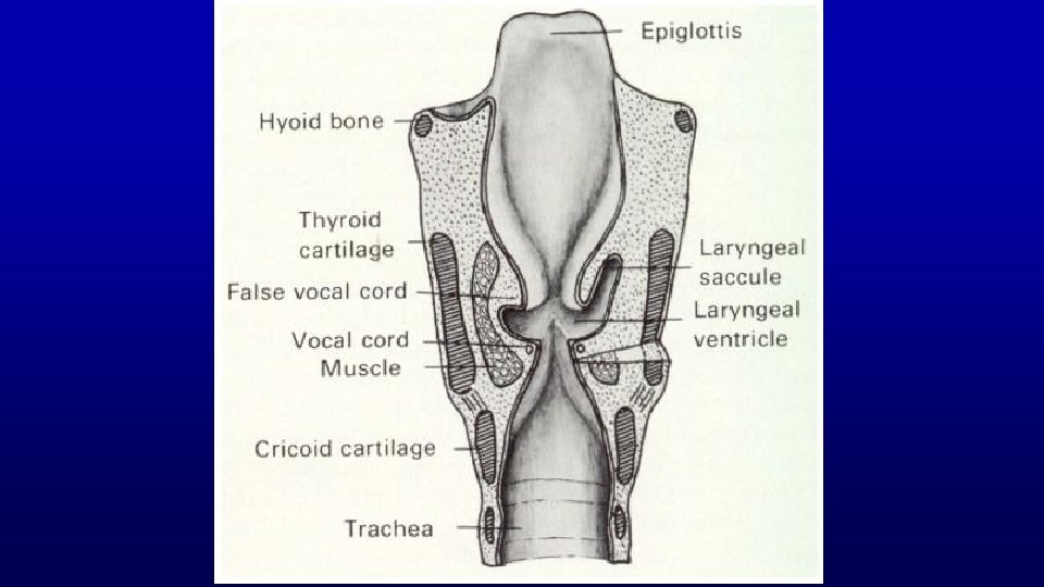
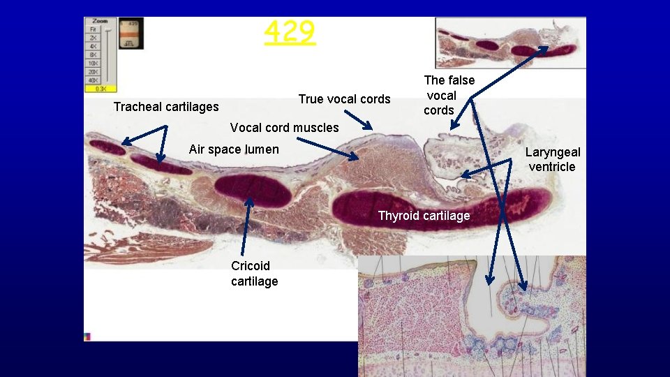
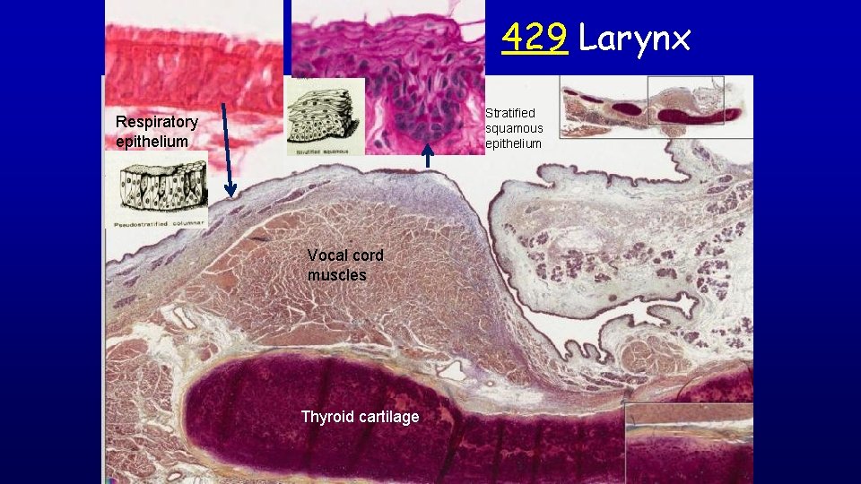
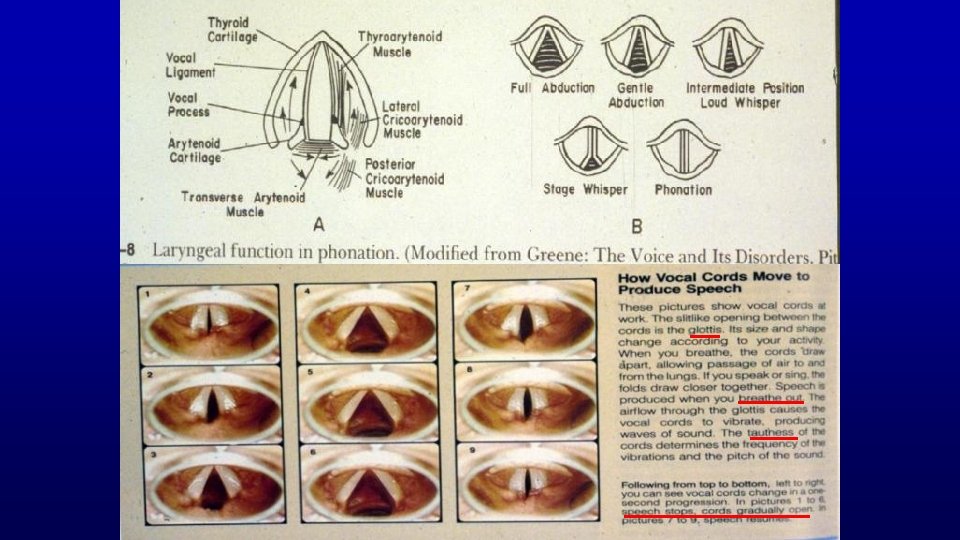
Larynx - Irregular Tube Connects Pharynx to Trachea Vocal cords – False – True • Vocal ligaments • Vocalis muscle skeletal muscle
404 Elastic cartilage in epiglottis Hyaline cartilage provides flexible support in the respiratory system to hold the air way open. 242 Hyaline cartilage
429 larynx True vocal cords Tracheal cartilages The false vocal cords Vocal cord muscles Air space lumen Laryngeal ventricle Thyroid cartilage Cricoid cartilage
429 Larynx Stratified squamous epithelium Respiratory epithelium Vocal cord muscles Thyroid cartilage
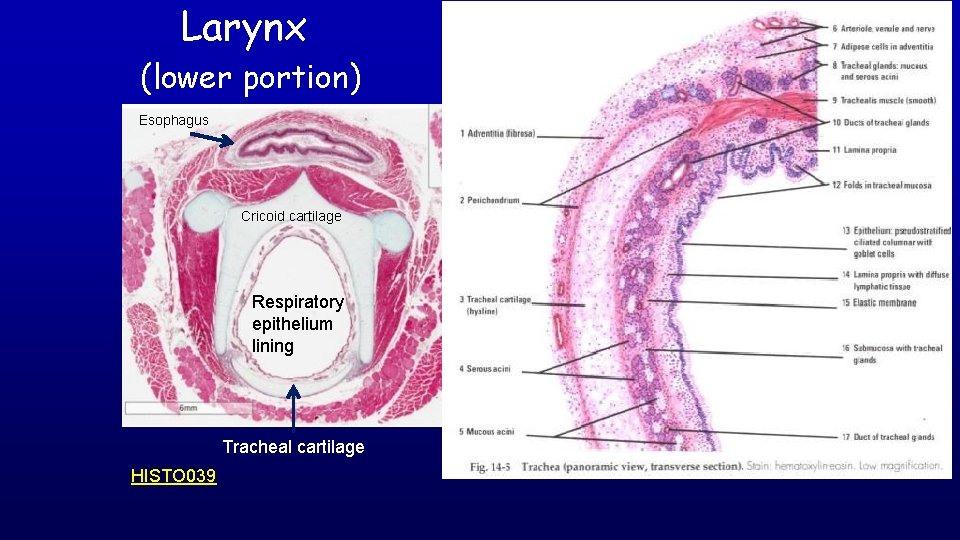
Larynx (lower portion) Esophagus Cricoid cartilage Respiratory epithelium lining Tracheal cartilage HISTO 039
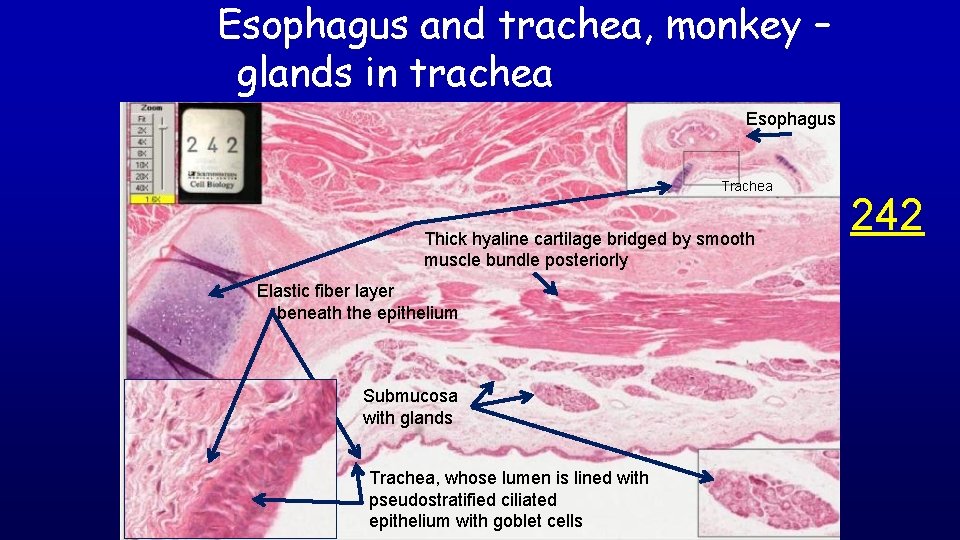
Esophagus and trachea, monkey – glands in trachea Esophagus Trachea Thick hyaline cartilage bridged by smooth muscle bundle posteriorly Elastic fiber layer beneath the epithelium Submucosa with glands Trachea, whose lumen is lined with pseudostratified ciliated epithelium with goblet cells 242
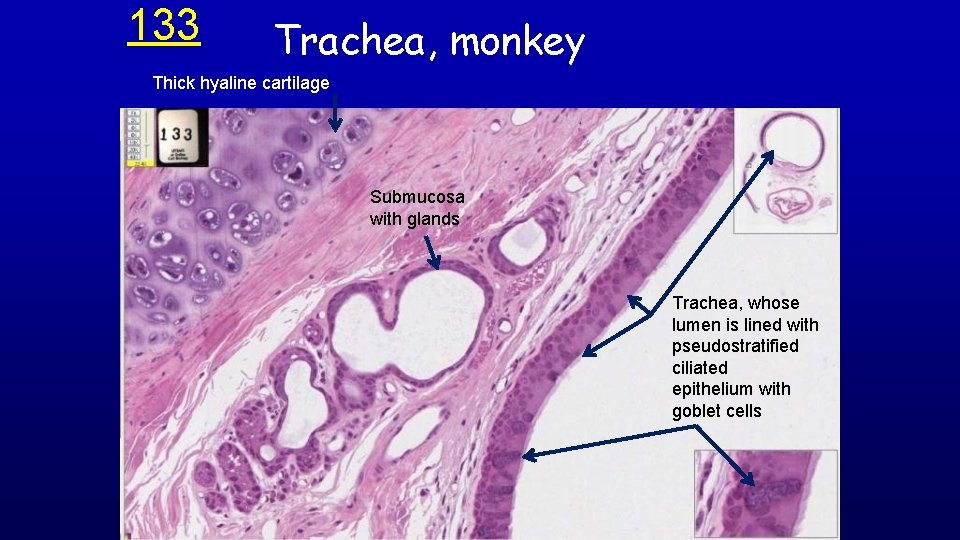
133 Trachea, monkey Thick hyaline cartilage Submucosa with glands Trachea, whose lumen is lined with pseudostratified ciliated epithelium with goblet cells
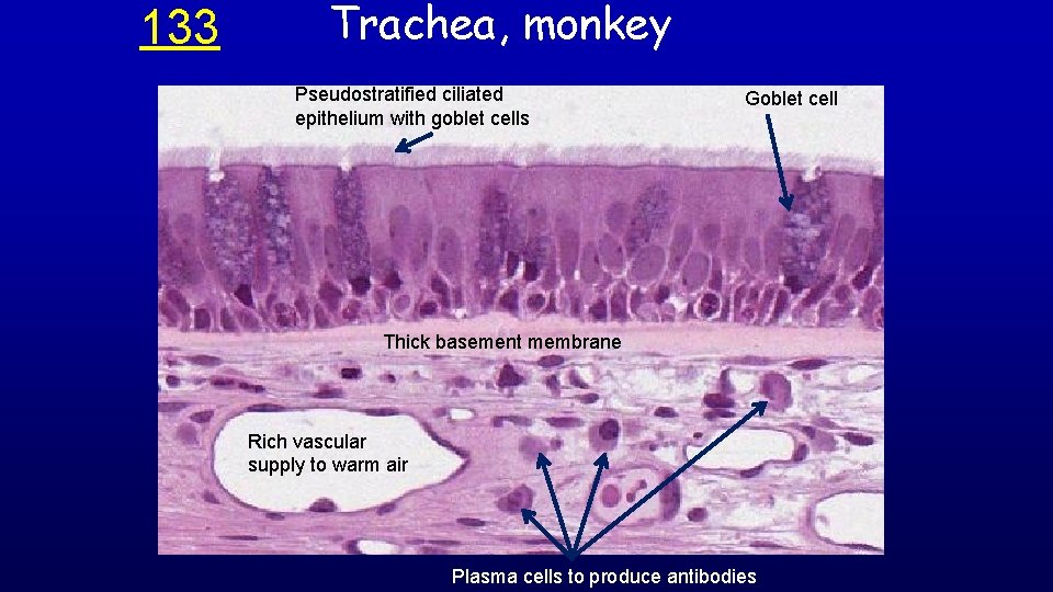
133 Trachea, monkey Pseudostratified ciliated epithelium with goblet cells Goblet cell Thick basement membrane Rich vascular supply to warm air Plasma cells to produce antibodies
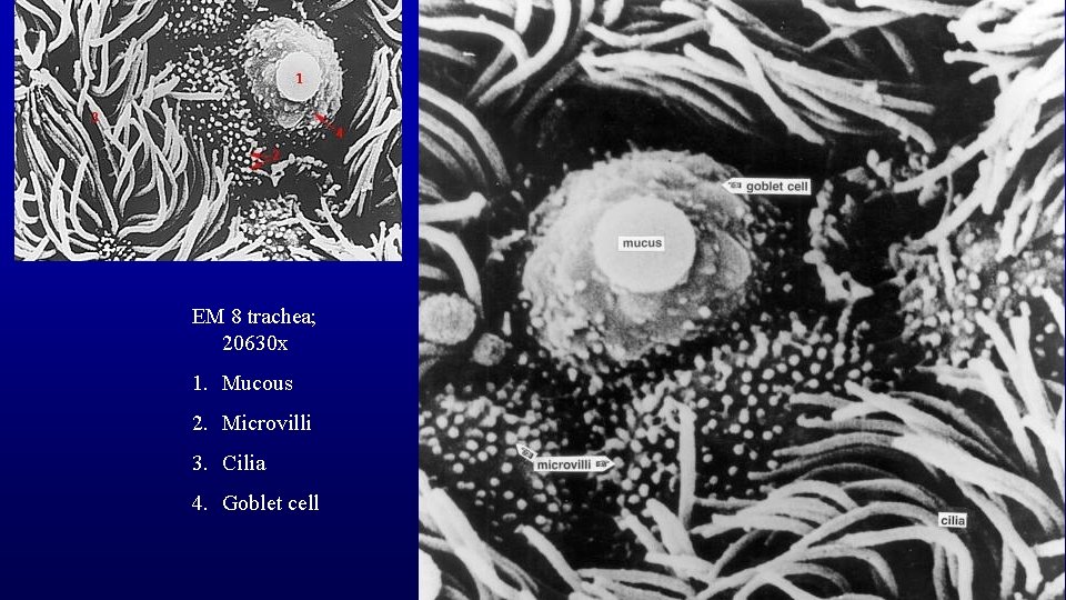
EM 8 trachea; 20630 x 1. Mucous 2. Microvilli 3. Cilia 4. Goblet cell
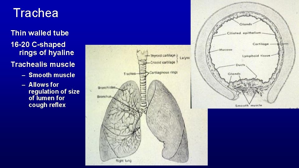
Trachea Thin walled tube 16 -20 C-shaped rings of hyaline Trachealis muscle – Smooth muscle – Allows for regulation of size of lumen for cough reflex
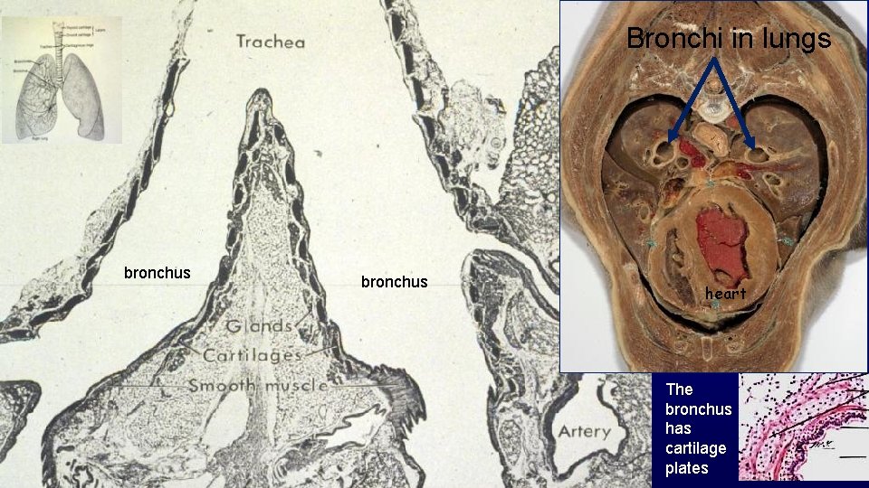
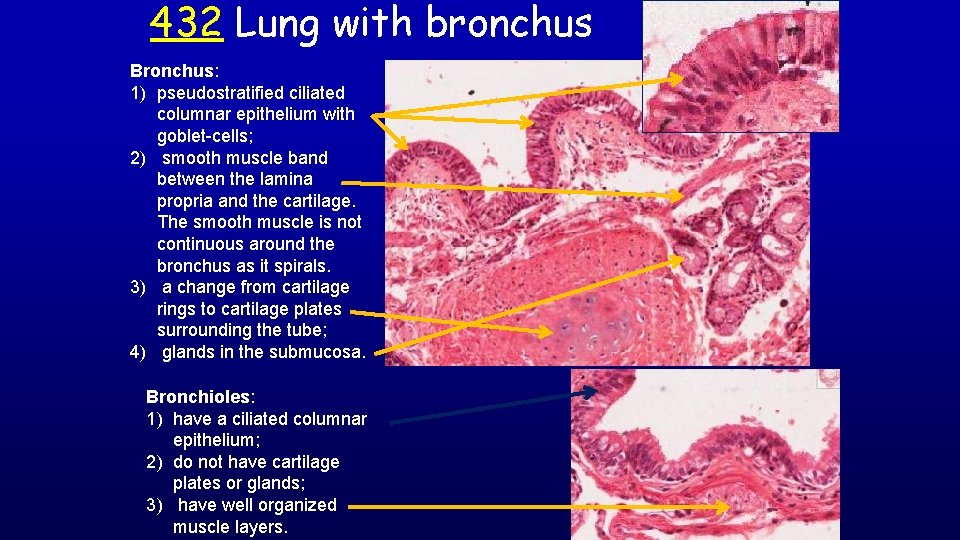
Bronchi in lungs bronchus heart The bronchus has cartilage plates
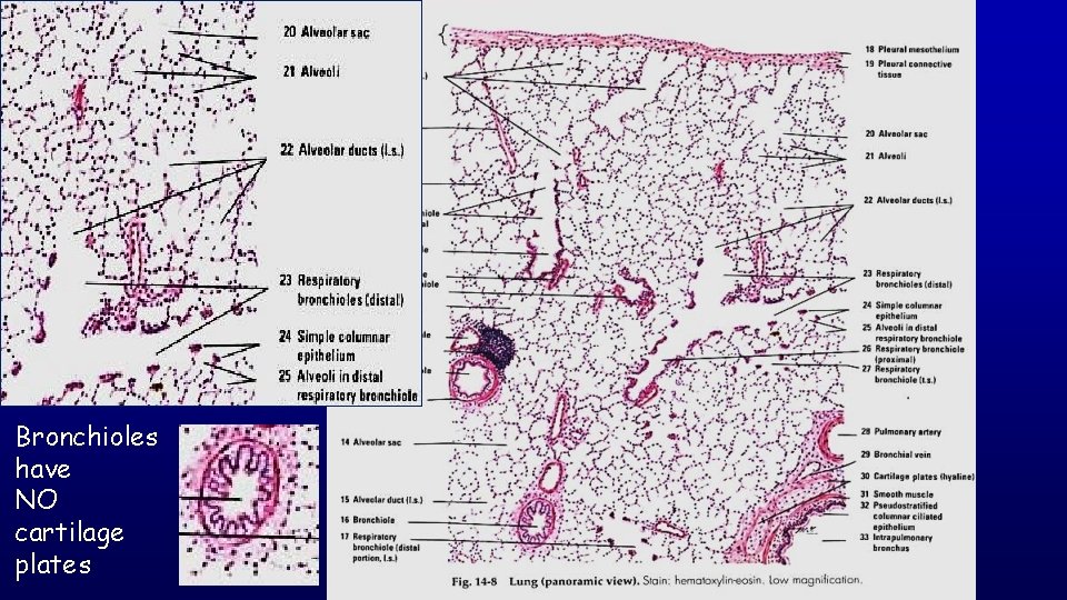
Bronchioles have NO cartilage plates
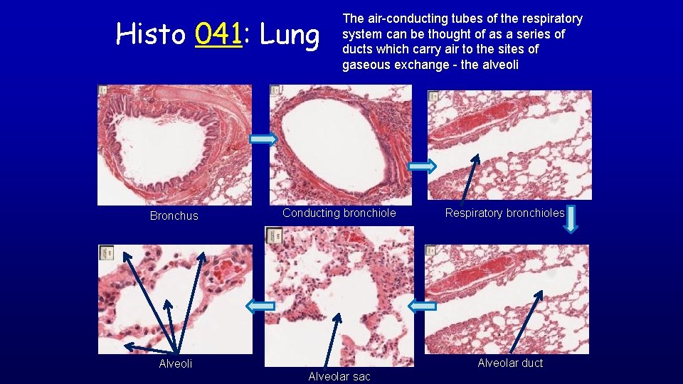
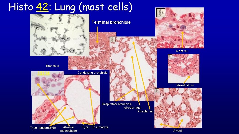
Histo 041: Lung Bronchus The air-conducting tubes of the respiratory system can be thought of as a series of ducts which carry air to the sites of gaseous exchange - the alveoli Conducting bronchiole Respiratory bronchioles Alveolar duct Alveoli Alveolar sac
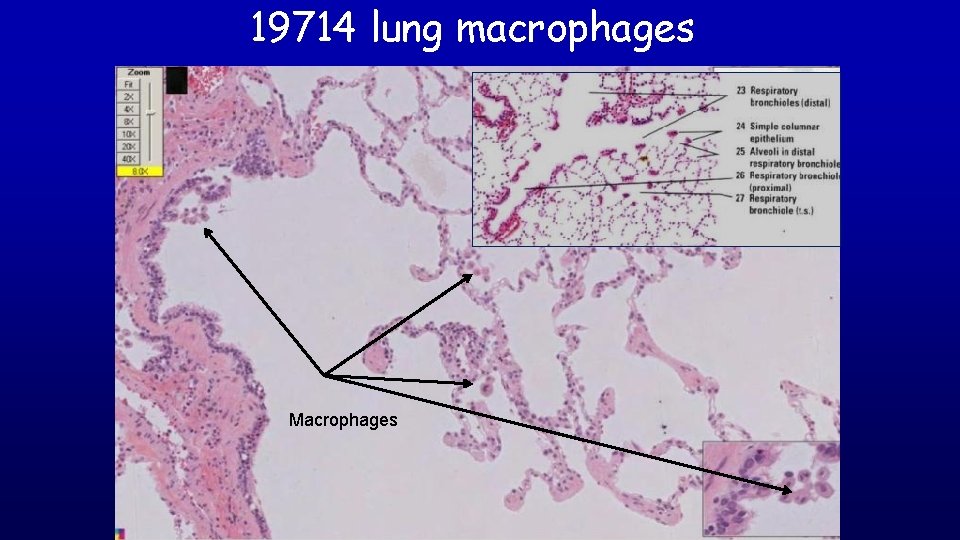
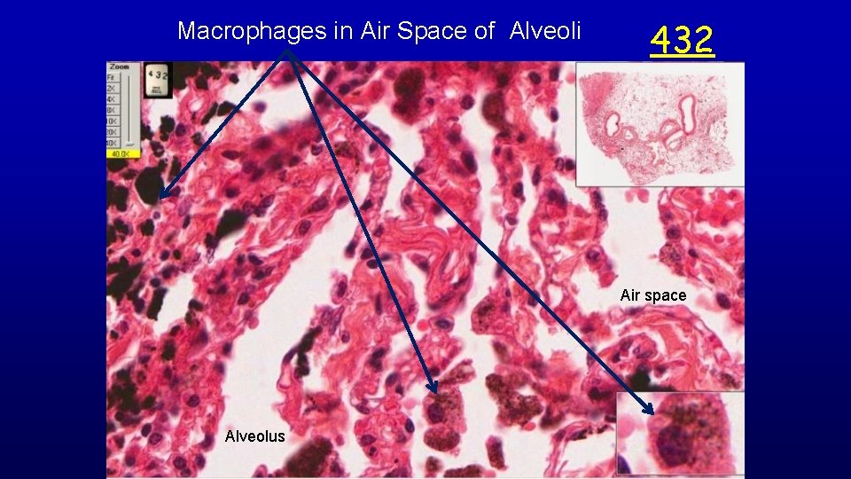
19714 lung macrophages Macrophages
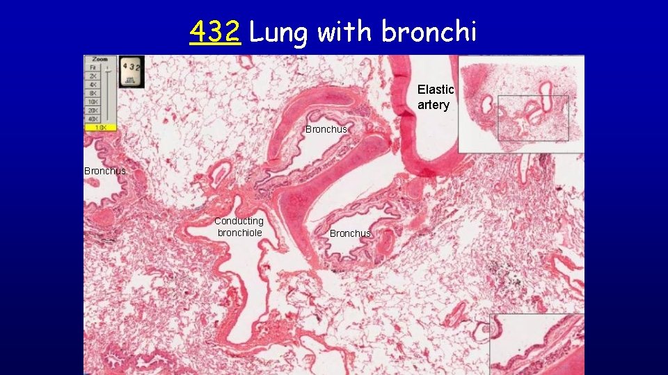
432 Lung with bronchi Elastic artery Bronchus Conducting bronchiole Bronchus
Macrophages in Air Space of Alveoli 432 Air space Alveolus
432 Lung with bronchus Bronchus: 1) pseudostratified ciliated columnar epithelium with goblet-cells; 2) smooth muscle band between the lamina propria and the cartilage. The smooth muscle is not continuous around the bronchus as it spirals. 3) a change from cartilage rings to cartilage plates surrounding the tube; 4) glands in the submucosa. Bronchioles: 1) have a ciliated columnar epithelium; 2) do not have cartilage plates or glands; 3) have well organized muscle layers.
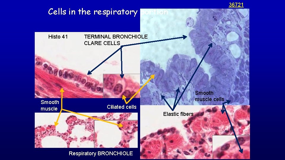
36721 Cells in the respiratory portion Histo 41 Smooth muscle TERMINAL BRONCHIOLE CLARE CELLS Smooth muscle cells Ciliated cells Elastic fibers Respiratory BRONCHIOLE
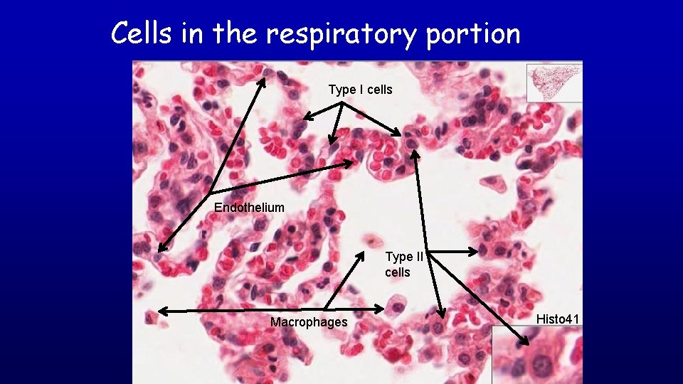
Cells in the respiratory portion BRONCHIOLE Type I cells Endothelium Type II cells Macrophages Histo 41
Slide Histo 41 and Histo 42: Lung 42 42 Mesothelium and connective tissue of lung capsule Type I & Type II pneumocytes 42 41 Capillary endothelial cells and fibroblasts Alveolar macrophage
Respiratory Portion - Site Of Gases Exchange Respiratory bronchioles Alveolar ducts Alveoli
Blood Air Blood
Histo 42: Lung (mast cells) Terminal bronchiole Mast cell Bronchus 19714 Conducting bronchiole Mesothelium Respiratory bronchiole Alveolar duct Alveolar sac Type I pneumocyte Alveolar macrophage Type II pneumocyte Alveoli
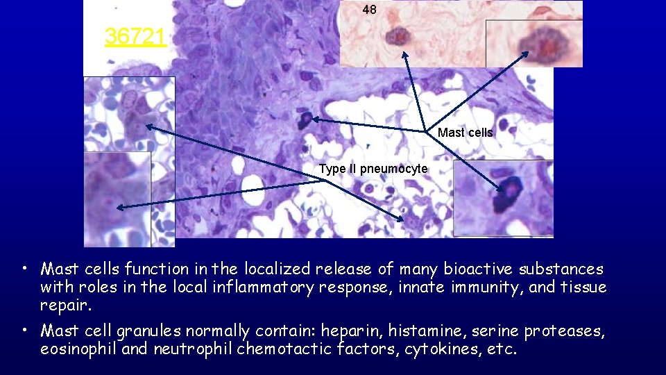
48 36721 Mast cells Type II pneumocyte • Mast cells function in the localized release of many bioactive substances with roles in the local inflammatory response, innate immunity, and tissue repair. • Mast cell granules normally contain: heparin, histamine, serine proteases, eosinophil and neutrophil chemotactic factors, cytokines, etc.
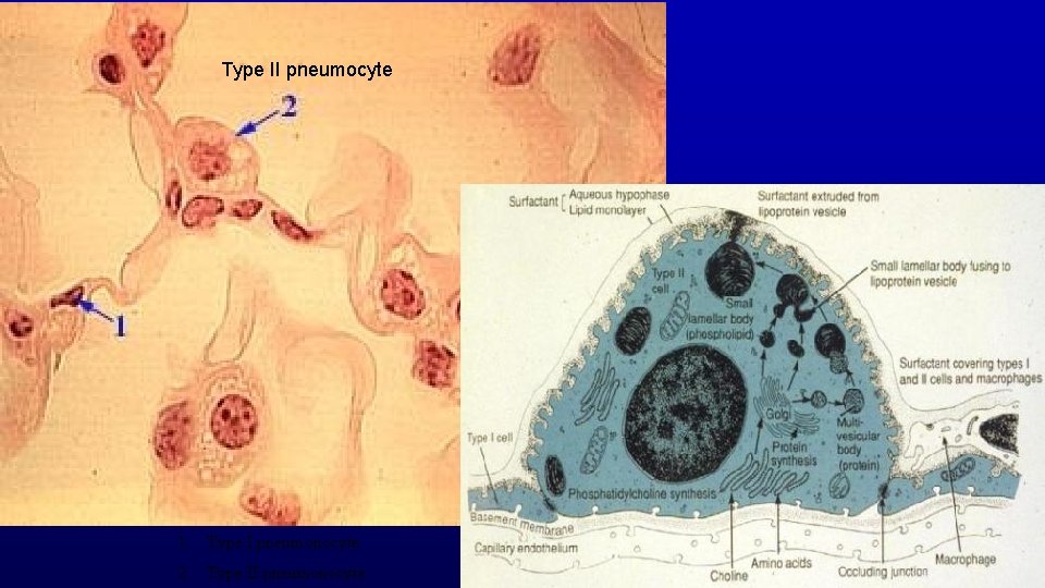
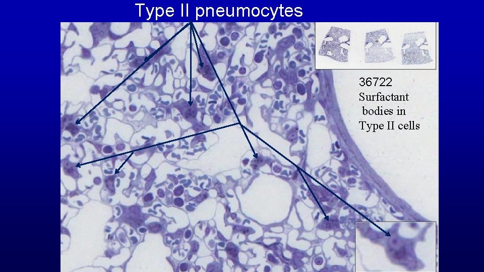
Type II pneumocyte 1. Type I pneumonocyte 2. Type II pneumonocyte
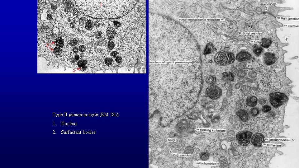
Type II pneumonocyte (EM 18 c). 1. Nucleus 2. Surfactant bodies
Type II pneumocytes 36722 Surfactant bodies in Type II cells
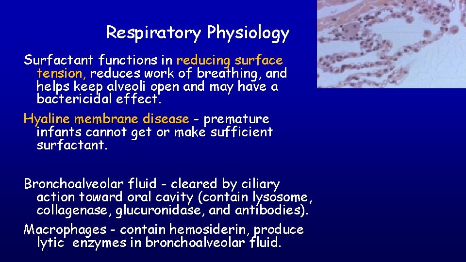
Respiratory Physiology Surfactant functions in reducing surface tension, reduces work of breathing, and helps keep alveoli open and may have a bactericidal effect. Hyaline membrane disease - premature infants cannot get or make sufficient surfactant. Bronchoalveolar fluid - cleared by ciliary action toward oral cavity (contain lysosome, collagenase, glucuronidase, and antibodies). Macrophages - contain hemosiderin, produce lytic enzymes in bronchoalveolar fluid.
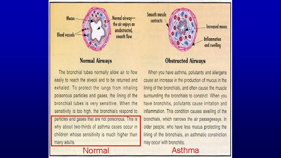
Normal Asthma
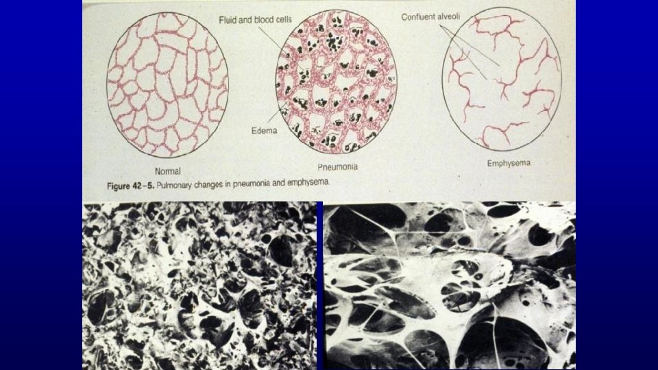
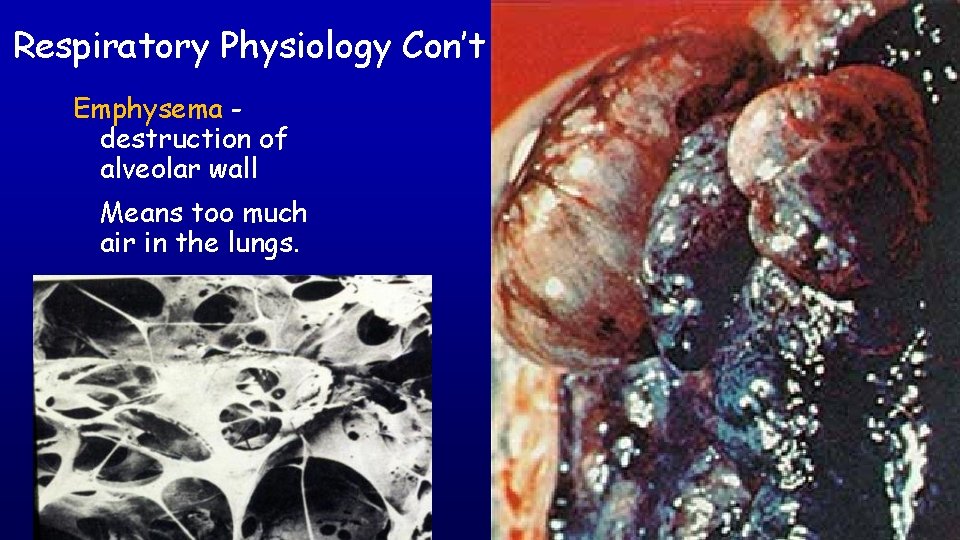
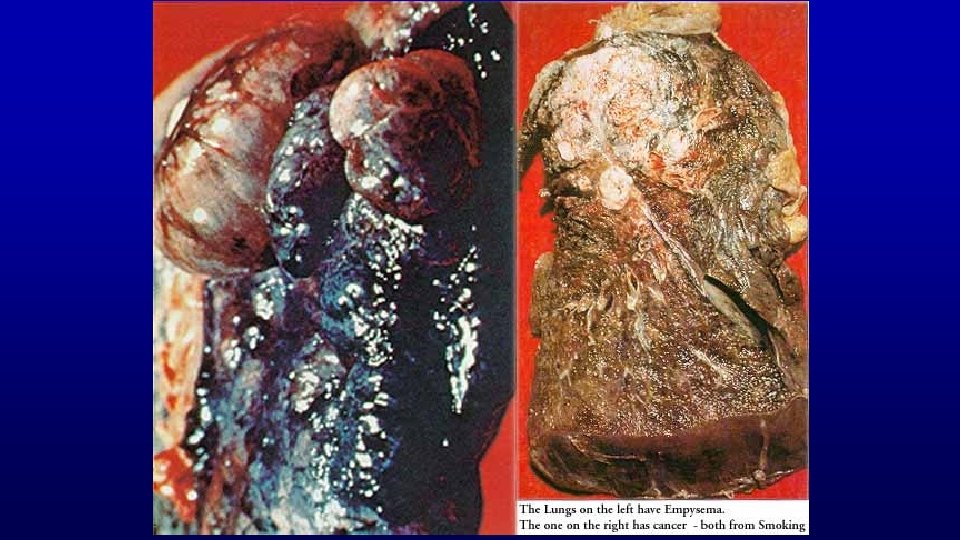
Respiratory Physiology Con’t Emphysema destruction of alveolar wall Means too much air in the lungs.
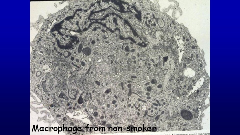
Macrophage from non-smoker
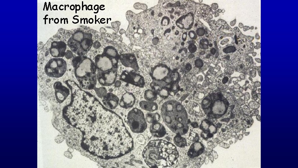
Macrophage from Smoker
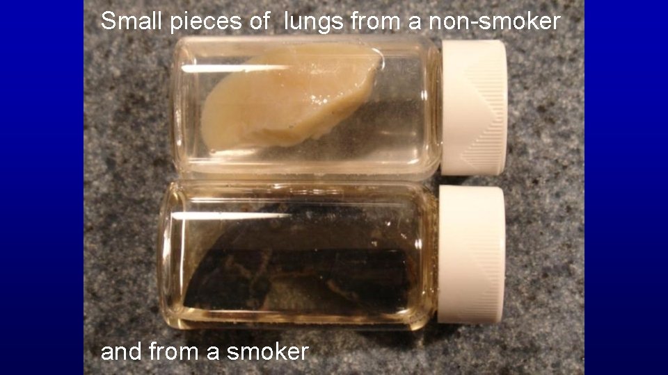
Small pieces of lungs from a non-smoker and from a smoker
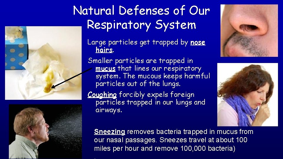
Natural Defenses of Our Respiratory System Large particles get trapped by nose hairs. Smaller particles are trapped in mucus that lines our respiratory system. The mucous keeps harmful particles out of the lungs. Coughing forcibly expels foreign particles trapped in our lungs and airways. Sneezing removes bacteria trapped in mucus from our nasal passages. Sneezes travel at about 100 miles per hour and remove 100, 000 bacteria)
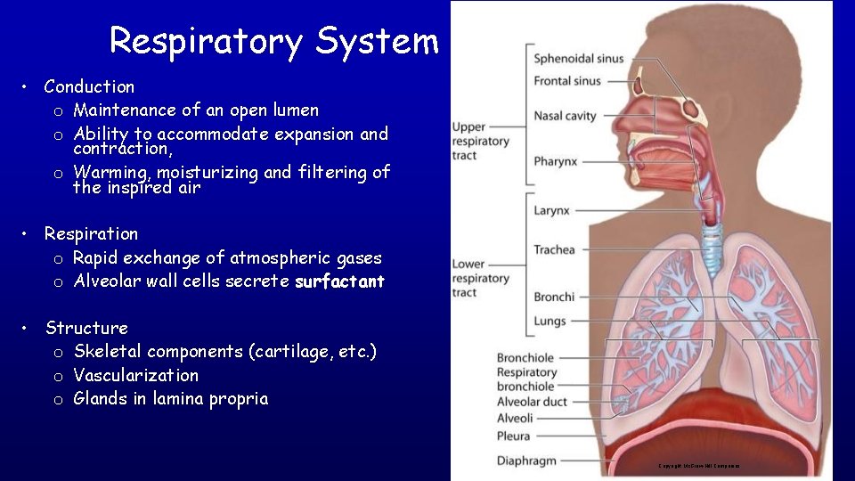
Respiratory System • Conduction o Maintenance of an open lumen o Ability to accommodate expansion and contraction, o Warming, moisturizing and filtering of the inspired air • Respiration o Rapid exchange of atmospheric gases o Alveolar wall cells secrete surfactant • Structure o Skeletal components (cartilage, etc. ) o Vascularization o Glands in lamina propria Copyright Mc. Graw-Hill Companies
In summary
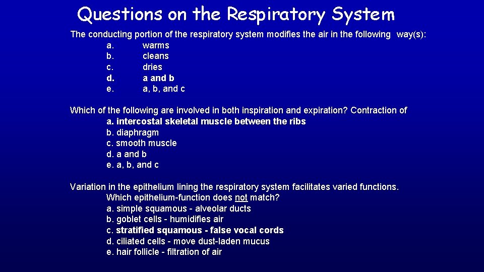
Questions on the Respiratory System The conducting portion of the respiratory system modifies the air in the following way(s): a. warms b. cleans c. dries d. a and b e. a, b, and c Which of the following are involved in both inspiration and expiration? Contraction of a. intercostal skeletal muscle between the ribs b. diaphragm c. smooth muscle d. a and b e. a, b, and c Variation in the epithelium lining the respiratory system facilitates varied functions. Which epithelium-function does not match? a. simple squamous - alveolar ducts b. goblet cells - humidifies air c. stratified squamous - false vocal cords d. ciliated cells - move dust-laden mucus e. hair follicle - filtration of air
• • • • Many illustrations in these VIBS Histology You. Tube videos were modified from the following books and sources: Many thanks to original sources! Bruce Alberts, et al. 1983. Molecular Biology of the Cell. Garland Publishing, Inc. , New York, NY. Bruce Alberts, et al. 1994. Molecular Biology of the Cell. Garland Publishing, Inc. , New York, NY. William J. Banks, 1981. Applied Veterinary Histology. Williams and Wilkins, Los Angeles, CA. Hans Elias, et al. 1978. Histology and Human Microanatomy. John Wiley and Sons, New York, NY. Don W. Fawcett. 1986. Bloom and Fawcett. A textbook of histology. W. B. Saunders Company, Philadelphia, PA. Don W. Fawcett. 1994. Bloom and Fawcett. A textbook of histology. Chapman and Hall, New York, NY. Arthur W. Ham and David H. Cormack. 1979. Histology. J. S. Lippincott Company, Philadelphia, PA. Luis C. Junqueira, et al. 1983. Basic Histology. Lange Medical Publications, Los Altos, CA. L. Carlos Junqueira, et al. 1995. Basic Histology. Appleton and Lange, Norwalk, CT. L. L. Langley, et al. 1974. Dynamic Anatomy and Physiology. Mc. Graw-Hill Book Company, New York, NY. W. W. Tuttle and Byron A. Schottelius. 1969. Textbook of Physiology. The C. V. Mosby Company, St. Louis, MO. Leon Weiss. 1977. Histology Cell and Tissue Biology. Elsevier Biomedical, New York, NY. Leon Weiss and Roy O. Greep. 1977. Histology. Mc. Graw-Hill Book Company, New York, NY. Nature (http: //www. nature. com), Vol. 414: 88, 2001. A. L. Mescher 2013 Junqueira’s Basis Histology text and atlas, 13 th ed. Mc. Graw Internet images and videos on biological presentations