Structure and development of the diaphragm Semmelweis University

 diaphrassein = separate (Greek)")

 Centrum tendineum 5")

4 body • Left: L")


 sternocostal triangle •")
 superior")


 (Cervical pl. )")


 2. aquired (hiatushernia)")

- Slides: 38
Structure and development of the diaphragm Semmelweis University, Faculty of Medicine 1 st year 2 nd semester Department of Anatomy, Histology and Embryology Katalin Kocsis 2018. 03. 09 -10.
Diaphragm (thoracic) diaphrassein = separate (Greek)
Diaphragm • • • 4 -5 mm thick striated muscle sheet Separates thoracic cavity from abdominal cavity Most important respiratory muscle Role in abdominal pressure Important structures pierceing it: – Esophagus – Aorta – Inferior vena cava – Nerves – Lymph vessel
Muscular-tendineous separating sheet between thoracic and abdominal cavities Thoracic cavity (Thorax) Centrum tendineum 5 th rib upper edge 5 th intercostal space Abdominal cavity (Abdomen)
Parts of diaphragm • Tendinuous part: centrum tendineum • Muscular part: • sternal part (xyphoid proc. ) • costal part (lower 6 ribs) • lumbal part • medial crus • (intermediate crus) • laterale crus
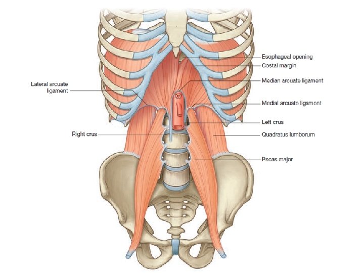
Lumbal part – Medial crus: • Right: L 1 -L(3)4 body • Left: L 1 -L(2)3 body (median arcuate lig. ) Aortic hiatus (L 1) Esophageal hiatus (Th 11) – (Intermediate crus: L 2 body) – Lateral crus: • Medial arcuate lig. - L 2 body- L 2 Costal proc. (M. psoas major) • Lateral arcuate lig. - L 2 Costal proc. - 12 th rib (M. quadratus lumborum)
Lumbal part Medial crus Lateral and medial arcuate ligaments Median arcuate ligament
Diaphragm superior view inf. vena cava esophagus aorta thoracica centrum tendineum pericardium costal part abdominal muscles liver stomach
Diaphragm inferior view inf. vena cava sternal part costal part centrum tendineum esophageal hiatus esophagus aortic hiatus lumbal part m. quadratus lumborum m. psoas minor m. psoas major vertebral column
Diaphragm inferior view
Sternocostal triangle Hiatus of inferior vena cava Openings Esophageal hiatus Aortic hiatus Lumbocostal triangle
Structures passing through • internal thoracic a. (superior epigastric a. ) sternocostal triangle • inferior vena cava (right phrenic n. , pericardiacophrenic a. ) inf. v. cava hiatus • esophagus • n. vagus • (left gastric a. (v. ), esoph. branches) esophageal hiatus • aorta • thoracicus duct aortic hiatus • v. azygos , v. hemiazygos • greater splanchnic n. medial crus • truncus sympathicus • lesser splanchnic n. mediale crus - laterale crus
Structures passing through internal thoracic a. , v. Sternocostal triangle (Larrey, Morgagni foramen) superior epigastric a. , v.
Structures passing through
Structures passing through Aortic arch Sternum manubrium Sternum body heart Thoracic aorta xyphoid process Abdominal aorta 17
Structures passing through
Structures passing through right vagus nerve esophagus inf. vena cava left vagus n. Esophageal hiatus Aortic hiatus Thoracic duct Cysterna chyli Descending aorta
Structures passing through sympathetic trunk azygos v. hemiazygos v. greater splanchnic n. Between the fibers of medial crus: greater splanchnic nerve and azygos/hemiazygos vein (ascending lumbar veins) Between medial and lateral crus: sympathetic trunk and lesser splanchnic nerve
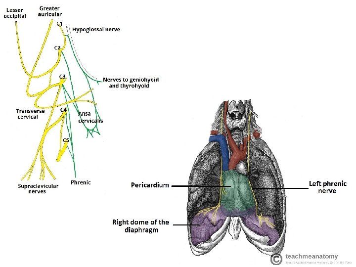
Blood supply, innervation Motor innervation: phrenic n. (C 3 -4 -5) (Cervical pl. ) Sensory innervation: • phrenic n. : centrum tendineum (diaphragmatica pleura, parietal peritoneum) • intercostal nerves (7 -12): muscular part Blood supply • • pericardiacophrenic a. musculophrenic a. interna thoracica a. (subclavian a. ) • • sup. phrenic a. inf. phrenic a. thoracic aorta abdominal aorta
http: //www. flspinalcord. us/innervation-of-diaphragm-studydroid-flashcards-on-the-web-and-in-your-hand/
Blood supply From internal thoracic artery: -pericardicophrenic artery -musculophrenic artery From descending aorta: -superior phrenic artery -inferior phrenic artery
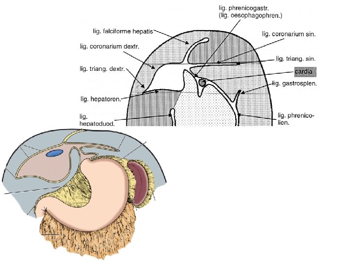
Serous membranes on the diaphragm fibrous pericardium pleura serous pericardium peritoneum diaphragm
Paralysis of the phrenic nerve – paradox movement of diaphragm Normal inspiration Paralysis of right phrenic nerve
Development of diaphragm
Development of diaphragm develops from 4 parts: 1. 2. 3. 4. Septum transversum (C 3 -C 5 cervical somites) Dorsalis mesentery of esophagus Pleuroperitoneal membrane Lateral body wall Szív Máj Septum transversum
Development of diaphragm 4 th week 5 th week
Development of diaphragm
Development of diaphragm
Development of diaphragm
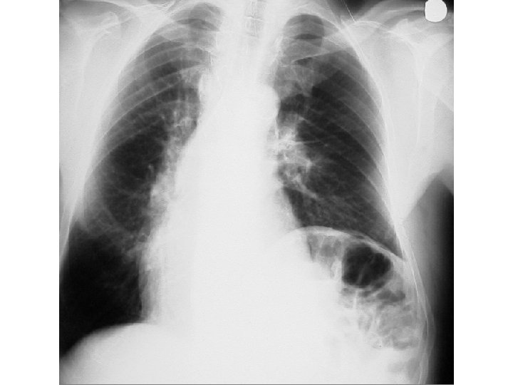
Diaphragmatic hernia
Diaphragmatic hernia 1. congenital (CDH) 2. aquired (hiatushernia)
References