DIAPHRAGM THORACIC DIAPHRAGM Dome shaped Musculoaponeurotic structure Separates


- Slides: 21

DIAPHRAGM

THORACIC DIAPHRAGM • Dome shaped • Musculoaponeurotic structure • Separates thorax from abdomen • Forms roof of abdominal cavity & floor of thoracic cavity

Parts of thoracic diaphragm • Thoracic surface: convex on right & left side but depressed in centre • Summits of the convexities are c/as Cupolae • Right cupola is higher than left due to presence of liver • Abdominal surface : concave • Peripheral part : muscular • Central part : tendinous

THORACIC DIAPHRAGM(contd…. . ) • Sternal heads: 2 fleshy slips from back of xiphoid process • Costal heads : From inner surface of lower 6 ribs & their costal cartilages • Vertebral heads: A pair of crura A pair of medial arcuate ligament A pair of lateral arcuate ligament

THORACIC DIAPHRAGM(contd…. . ) • Right Crus of diaphragm: Right crus is longer & more muscular than left one. Origin is from front of the bodies of L₁ - L₃ & interveining intervertebral discs.

THORACIC DIAPHRAGM(contd…. . ) • Left crus of diaphragm: shorter than right crus origin is from bodies & discs of L₁- L₂ left crus is attached to bare area of stomach by Gastro phrenic ligament both crura are connected to each other by a Median arcuate ligament

THORACIC DIAPHRAGM(contd…. . ) • Medial Arcuate Ligament/ Medial Lumbocostal Arch Bridges in front of psoas major Formed by thickening of psoas fascia Attached med to body of L₁ - L₂ Attached lat to transverse process of L₁ • Lateral Arcuate Ligament/ Lateral Lumbocostal Arch Bridges in front of Quadratus lumborum Formed by thickening of ant. Layer of thoracolumbar fascia Attachments : med… transverse process of L₁ lat…. . Tip of 12 th rib

THORACIC DIAPHRAGM(contd…. . ) Central Tendon • Present in central depressed part • Trefoil leaf shaped with median, right & left leaflets • IVC opening present at junction of median & right leaflet • Left to IVC opening is central point of decussation • Sternal fibers are shortest • Fibers from 9 th costal cartilage are longest

Openings in diaphragm Venecaval opening(T₈) • Quadrilateral in shape • IVC • Branches of Phrenic nerve • Lymphatics from liver Oesophageal Opening (T₁₀) • Elliptical in shape • Oesophagus • Ant. & post. Vagal trunks • Oesophageal branches of left gastric artery • Lymphatics from liver • Phrenoesophageal ligament

Openings in diaphragm Aortic Opening(T₁₂) • Rounded in shape • osseoaponeurotic • Boundaries : median arcuate ligament (ant), body of T₁₂, both crura on sides • Abdominal aorta • Thoracic duct • Azygos vein (when its lumbar azygos vein )

Openings in diaphragm Other Openings • Space of Larrey : Sup. Epigastric vessels & lymphatics • Between costal origin & T. abdominis : lower 5 intercostal nerves & vessels • Between costal origin from 7 th & 8 th costal cartilage : musculophrenic vessels • Behind Lat. Arcuate ligament : subcostal nerve & vessels • Behind medial arcuate ligament : sympathetic trunk & least splanchnic nerve

Openings in diaphragm Other Openings • Each crus is pierced by greater & lesser splanchnic nerves • Left crus is also pierced by hemiazygos vein • Left phrenic nerve pierces left cupola in front of central tendon

Relations of diaphragm • Superior : Pleurae Lungs Pericardium Heart • Inferior : Peritoneum Liver Fundus of stomach Spleen Kidneys Suprarenal glands

Nerve Supply of Diaphragm • Motor supply: Phrenic nerve (C₃‚₄‚₅) Anteromedial Anterolateral Posteromedial Posterolateral • Sensory supply: Central – phrenic nerve Peripheral – lower 6 intercostal nerves • Sympathetic supply: Coeliac plexus via inferior phrenic plexus

Blood supply of diaphragm • Musculophrenic artery • Pericardiophrenic artery • Lower 5 -6 post. Intercostal arteries • Sup. Phrenic artery • Inf. Phrenic artery

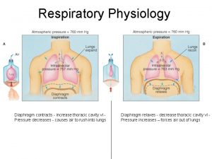
Actions of diaphragm • Principal muscle of inspiration • Contraction descent vertical diameter of thorax increases • Upper abdominal viscera displaced below ant. Abdominal wall bulges • Central tendon then becomes fixed lower ribs are elevated increase in AP diameter

Actions of diaphragm • Muscle of Abdominal Straining: Supports expulsive acts like sneezing , coughing , vomiting , crying, defecation , micturation, etc. • Sphincteric action at lower end of esophagus • Thoracoabdominal pump for blood & lymph • Effect of contraction of diaphragm on 3 major openings in diaphragm 1. Venacaval opening ---- dilates 2. Oesophageal opening -------constricts 3. Aortic opening ---- no change

Development of diaphragm • Develops at level of neck & derives its muscular components from cervical myotomes • Septum transversum central tendon • Pleuroperitoneal membrane dorsal paired portion • Lateral thoracic wall circumferential portion of diaphragm • Dorsal mesentery of esophagus dorsal unpaired portion (Crura )

Applied • Hiccups /hiccough occurs due to spamodic contraction of diaphragm 1. Central hiccups due to hiccup centre in medulla (MC cause is uremia ) 2. Peripheral local irritation of diaphragm or its nerve • Shoulder tip pain irritation of diaphragm leads to pain in shoulder because of the common root value of supraclavicular nerve & phrenic nerve. • Unilateral paralysis of diaphragm due to lesion of phrenic nerve. Paralyzed side shows paradoxical movement. • Eventration is congenital defect in the musculature of diaphragm (fibrous membrane present )

Diaphragmatic hernias • Congenital diaphragmatic hernias 1. Morgagni hernia /Retrosternal hernia (anterior right) 2. Bochdalek hernia /posterolateral hernia (posterior left) Occurs through pleuroperitoneal hiatus Fatal condition(lung hypoplasia) S/S: scaphoid abdomen respiratory distress 3. Central hernia 4. Posterior hernia (posterior defect in diaphragm)

Hiatus hernia • Congenital / rolling / paraesophageal hiatus hernia : stomach rolls upwards & lies in post. Mediastinum GE junction is normal • S/S : nausea, dysphagia, pain • Acquired / sliding hiatus hernia : weakness of phrenicoesophageal membrane. • S/S: GERD, heart burn
 Diaphragm easy diagram
 Saddle ridge lap pontic
 Saddle or ridge lap
 Soft spongy cone shaped organs in the thoracic cavity
 Type of writing
 Pouchlike structure at the start of the thoracic duct
 What separates texas from mexico
 Round yellow sign with black markings
 Rekening tamu untuk rombongan disebut
 I am malleable, but i do not have a shiny luster.
 Lateral sulcus separates which lobes
 Lines of latitude
 Why dna is negatively charged
 5 major peninsulas of europe
 Gel electrophoresis separates dna by
 Superior frontal gyri
 What separates the inner and outer planets
 What separates inner and outer planets
 To separate items in a list
 What separates watersheds
 Separating mixtures worksheet grade 8
 What is the process that separates components h&o of water
