Respiratory System Chapter 16 Chapter 16 1 Introduction


- Slides: 24
Respiratory System Chapter #16
Chapter 16. 1 Introduction n 1. 2. 3. 4. 5. Obtaining oxygen and removing carbon dioxide are the primary functions of the Respiratory System. The Respiratory system includes tubes that: Remove particles from incoming air Help control the temperature & water content Produce vocal sounds Transport air into and out of the lungs Play an important role in the sense of smell and blood p. H
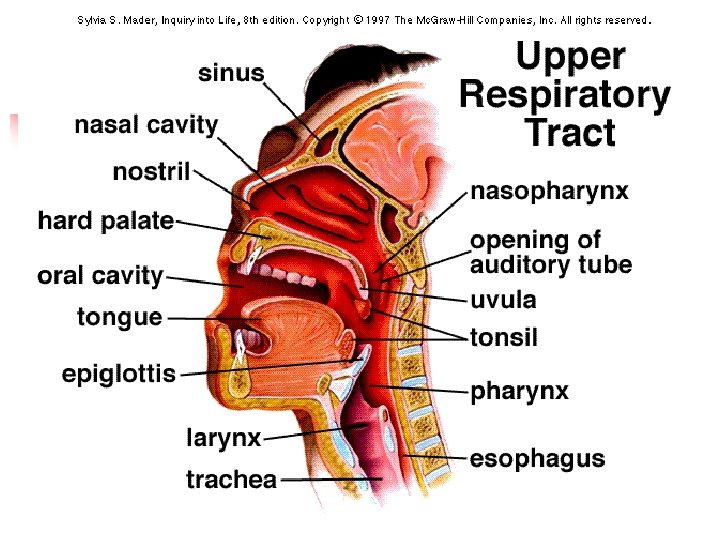
Chapter 16. 2 Organs of the Respiratory System n 1. 2. The organs of the respiratory system can be divided into 2 groups. Upper respiratory tract include the nose, nasal cavity, paranasal sinuses, and pharynx. Lower respiratory tract (larynx, trachea, bronchial tree, and lungs)
Nose n n Two nostrils or openings Internal hairs that guard against large particles entering the nasal cavity.
Nasal Cavity n n n Is a hollow space behind the nose. The nasal septum, composed of bone and cartilage, divides the nasal cavity into right and left portions. Nasal conchae are bones that curl out from the lateral walls of the nasal cavity on each side, dividing the cavity into passageways. They also support the mucous membrane that lines the nasal cavity and help increase its surface area.
Paranasal Sinuses n 1. 2. Are air-filled spaces located within the maxillary, frontal, ethmoid, and sphenoid bones of the skull and opening into the nasal cavity. They reduce the weight of skull They are resonant chambers that affect the quality of the voice.
Pharynx n n n Or throat is behind the oral cavity and between the nasal cavity and larynx. It is a passageway for food traveling from the oral cavity to the esophagus. Passageway for air passing between the nasal cavity and larynx. Helps produce the sounds of speech. Pharynx is divided into the nasopharynx, oropharynx, laryngopharynx.
Larynx n n n Is an enlargement in the airway at the top of the trachea and below the pharynx. It conducts air in and out of the trachea and prevents foreign objects from entering the trachea. It houses the vocal cords. False vocal cords upper folds they don’t produce sound. True vocal cords lower folds that produce sound. Glottis is a triangular slit opening between the vocal cords. When you swallow the false vocal cords close the glottis Epiglottis is a flap like structure that covers the larynx preventing food from going into the trachea.
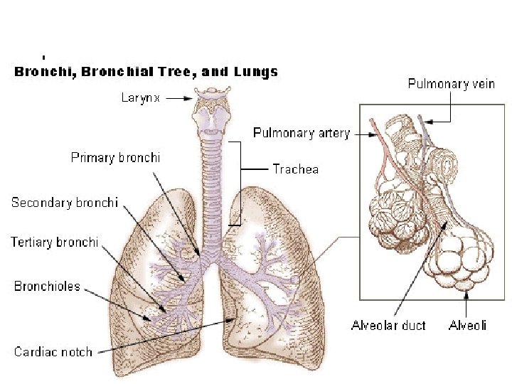
Trachea n n n Or windpipe is a flexible, cylindrical tube about 2. 5 cm in diameter and 12. 5 cm long. It extends downward in front of the esophagus and into the thoracic cavity where it splits into right and left bronchi. Has cartilage rings to prevent the trachea from collapsing.
Bronchial Tree n n Consists of branched airways leading from the trachea to the air sacs (Alveoli) in the lungs. Primary bronchi right and left that branch from the trachea at the 5 th thoracic vertebrae. These then branch into secondary bronchi. Bronchioles are even smaller tubes. As the branches become smaller, there are fewer muscle fibers in their walls.
Lungs n n Are soft, spongy, cone-shaped organs in the thoracic cavity. The mediastinum separates the right and left lung. The diaphragm separates the lungs from the abdominal organs and aids in breathing. The right lung (3 lobes) is larger than the left lung (2 lobes).
16. 3 Breathing Mechanism n Breathing, or ventilation is the movement of air from outside the body into and out of the bronchial tree and alveoli.
Inspiration n n 1. 2. 3. Inspiration or inhalation Atmospheric pressure due to the weight of air is the force that moves air into the lungs. v When you breath in: Your diaphragm contracts (moves downward) the thoracic cavity enlarges. The ribs raise and elevates the sternum The pressure within the cavity decreases…and the lungs fill.
Expiration n n 1. 2. Expiration or exhalation The forces responsible for normal expiration come from the elastic recoil of tissues and from surface tension. v When you breath out Your diaphragm relaxes (moves upward) the thoracic cavity compresses. The ribs lower and lower the sternum
n 1. 2. 3. 4. n Spirometry measures air volumes, revealing 4 distinct respiratory volumes. Tidal Volume the air that enters and leaves the lungs during one resting respiratory cycle. Vital Capacity is the maximum amount of air a person can exhale after taking the deepest breath possible. Functional Residual Capacity is the amount of air that remain in the lungs after a resting expiration. Total Lung Capacity- is the vital capacity + residual capacity. Spirometer is an instrument used to measure air volumes. Such measurements are used to evaluate the course of emphysema, pneumonia, and lung cancer, conditions in which functional lung tissue is lost.
16. 4 Control of Breathing n n n Normal breathing is a rhythmic, involuntary act that continues even when a person is unconscious. The respiratory muscles, however are under voluntary control. Respiratory center in the brain stem control both inspiration and expiration it responds to the concentrations of CO 2 and p. H (H+).
n Hyperventilation is a condition due to a lowered carbon dioxide concentration followed by a rise in p. H, a localized vasoconstriction of cerebral arterioles, which causes a decreased blood flow to brain cells. Causing you to pass out!!! Swimmers shouldn’t do!!
16. 5 Alveolar Gas Exchange n n Alveoli are microscopic air sacs clustered at the distal ends of the narrowest respiratory tubes. Simple squamous epithelium cells comprise the alveoli membrane through which gases can easily be exchanged. O 2 diffuses through the alveolar walls and enters the blood in nearby capillaries. CO 2 diffuses from the blood within the capillaries into the alveoli.
16. 6 Gas Transport n n Blood transport oxygen and carbon dioxide between the lungs and cells. Gases enter blood and they dissolve in the plasma or combine chemically with blood components. Almost all the oxygen that blood transports combines with the iron-containing protein hemoglobin in red blood cells. Carbon dioxide is carried in the blood dissolved in plasma, as part f a bicarbonate ion bound to the amino groups of hemoglobin.
n n n In the lungs oxygen dissolved and combines with oxyhemoglobin. More oxygen is released from oxyhemoglobin in an acid environment. The attraction of carbon monoxide to hemoglobin is stronger than for oxygen, therefore the person may die of hypoxia. Carbon monoxide is toxic. Hypoxia is the deficiency of oxygen reaching tissue.
Work Cited n n “Respiratory System”. May 23, 2007. http: //www. medem. com/MEDEM/images/AMA/ama_i d_respiratoryinfections_lev 20_respiratorysystemstruc turedetail_01. gif “Trachea”. May 23, 2007. http: //www. asdonline. com/piceng/trachea. jpg “Lungs”. May 23, 2007. http: //www. webbooks. com/e. Library/Medicine/Physiology/Respiratory/ bronchi_lungs. jpg “Breathing”. May 23, 2007. http: //www. fda. gov/Fdac/graphics/1999 graphics/bre athing. gif