Early development cleavage GASTRULATION Semmelweis University Department of


 test (yellow line: exam test material) • Mitotic")


")


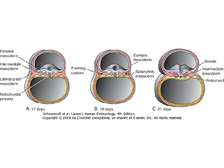
 intermediate mesoderm (nephrotom)")


- Slides: 36
Early development, cleavage, GASTRULATION Semmelweis University, Department of Anatomy, Histology and Embryology
The zygote =fertilized egg The new diploid cell is created by the fusion of the male and female pronucleus ( )
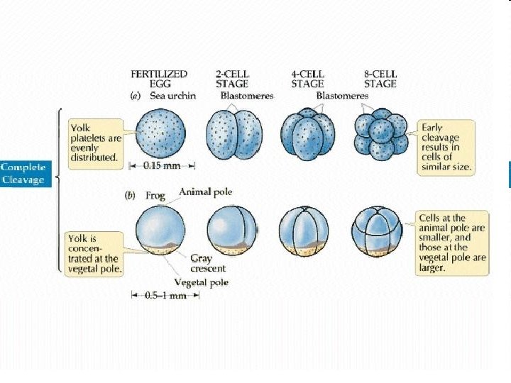
purple dashed double line: probe (mock) test (yellow line: exam test material) • Mitotic division process • Cleavage stage cells: blastomeres • The volume of egg’s cytoplasm divides into numerous smaller, nucleated cells → in humans whole cytoplasmatic volume of the egg packs into smaller and smaller blastomeres • The human cleavage is equal, but asynchronous Cleavage
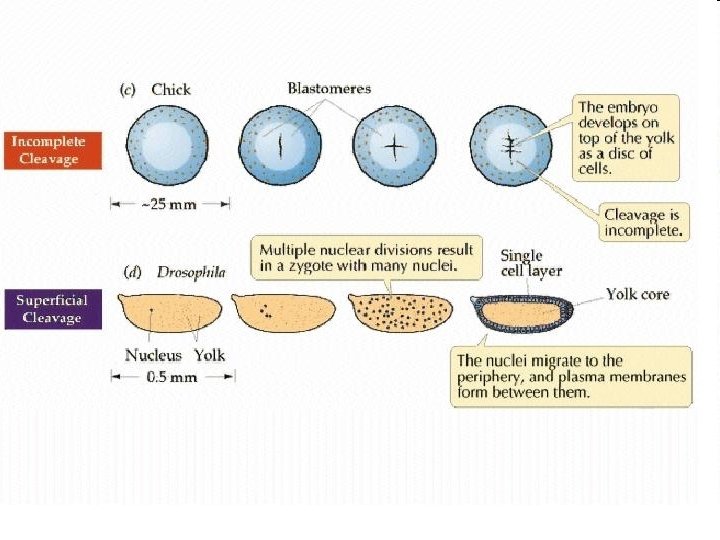
Different type of cleavage • The cleavage process is influenced by the yolk contents of the egg. Incomplete (Meroblasticus) fish, reptiles, birds • The egg may contain large or small amount of yolk • The distribution of the yolk: • uniform • concentrated on one of pole of the egg. Complete • Due to the asymmetric yolk (Holoblasticus) distribution: • Only a part of the egg is packed into blastomeres • blastomeres have different size • Complete and incomplete types of cleavage can be distinguished Frog Human
Compaction -in the 8 -cell-stage cleavage. -The blastomeres, which show loose arrangement, suddenly form a compact ball -Before the compaction blastomeres loosly adhere to each other by microvilli on their surface. -After compaction blastomeres tightly adhere with each other through intercellular junctions before compaction and after compaction
• Compaction is a membrane polarization process → well-defined apical, basal and lateral side is developing. • Different components of the cell membrane concentrate at different regions of the cell causing the polarization of the cells. • Membrane polarization is influenced by cell -cell interactions • This polarization process takes place only in those parts of the cell membrane where the cell is in contact with other blastomeres. • E-cadherin plays a main role in compaction. • At 2 -cell stage, E-cadherin is uniformly spread throughout the cell membrane. During compaction E-cadherin becomes restricted to those sites of cell membrane where adjacent blastomeres are in contact with each other. • E-cadherin molecules accumulate and form zonula adherens
16 -64 cells stage
Blastula formation • Newly formed structures between the outer blastomeres: -tight junctions (apical part) -gap juction (lateral part) -membrane transport molecules on basal part: mainly sodium pumps • Due to the activated sodium pumps the sodium concentration increase in the intercellular space and parallel the water flows into the intercelullar space by osmosis → forming fluid filled cavity called blastocoel. • The blastocoel is expanded gradually by the increasing amount of the fluid
The expanding blastocoel pushes the internal cells to one side of the blastocyst → inner cell mass (ICM) In mice: • trophoblast cells differentiate into the fetal membrane system • inner cell mass forms the whole body of the embryo and extraembryonic (primitive) endoderm
Twins • Monozygotic twins arise from one zygote • Dizygote twins arise two different eggs, that are fertilised by two different sperms . Monozygotic twins • At cleavage: after the first cell division the newly formed two blastomeres completely separete from each other. • At blastulation: the subdivision of the inner cell mass within the blastocyst.
The differentiation of the ICM cells into a bilaminar structure containing epiblast and hypoblast (primitive endoderm) layer. The sorting model At the beginning, the epiblast and hypoblast cells distribute within the ICM showing „salt and pepper pattern”. The sorting event, in which the bilaminar disc will be developed, is caused by two main ways. 1. : the different strenghts of adhesion between the two cells type (adhesion is stronger between same type cells than different type cells) and 2. : signals coming from either the blastocoel or from trophoblast (the epiblast cells express nanog while hypoblast cells express GATA 6 and this expression pattern is caused by the different FGF signaling.
Why is gastrulation so important? §Generation of the basic body plan. §Specification of the axes: §Anterior and posterior §Dorsal and ventral §Left and right §Generation of the three germ layers §Ectoderm, mesoderm, and endoderm
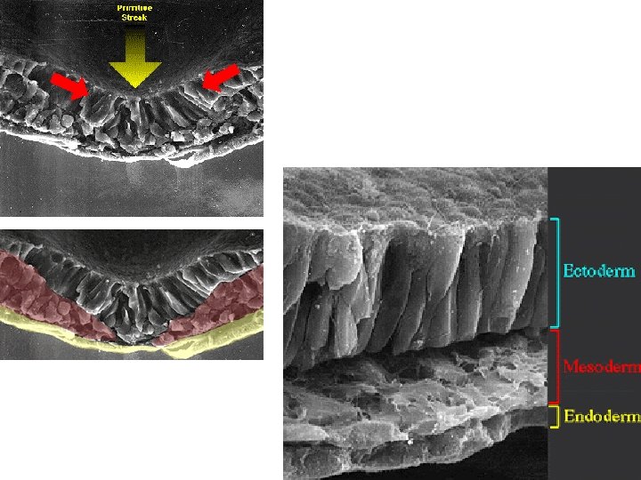
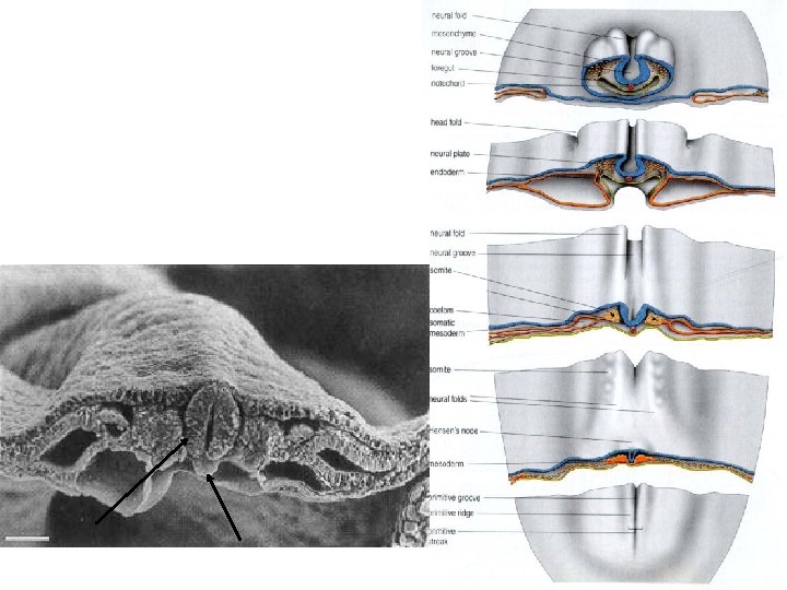
Primitive streak, groove Anterior • Gastrulation begins with the formation of primitive streak. Epiblast proliferate and they are pushed toward the midline of the embryo → they are jammed in the midline forming the primitive streak that first appear in the posterior part of the embryo. • Cells, which are located in the middle of the primitive streak, migrate into the interior of the embryo resulting the formation of primitive groove in the middle of the primitive streak. Posterior
Hensen’s node • The primitive streak with the primitive groove is growing gradually anteriorly • At the anterior end of the primitive streak there is a small but well-defined accumulation of cells, called primitive node or Hensen’s node. cranial side caudal side Cranio-caudal, left-right axes well-defined!
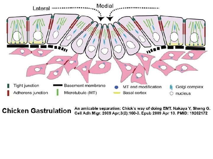
What happens to the cells in the primitive groove? • The movements of the cells are accompanied by major changes in their structure. • When epiblast cells enter the primitive streak, they become elongated and lose their connection with the adjacent cells → their morphology change forming bottle cells. • Within the primitive groove these bottle cells lose their connection with the basal lamina and become free from the epiblast layer. • Bottle cells undergo an epithelio-mesenchymal transformation within the primitive groove and the newly formed mesenchymel cells are able to migrate as individual cells. • During the epithelio-mesenchymal transformation the E-cadherin synthesis is downregulated within the bottle cells, Epithelial cell E-cadherin epiblast „slug” FGF 8 Ecadherin mesoderm endoderm
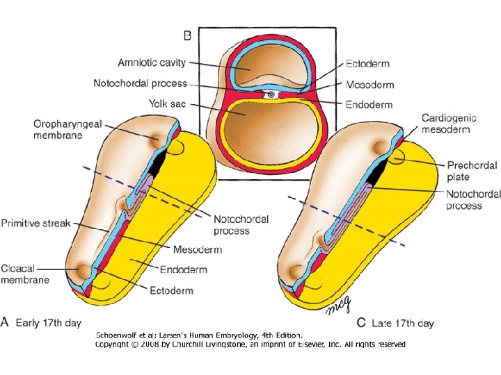
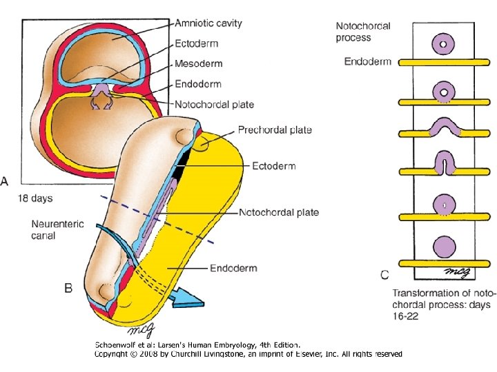
1. : At the biginnig of the notochord process first form a notochord canal with a central lumen Human notochord 3. : The notochord plate starts to infold later form the rope like notochord 2. : The floor of the notochordal canal disappears remaining a flatten plate (notochordal plate) which incorporate into the definitiv endoderm
D 1415 Development of the definitive endoderm Bi-laminar embryonic disk Hypoblast cells develop only into extraembryonal mesoderm Primitive streak Epiblast Hypobla st First entering epiblast-cells migrate and replace the hypoblast-cells forming the definitive endoderm Endoder m Epiblast cells give rise to the three germ layers of the embryo!!
Development of the Intraembryonal Mesoderm Primitive streak D 16 epitheliomesenchymal transformation Intraembryonal mesoderm Epiblast-cells migrate in the interlaminar space and forming intraembryonal mesoderm Definitive entoderm
Migration of the Mesodermal Cells • The newly form mesenchymal cells migrate and spread bilaterally. Epiblast Mesoder m Entoderm • Those cells, which pass through at the level of Hensen’s node, migrate directly cranially and form the precordal plate and later take part in the formation of the notochord. Epiblast Entoder m
Differentation of the mesoderm, convergent extension membrana buccopharyngea paraxial mesoderm (somites) intermediate mesoderm (nephrotom) lateral mesoderm (somatopleura, splanchnopleura) extraembryonal mesoderm primitivenode strike
Fate map GE: gut endoderm PP: prechordal plate PS: primitive streak CM: cardiac mesoderm PEEM: extraembryonic mesoderm HM: head mesoderm S: somitic mesoderm IM: intermedier mesoderm LPM: lateral plate mesoderm SE: surface ectoderm NP: neural plate PE: placod ectoderm NC: neural crest
Primitiv streak regression • At the beginning of gastrulation the primitive streak grows cranially • The primitive streak growing changes for regression. → its length is decreasing toward caudally • This regression process is related with the elongatoin of the notochord • The notochord is formed by the addition of cells to its caudal end while the primitive streak becomes shorter and shorter
Figure 5 -7 Summary of major genes involved in various stages of early embryonic development. A, Preprimitive streak (sagittal section). B, Early formation of the primitive streak. C, Gastrulation (period of germ layer formation). D, Late gastrulation and neural induction. The molecules in red are signaling molecules, and the molecules in blue are transcription factors. Names of specific molecules (bold) are placed by the structures in which they are expressed. Downloaded from: Student. Consult (on 29 September 2010 03: 06 PM) © 2005 Elsevier
„Shh, RS, FGF-8”
Figure 3 -2. Diagram illustrating a simplified scheme of key genes involved in establishing left-right asymmetry. The primitive streak, node, and early floor plate of the neural tube are viewed from the ventral side. Motor proteins (Lrd, Kif 3 A, B) expressed by the node regulate leftward (dashed arrow) nodal flow. Secreted factors (Shh, Fgf 8, Nodal) expressed by the node result in signaling to the lateral plate mesoderm, thereby resulting in asymmetric gene expression in the lateral plate mesoderm (e. g. , Nodal, Lefty 2 in left lateral plate). This in turn results in expression of Pitx 2 in the left lateral plate and changes in cell behaviors that result in asymmetric morphogenesis. Lefty 1 is expressed in the left floor plate of the neural tube. It is believed to serve a barrier function, allowing information that specifies left and right sides to remain separate.
The mechanosensory model of nodal flow. Model showing that nodal flow, generated by motile monocilia in cells expressing Lrd, stimulates calcium flux in cells containing nonmotile cilia that sense flow on the left side. Model showing the transport of nodal vesicular parcels by motile cilia and the stimulation of calcium signaling (blue) at the left side of the node by nonmotile cilia.
Gastrulation wall of amnion bi-laminar epiblast three-laminar embryonic ectoderm definitive ectoderm skin, nervous system, sensory organs roof of blastocyst hypoblast mesoblast (mesenchyma) extraembryonic mesoderm embryonic entoderm intraembryonic mesoderm primary mesoderm connective tissue, circulatory system, smooth and skeletal muscle, blood cells, skeleton, urogenital organs definitive entoderm lining epithelium and the glands of the respiratory and gastrointestinal organs, branchial pouches