PSYCHOPHYSIOLOGICAL INTERACTIONS Darren Gitelman MD Northwestern University dgitelmannorthwestern
- Slides: 91
PSYCHOPHYSIOLOGICAL INTERACTIONS Darren Gitelman, MD Northwestern University d-gitelman@northwestern. edu
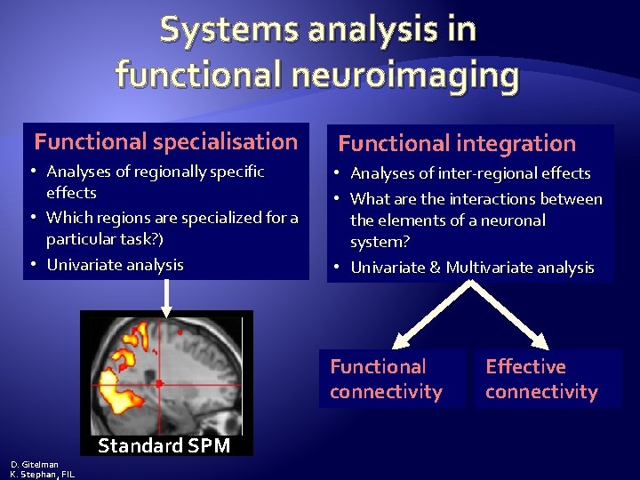
Systems analysis in functional neuroimaging Functional specialisation Functional integration • Analyses of regionally specific effects • Which regions are specialized for a particular task? ) • Univariate analysis • Analyses of inter-regional effects • What are the interactions between the elements of a neuronal system? • Univariate & Multivariate analysis Functional connectivity Standard SPM D. Gitelman K. Stephan, FIL Effective connectivity
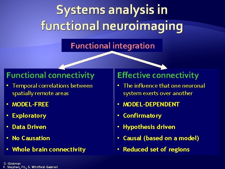
Systems analysis in functional neuroimaging Functional integration Functional connectivity Effective connectivity • Temporal correlations between spatially remote areas • The influence that one neuronal system exerts over another • MODEL-FREE • MODEL-DEPENDENT • Exploratory • Confirmatory • Data Driven • Hypothesis driven • No Causation • Causal (based on a model) • Whole brain connectivity • Reduced set of regions D. Gitelman K. Stephan, FIL; S. Whitfield-Gabrieli
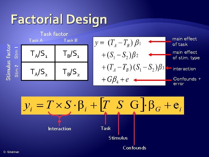
Factorial Design Task A Task B Stim 1 TA/S 1 TB/S 1 Stim 2 Stimulus factor Task factor TA/S 2 TB/S 2 Interaction main effect of task main effect of stim. type interaction Confounds + error Task Stimulus D. Gitelman Confounds
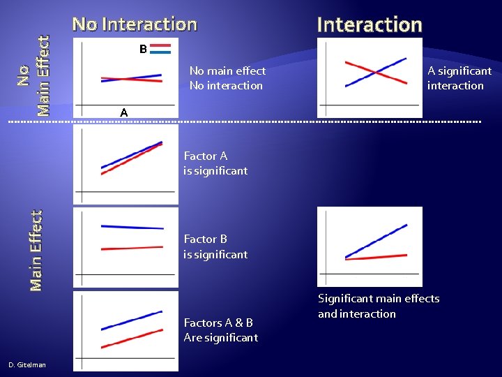
No Main Effect No Interaction B No main effect No interaction A significant interaction A Main Effect Factor A is significant Factor B is significant Factors A & B Are significant D. Gitelman Significant main effects and interaction
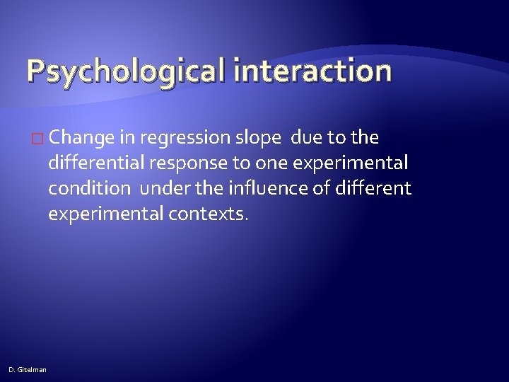
Psychological interaction � Change in regression slope due to the differential response to one experimental condition under the influence of different experimental contexts. D. Gitelman
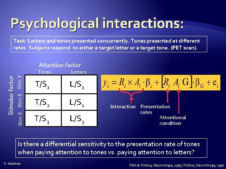
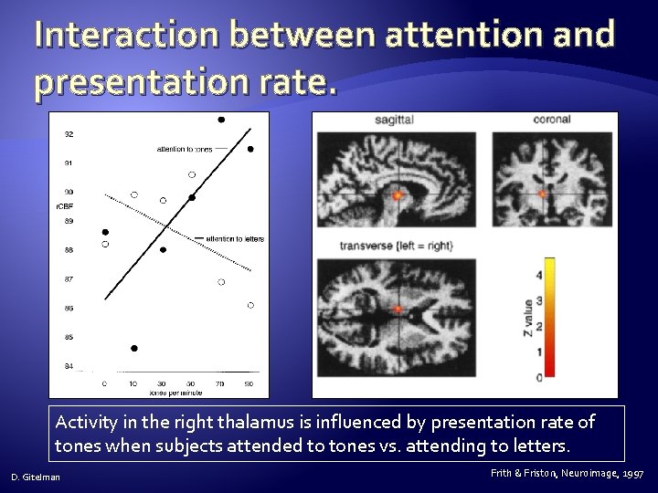
Psychological interactions: Task: Letters and tones presented concurrently. Tones presented at different rates. Subjects respond to either a target letter or a target tone. (PET scan). Attention factor Stim 2 Stim 1 Letters T/S 1 L/S 1 T/S 2 L/S 2 Stim 3 Stimulus factor Tones T/S 3 L/S 3 Interaction Presentation rates Attentional condition Is there a differential sensitivity to the presentation rate of tones when paying attention to tones vs. paying attention to letters? D. Gitelman Frith & Friston, Neuroimage, 1997; Friston, Neuroimage, 1997
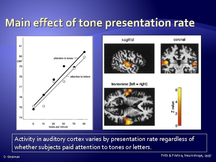
Main effect of tone presentation rate Activity in auditory cortex varies by presentation rate regardless of whether subjects paid attention to tones or letters. D. Gitelman Frith & Friston, Neuroimage, 1997
Interaction between attention and presentation rate. Activity in the right thalamus is influenced by presentation rate of tones when subjects attended to tones vs. attending to letters. D. Gitelman Frith & Friston, Neuroimage, 1997
Physiophysiological interaction � Change in regression slope due to the differential response of the signal (neural activity) from one region due to the signal from another (region). D. Gitelman
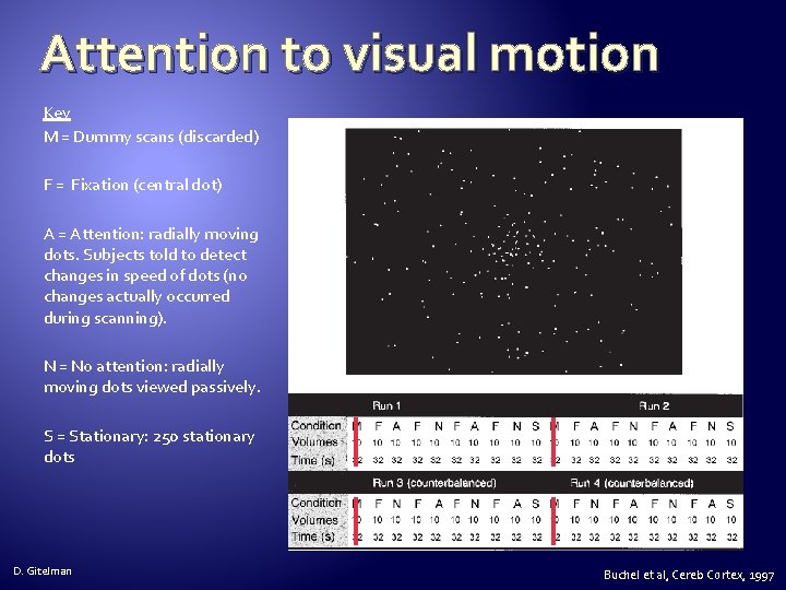
Attention to visual motion Key M = Dummy scans (discarded) F = Fixation (central dot) A = Attention: radially moving dots. Subjects told to detect changes in speed of dots (no changes actually occurred during scanning). N = No attention: radially moving dots viewed passively. S = Stationary: 250 stationary dots D. Gitelman Buchel et al, Cereb Cortex, 1997
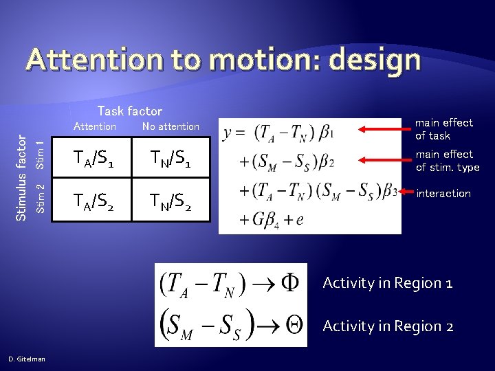
Attention to motion: design Attention No attention Stim 1 TA/S 1 TN/S 1 Stim 2 Stimulus factor Task factor TA/S 2 TN/S 2 main effect of task main effect of stim. type interaction Activity in Region 1 Activity in Region 2 D. Gitelman
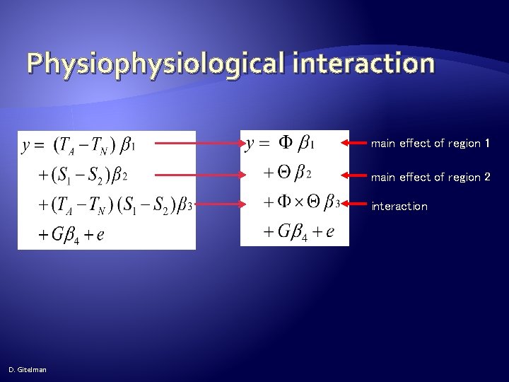
Physiophysiological interaction main effect of region 1 main effect of region 2 interaction D. Gitelman
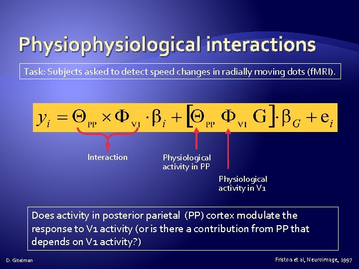
Physiophysiological interactions Task: Subjects asked to detect speed changes in radially moving dots (f. MRI). Interaction Physiological activity in PP Physiological activity in V 1 Does activity in posterior parietal (PP) cortex modulate the response to V 1 activity (or is there a contribution from PP that depends on V 1 activity? ) D. Gitelman Friston et al, Neuroimage, 1997
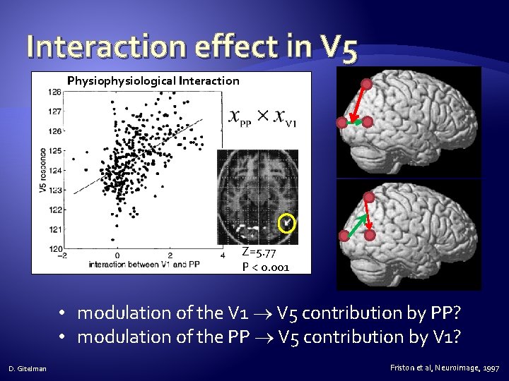
Interaction effect in V 5 Physiophysiological Interaction Z=5. 77 P < 0. 001 • modulation of the V 1 V 5 contribution by PP? • modulation of the PP V 5 contribution by V 1? D. Gitelman Friston et al, Neuroimage, 1997
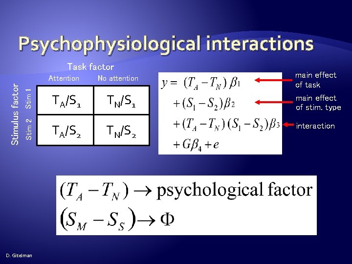
Psychophysiological interactions Task factor Stim 1 No attention TA/S 1 TN/S 1 Stim 2 Stimulus factor Attention TA/S 2 TN/S 2 D. Gitelman main effect of task main effect of stim. type interaction
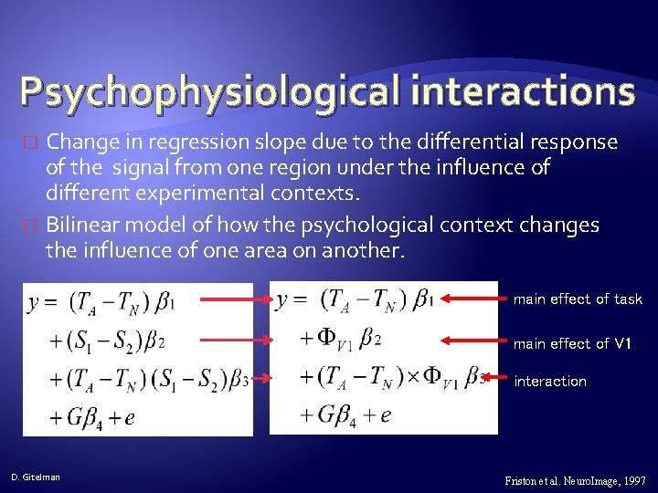
Psychophysiological interactions Change in regression slope due to the differential response of the signal from one region under the influence of different experimental contexts. � Bilinear model of how the psychological context changes the influence of one area on another. � main effect of task main effect of V 1 interaction D. Gitelman Friston et al. Neuro. Image, 1997
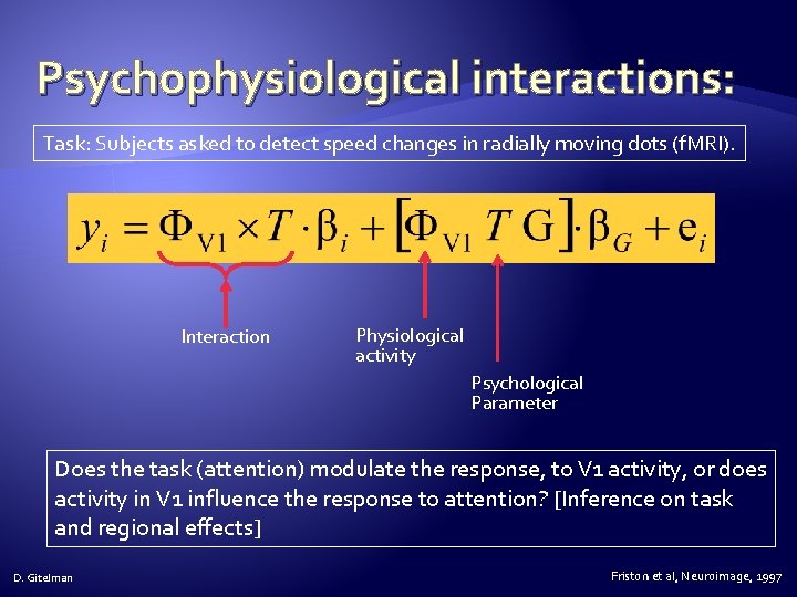
Psychophysiological interactions: Task: Subjects asked to detect speed changes in radially moving dots (f. MRI). Interaction Physiological activity Psychological Parameter Does the task (attention) modulate the response, to V 1 activity, or does activity in V 1 influence the response to attention? [Inference on task and regional effects] D. Gitelman Friston et al, Neuroimage, 1997
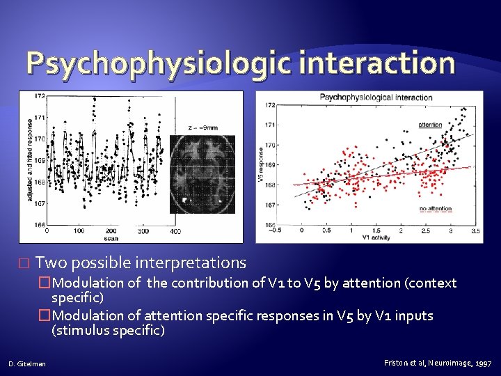
Psychophysiologic interaction � Two possible interpretations �Modulation of the contribution of V 1 to V 5 by attention (context specific) �Modulation of attention specific responses in V 5 by V 1 inputs (stimulus specific) D. Gitelman Friston et al, Neuroimage, 1997
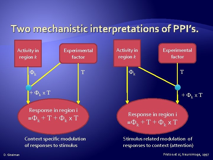
Two mechanistic interpretations of PPI’s. Activity in region k Experimental factor Фk T + Фk x T Response in region i =Фk + T + Фk x T Context specific modulation of responses to stimulus D. Gitelman + Фk x T Response in region i =Фk + T + Фk x T Stimulus related modulation of responses to context (attention) Friston et al, Neuroimage, 1997
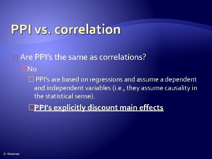
PPI vs. correlation � Are PPI’s the same as correlations? �No � PPI’s are based on regressions and assume a dependent and independent variables (i. e. , they assume causality in the statistical sense). �PPI’s explicitly discount main effects D. Gitelman
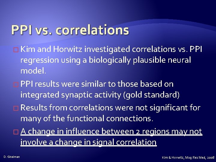
PPI vs. correlations � Kim and Horwitz investigated correlations vs. PPI regression using a biologically plausible neural model. � PPI results were similar to those based on integrated synaptic activity (gold standard) � Results from correlations were not significant for many of the functional connections. � A change in influence between 2 regions may not involve a change in signal correlation D. Gitelman Kim & Horwitz, Mag Res Med, 2008
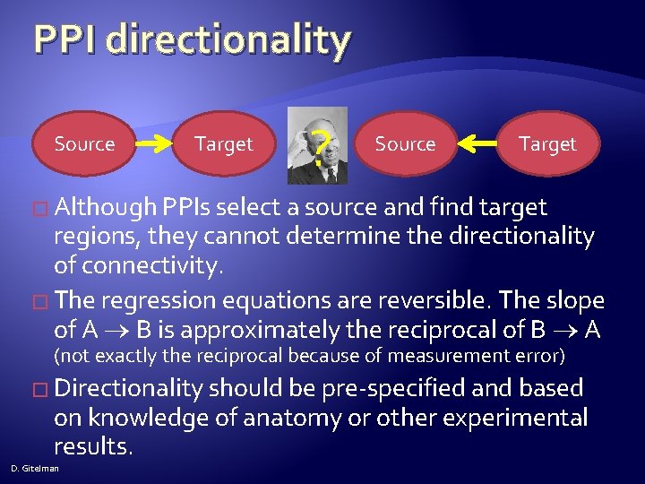
PPI directionality Source Target ? Source Target � Although PPIs select a source and find target regions, they cannot determine the directionality of connectivity. � The regression equations are reversible. The slope of A B is approximately the reciprocal of B A (not exactly the reciprocal because of measurement error) � Directionality should be pre-specified and based on knowledge of anatomy or other experimental results. D. Gitelman
D. Gitelman
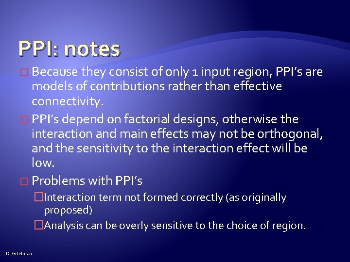
PPI: notes � Because they consist of only 1 input region, PPI’s are models of contributions rather than effective connectivity. � PPI’s depend on factorial designs, otherwise the interaction and main effects may not be orthogonal, and the sensitivity to the interaction effect will be low. � Problems with PPI’s �Interaction term not formed correctly (as originally proposed) �Analysis can be overly sensitive to the choice of region. D. Gitelman
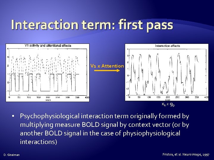
Interaction term: first pass V 1 x Attention xk x g p • Psychophysiological interaction term originally formed by multiplying measure BOLD signal by context vector (or by another BOLD signal in the case of physiological interactions) D. Gitelman Friston, et al. Neuroimage, 1997
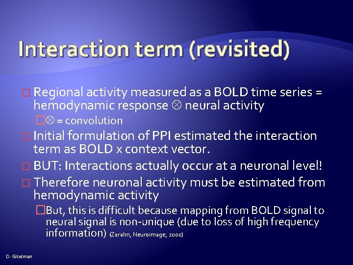
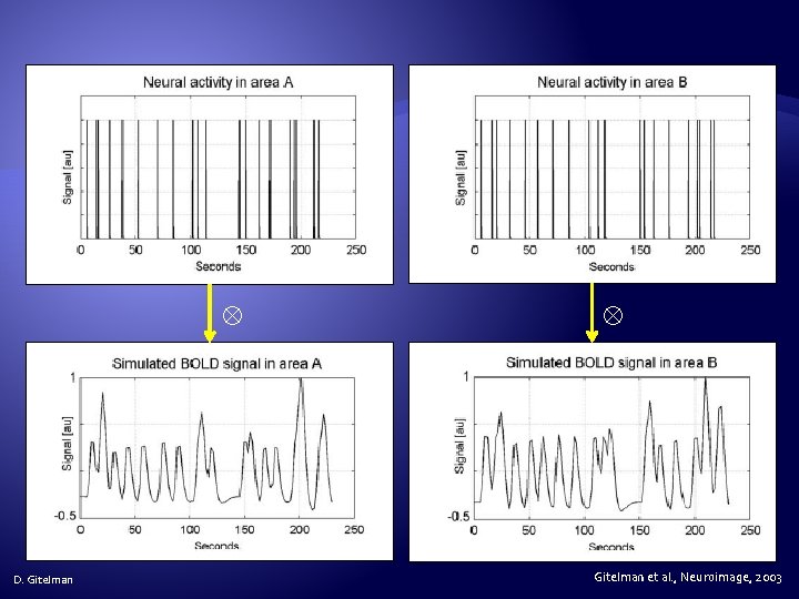
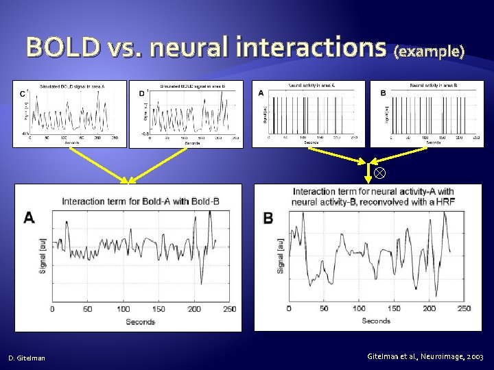
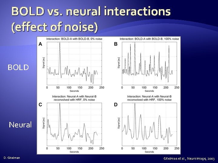
Interaction term (revisited) � Regional activity measured as a BOLD time series = hemodynamic response neural activity � = convolution � Initial formulation of PPI estimated the interaction term as BOLD x context vector. � BUT: Interactions actually occur at a neuronal level! � Therefore neuronal activity must be estimated from hemodynamic activity �But, this is difficult because mapping from BOLD signal to neural signal is non-unique (due to loss of high frequency information) (Zarahn, Neuroimage, 2000) D. Gitelman
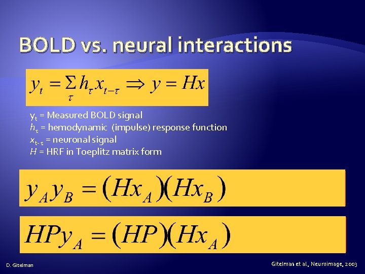
BOLD vs. neural interactions yt = Measured BOLD signal h = hemodynamic (impulse) response function xt- = neuronal signal H = HRF in Toeplitz matrix form D. Gitelman et al. , Neuroimage, 2003
D. Gitelman et al. , Neuroimage, 2003
BOLD vs. neural interactions (example) D. Gitelman et al. , Neuroimage, 2003
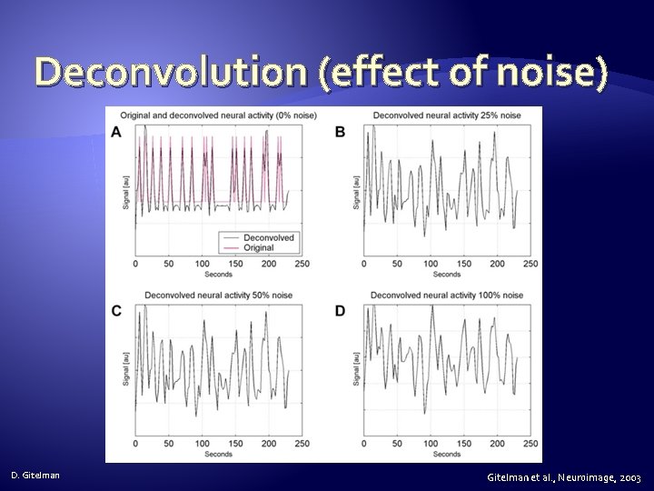
Deconvolution of BOLD signal � Can try using a maximum likelihood estimator (i. e. , least squares) but this runs into trouble with highfrequency components. �Zarahn constrained the estimates to particular temporal intervals. (Zarahn, Neuroimage, 2000) � Can try using a Weiner filter, but this requires high SNR and an estimate of the noise spectral density. (Glover, Neuroimage, 1999) � D. Gitelman Use empirical Bayes deconvolution to finesse the noise estimates by setting the prior precisions on the high frequencies to 0. (Gitelman, Neuroimage, 2003) Gitelman et al. , Neuroimage, 2003
Deconvolution (effect of noise) D. Gitelman et al. , Neuroimage, 2003
BOLD vs. neural interactions (effect of noise) BOLD Neural D. Gitelman et al. , Neuroimage, 2003
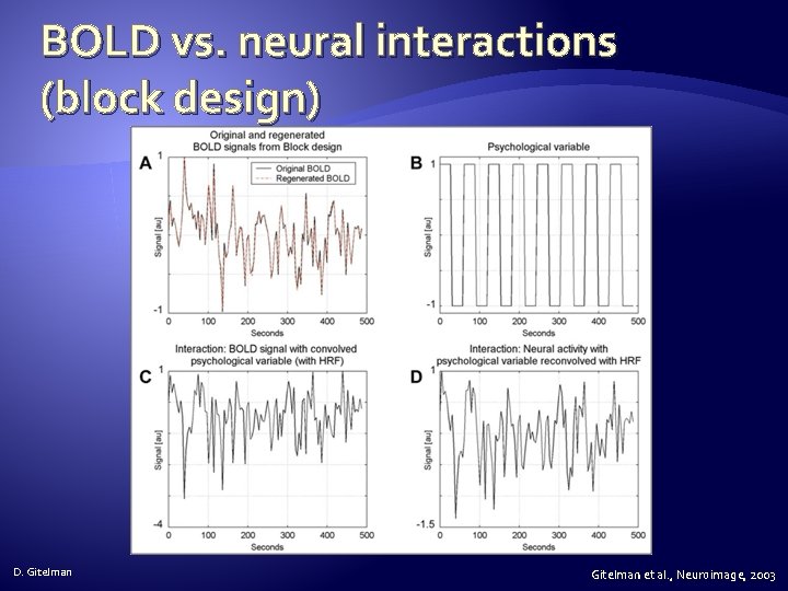
BOLD vs. neural interactions (block design) D. Gitelman et al. , Neuroimage, 2003
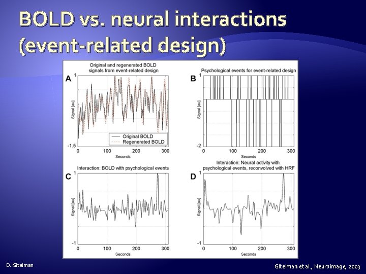
BOLD vs. neural interactions (event-related design) D. Gitelman et al. , Neuroimage, 2003
PPI EXAMPLES D. Gitelman
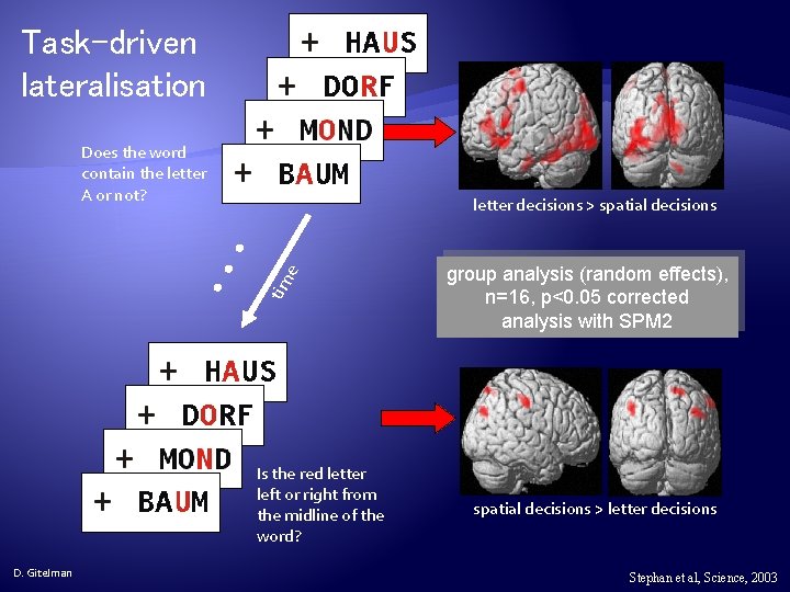
Task-driven lateralisation Does the word contain the letter A or not? • • • tim e letter decisions > spatial decisions Is the red letter left or right from the midline of the word? D. Gitelman group analysis (random effects), n=16, p<0. 05 corrected analysis with SPM 2 spatial decisions > letter decisions Stephan et al, Science, 2003
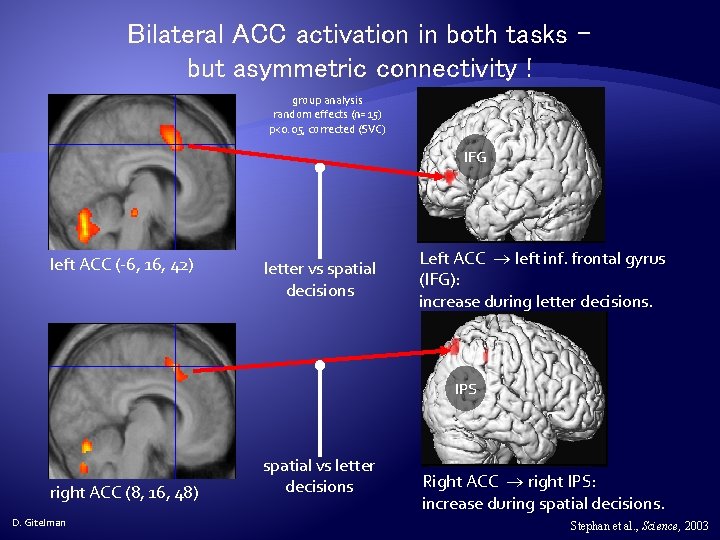
Bilateral ACC activation in both tasks – but asymmetric connectivity ! group analysis random effects (n=15) p<0. 05, corrected (SVC) IFG left ACC (-6, 16, 42) letter vs spatial decisions Left ACC left inf. frontal gyrus (IFG): increase during letter decisions. IPS right ACC (8, 16, 48) D. Gitelman spatial vs letter decisions Right ACC right IPS: increase during spatial decisions. Stephan et al. , Science, 2003
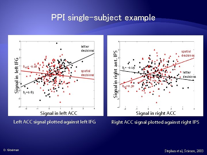
Signal in left IFG letter decisions b. VS= -0. 16 spatial decisions b. L=0. 63 Signal in left ACC Left ACC signal plotted against left IFG D. Gitelman Signal in right ant. IPS PPI single-subject example spatial decisions b. L= -0. 19 letter decisions b. VS=0. 50 Signal in right ACC Right ACC signal plotted against right IPS Stephan et al, Science, 2003
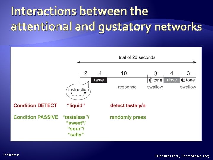
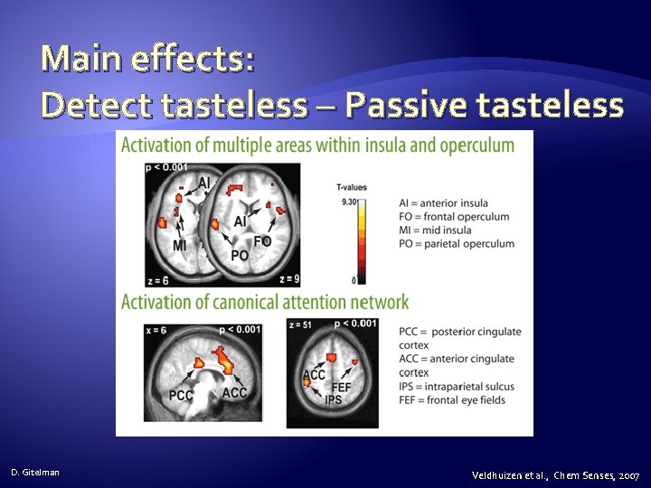
Interactions between the attentional and gustatory networks D. Gitelman Veldhuizen et al. , Chem Senses, 2007
Main effects: Detect tasteless – Passive tasteless D. Gitelman Veldhuizen et al. , Chem Senses, 2007
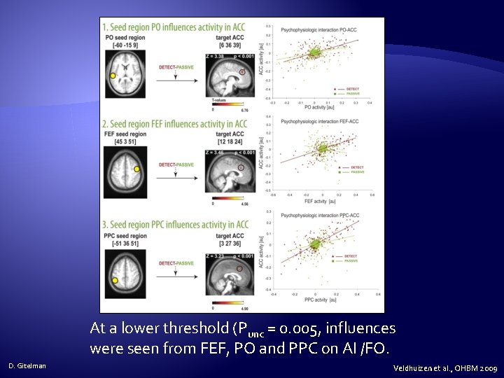
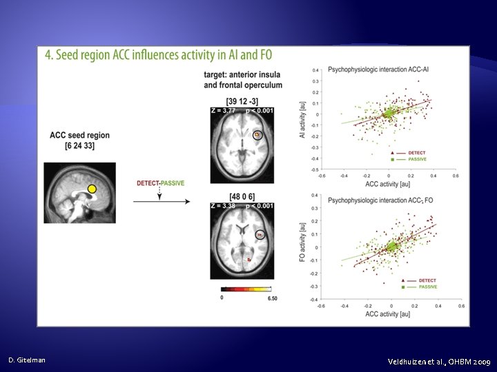
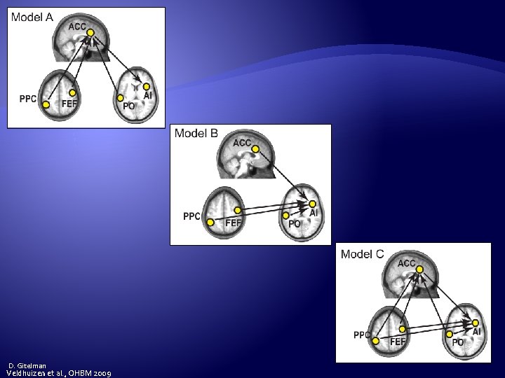
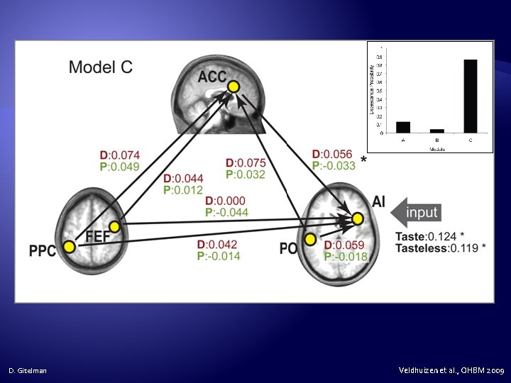
At a lower threshold (Punc = 0. 005, influences were seen from FEF, PO and PPC on AI /FO. D. Gitelman Veldhuizen et al. , OHBM 2009
D. Gitelman Veldhuizen et al. , OHBM 2009
D. Gitelman Veldhuizen et al. , OHBM 2009
D. Gitelman Veldhuizen et al. , OHBM 2009
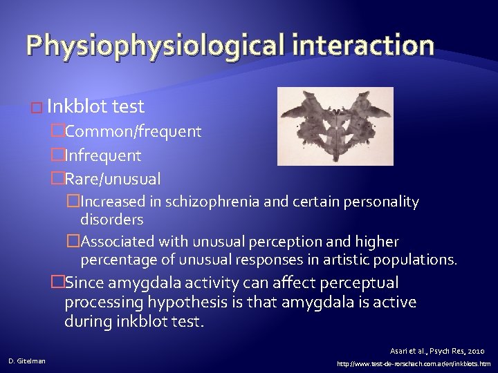
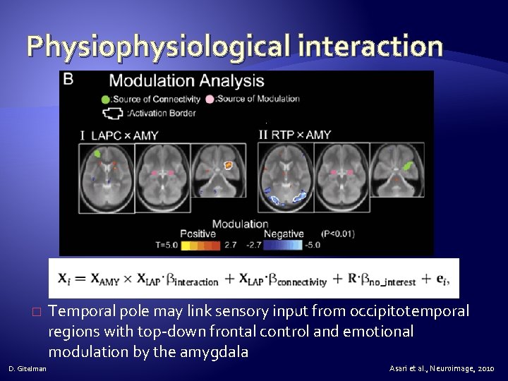
Physiophysiological interaction � Inkblot test �Common/frequent �Infrequent �Rare/unusual �Increased in schizophrenia and certain personality disorders �Associated with unusual perception and higher percentage of unusual responses in artistic populations. �Since amygdala activity can affect perceptual processing hypothesis is that amygdala is active during inkblot test. Asari et al. , Psych Res, 2010 D. Gitelman http: //www. test-de-rorschach. com. ar/en/inkblots. htm
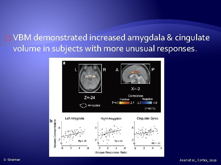
� VBM demonstrated increased amygdala & cingulate volume in subjects with more unusual responses. D. Gitelman Asari et al. , Cortex, 2010
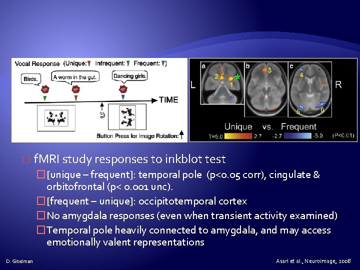
* � f. MRI study responses to inkblot test �[unique – frequent]: temporal pole (p<0. 05 corr), cingulate & orbitofrontal (p< 0. 001 unc). �[frequent – unique]: occipitotemporal cortex �No amygdala responses (even when transient activity examined) �Temporal pole heavily connected to amygdala, and may access emotionally valent representations D. Gitelman Asari et al. , Neuroimage, 2008
Physiophysiological interaction � D. Gitelman Temporal pole may link sensory input from occipitotemporal regions with top-down frontal control and emotional modulation by the amygdala Asari et al. , Neuroimage, 2010
Pros & Cons of PPIs � Pros: �Given a single source region, we can test for its contextdependent connectivity across the entire brain �Simple to perform � Cons: �Very simplistic model: only allows modelling contributions from a single area �Ignores time-series properties of data (can do PPI’s on PET and f. MRI data) �Inputs are not modelled explicitly �Interactions are instantaneous D. Gitelman K. Stephan, FIL
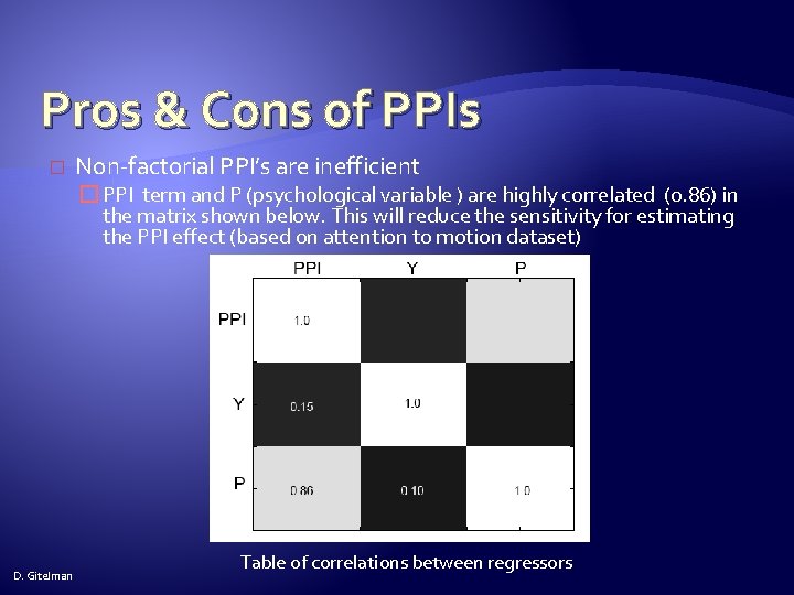
Pros & Cons of PPIs � Non-factorial PPI’s are inefficient � PPI term and P (psychological variable ) are highly correlated (0. 86) in the matrix shown below. This will reduce the sensitivity for estimating the PPI effect (based on attention to motion dataset) D. Gitelman Table of correlations between regressors
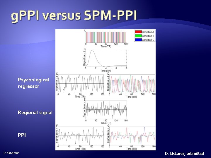
g. PPI versus SPM-PPI Psychological regressor Regional signal PPI D. Gitelman D. Mc. Laren, submitted
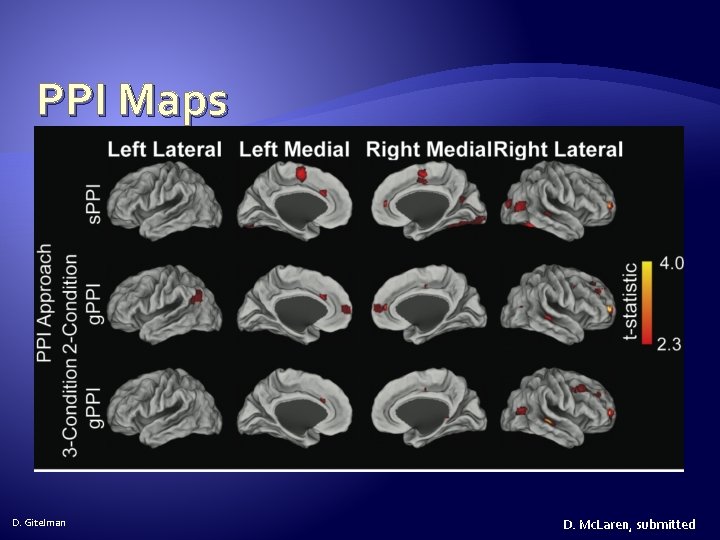
PPI Maps D. Gitelman D. Mc. Laren, submitted
PPI analysis: overview � Run a standard GLM analysis �Include conditions and any nuisance effects (motion, etc. ) �For ease of analysis combine multiple sessions into a single session and include block effects � Display contrast of interest � Extract VOI (volume of interest), adjusting for effects of interest (i. e. , exclude any nuisance regressors) � PPI button (makes PPI regressors) �SPM. mat file from standard GLM analysis �Setup contrast of Psych conditions D. Gitelman
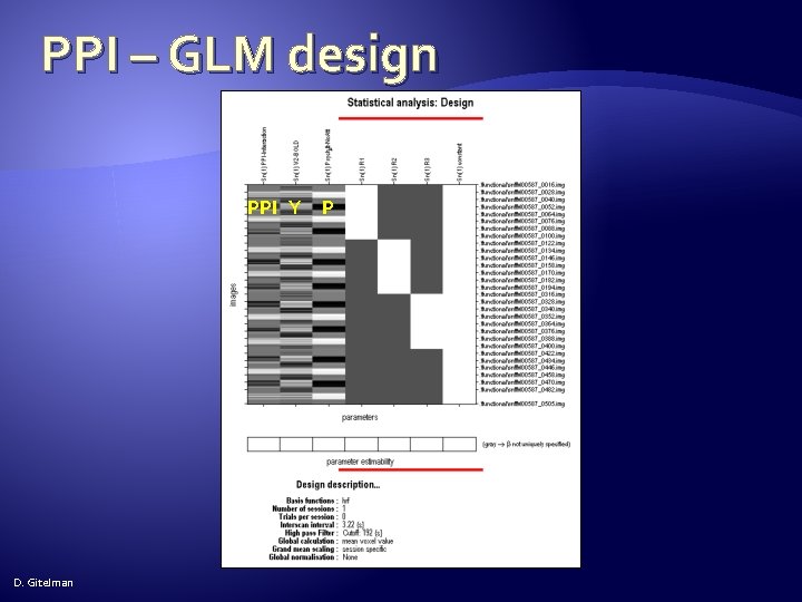
PPI analysis: overview � Run PPI – GLM analysis �PPI. ppi (interaction) �PPI. Y (main effect: source region BOLD data) �PPI. P (main effect: Psych conditions that formed PPI) � Contrast is [1 0 0] for positive effect of interaction and [-1 0 0 ] for negative effect (assuming conditions entered in order listed) D. Gitelman
Standard GLM: design setup and estimation � The analysis directory should include: �Directory named functional which includes the preprocessed f. MRI volumes �Directory name structural, which includes a T 1 image �Files: factors. mat, block_regressors. mat, multi_condition. mat and multi_block_regressors. mat �Make 2 empty directories called GLM and PPI D. Gitelman
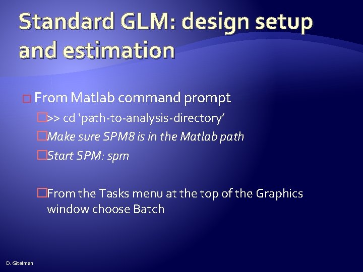
Standard GLM: design setup and estimation � From Matlab command prompt �>> cd ‘path-to-analysis-directory’ �Make sure SPM 8 is in the Matlab path �Start SPM: spm �From the Tasks menu at the top of the Graphics window choose Batch D. Gitelman
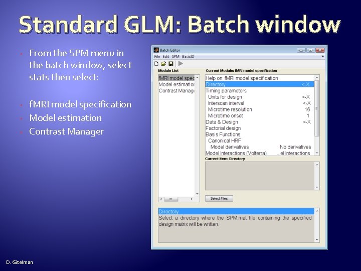
Standard GLM: Batch window • From the SPM menu in the batch window, select stats then select: • f. MRI model specification Model estimation Contrast Manager • • D. Gitelman
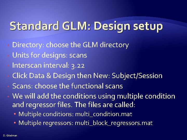
Standard GLM: Design setup • • • Directory: choose the GLM directory Units for designs: scans Interscan interval: 3. 22 Click Data & Design then New: Subject/Session Scans: choose the functional scans We will add the conditions using multiple condition and regressor files. The files are called: • Multiple conditions: multi_condition. mat • Multiple regressors: multi_block_regressors. mat D. Gitelman
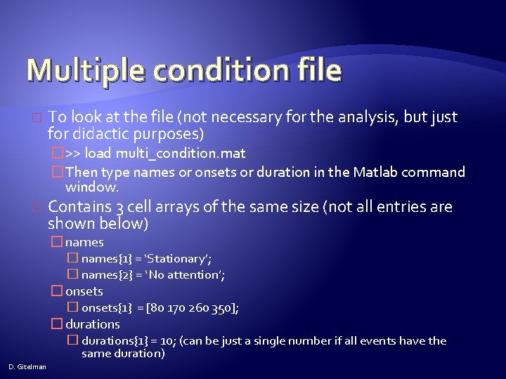
Multiple condition file � To look at the file (not necessary for the analysis, but just for didactic purposes) �>> load multi_condition. mat �Then type names or onsets or duration in the Matlab command window. � Contains 3 cell arrays of the same size (not all entries are shown below) � names{1} = ‘Stationary’; � names{2} = ‘No attention’; � onsets{1} = [80 170 260 350]; � durations{1} = 10; (can be just a single number if all events have the same duration) D. Gitelman
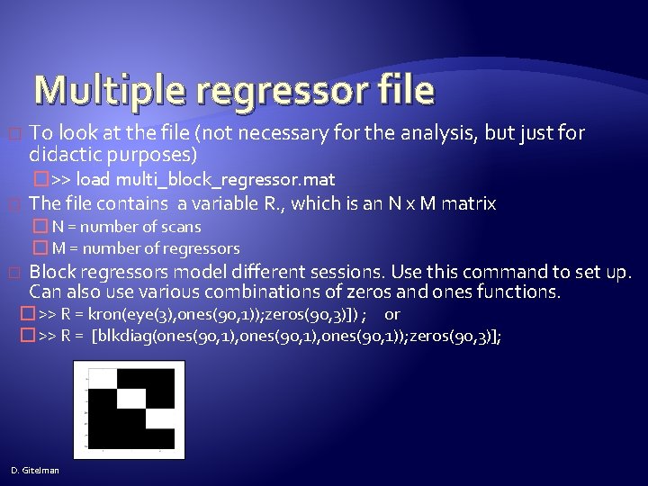
Multiple regressor file � � To look at the file (not necessary for the analysis, but just for didactic purposes) �>> load multi_block_regressor. mat The file contains a variable R. , which is an N x M matrix � N = number of scans � M = number of regressors � Block regressors model different sessions. Use this command to set up. Can also use various combinations of zeros and ones functions. � >> R = kron(eye(3), ones(90, 1)); zeros(90, 3)]) ; or � >> R = [blkdiag(ones(90, 1), ones(90, 1)); zeros(90, 3)]; D. Gitelman
Standard GLM: Design setup � High-Pass Filter : 192 �Note: most designs will use a high-pass filter value of 128. This dataset requires a longer high-pass filter in order not to lose the low frequency components of the design. � The default values on the rest of the entries are correct � Click Model Estimation Select SPM. mat Click Dependency and choose f. MRI model specification: SPM. mat File D. Gitelman
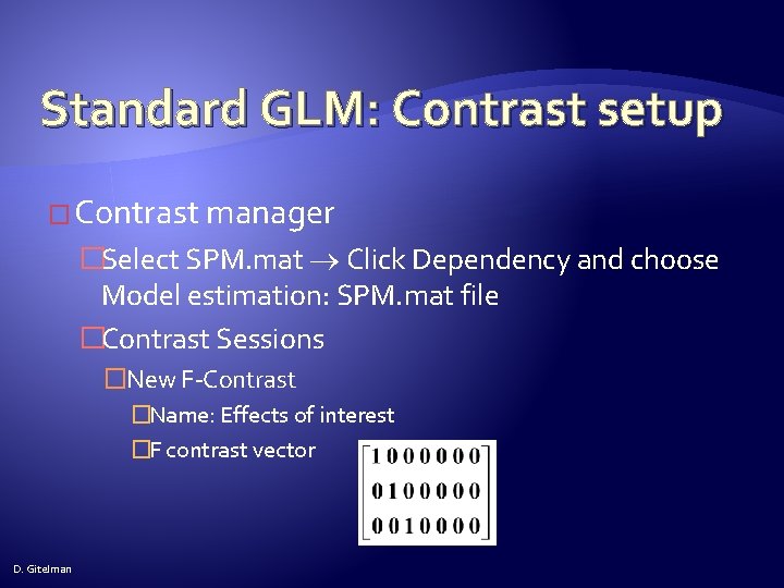
Standard GLM: Contrast setup � Contrast manager �Select SPM. mat Click Dependency and choose Model estimation: SPM. mat file �Contrast Sessions �New F-Contrast �Name: Effects of interest �F contrast vector D. Gitelman
Standard GLM: Contrast setup � Contrast Sessions �New T-Contrast �Name: Attention �T contrast vector: [0 -1 1 0 0] �New T-Contrast �Name: Motion �T contrast vector: [-2 1 1 0 0] � Click Save button and save the batch file � Click Run button (green arrow) D. Gitelman
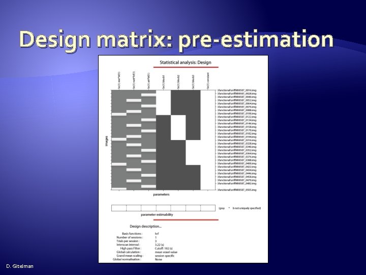
Design matrix: pre-estimation D. Gitelman
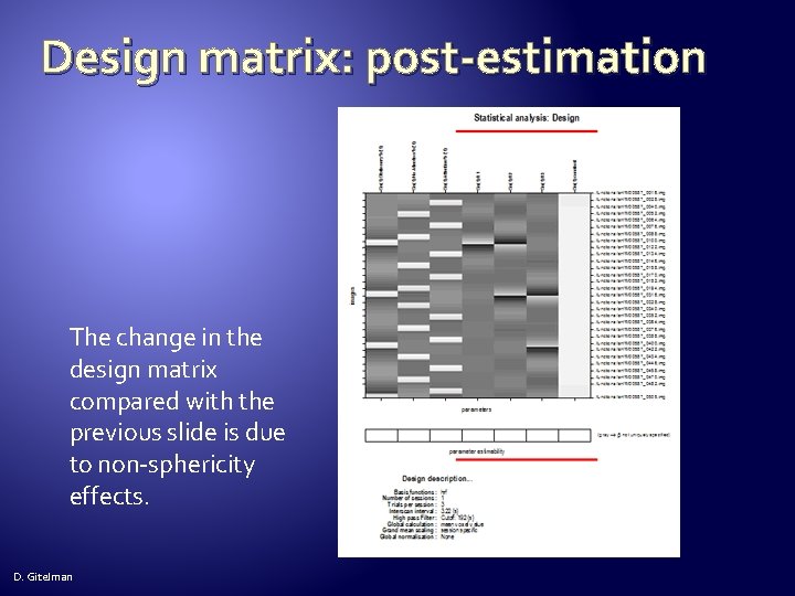
Design matrix: post-estimation The change in the design matrix compared with the previous slide is due to non-sphericity effects. D. Gitelman
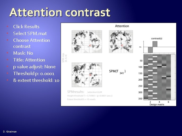
Attention contrast • Click Results • Select SPM. mat • Choose Attention contrast • Mask: No • Title: Attention • p value adjust: None • Threshold p: 0. 0001 • & extent threshold: 10 D. Gitelman
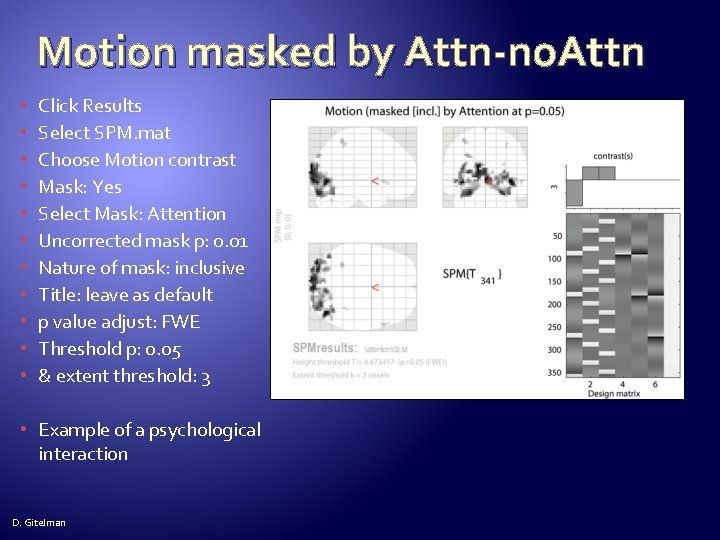
Motion masked by Attn-no. Attn • • • Click Results Select SPM. mat Choose Motion contrast Mask: Yes Select Mask: Attention Uncorrected mask p: 0. 01 Nature of mask: inclusive Title: leave as default p value adjust: FWE Threshold p: 0. 05 & extent threshold: 3 • Example of a psychological interaction D. Gitelman
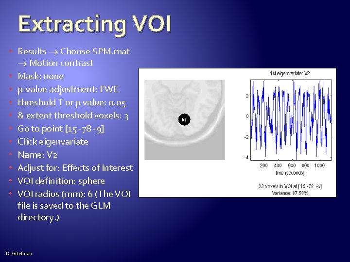
Extracting VOI • Results Choose SPM. mat Motion contrast • Mask: none • p-value adjustment: FWE • threshold T or p value: 0. 05 • & extent threshold voxels: 3 • Go to point [15 -78 -9] • Click eigenvariate • Name: V 2 • Adjust for: Effects of Interest • VOI definition: sphere • VOI radius (mm): 6 (The VOI file is saved to the GLM directory. ) D. Gitelman
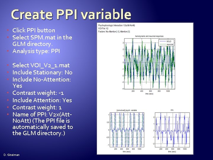
Create PPI variable • Click PPI button • Select SPM. mat in the GLM directory. • Analysis type: PPI • Select VOI_V 2_1. mat • Include Stationary: No • Include No-Attention: Yes • Contrast weight: -1 • Include Attention: Yes • Contrast weight: 1 • Name of PPI: V 2 x(Att. No. Att) (The PPI file is automatically saved to the GLM directory. ) D. Gitelman
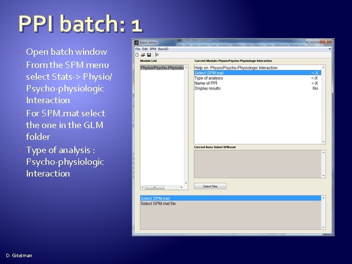
PPI batch: 1 • • Open batch window From the SPM menu select Stats-> Physio/ Psycho-physiologic Interaction For SPM. mat select the one in the GLM folder Type of analysis : Psycho-physiologic Interaction D. Gitelman
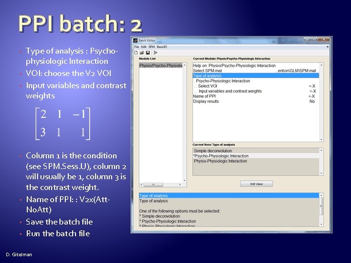
PPI batch: 2 • • Type of analysis : Psychophysiologic Interaction VOI: choose the V 2 VOI Input variables and contrast weights Column 1 is the condition (see SPM. Sess. U), column 2 will usually be 1, column 3 is the contrast weight. Name of PPI: : V 2 x(Att. No. Att) Save the batch file Run the batch file D. Gitelman
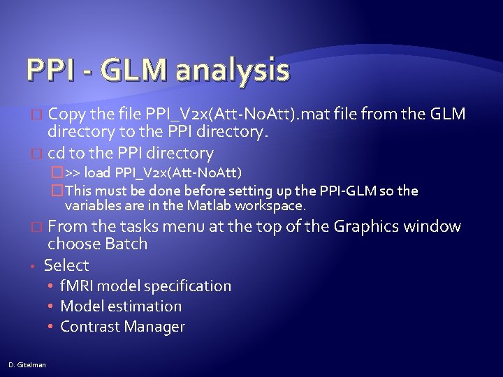
PPI - GLM analysis Copy the file PPI_V 2 x(Att-No. Att). mat file from the GLM directory to the PPI directory. � cd to the PPI directory � �>> load PPI_V 2 x(Att-No. Att) �This must be done before setting up the PPI-GLM so the variables are in the Matlab workspace. From the tasks menu at the top of the Graphics window choose Batch Select � • • f. MRI model specification • Model estimation • Contrast Manager D. Gitelman
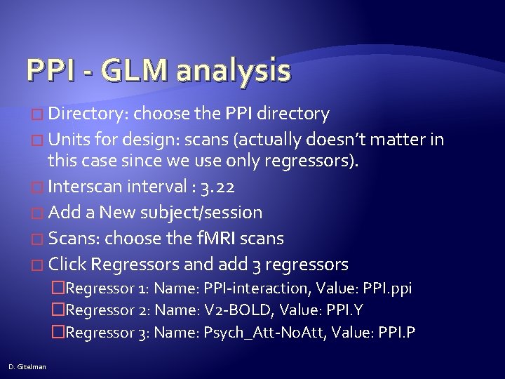
PPI - GLM analysis � Directory: choose the PPI directory � Units for design: scans (actually doesn’t matter in this case since we use only regressors). � Interscan interval : 3. 22 � Add a New subject/session � Scans: choose the f. MRI scans � Click Regressors and add 3 regressors �Regressor 1: Name: PPI-interaction, Value: PPI. ppi �Regressor 2: Name: V 2 -BOLD, Value: PPI. Y �Regressor 3: Name: Psych_Att-No. Att, Value: PPI. P D. Gitelman
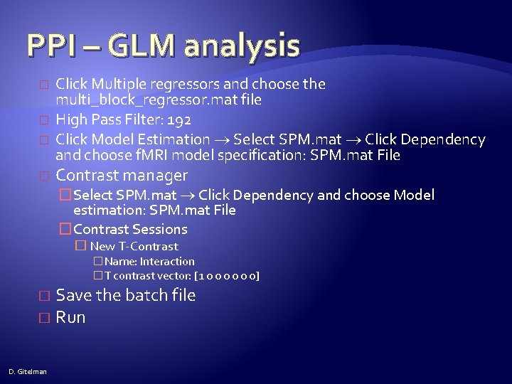
PPI – GLM analysis � � Click Multiple regressors and choose the multi_block_regressor. mat file High Pass Filter: 192 Click Model Estimation Select SPM. mat Click Dependency and choose f. MRI model specification: SPM. mat File Contrast manager �Select SPM. mat Click Dependency and choose Model estimation: SPM. mat File �Contrast Sessions � New T-Contrast �Name: Interaction �T contrast vector: [1 0 0 0] Save the batch file � Run � D. Gitelman
PPI – GLM design PPI Y D. Gitelman P
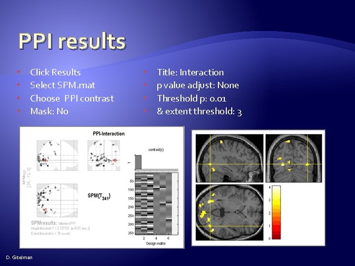
PPI results • • Click Results Select SPM. mat Choose PPI contrast Mask: No D. Gitelman • • Title: Interaction p value adjust: None Threshold p: 0. 01 & extent threshold: 3
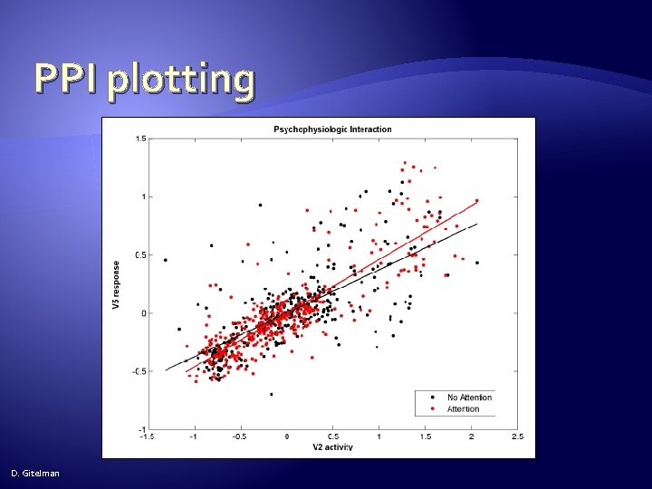
PPI plotting 1/8 � Click Results, select the original GLM analysis SPM. mat file � Choose the Motion contrast � Mask: No � Title: Motion � p value adjustment to control: None � Threshold T or p value: 0. 001 � & extent threshold voxels: 3 D. Gitelman
PPI plotting � Go to point: [39 -72 0] � Press eigenvariate � Name of region: V 5 � Adjust data for: effects of interest � VOI definition: sphere � VOI radius: 6 �The VOI file is saved to the GLM directory. D. Gitelman 2/8
PPI plotting 3/8 � Create 4 PPIs (Click PPI button , then choose psychophysiological interaction as the task to perform. ) D. Gitelman �V 2 x. No. Attention (Use the V 2 VOI and include No-Attention with a contrast weight of 1, do not include Stationary, Attention) �V 2 x. Attention (Use the V 2 VOI and include Attention with a contrast weight of 1, do not include Stationary, No. Attention) �V 5 x. No. Attention (Use the V 5 VOI and include No-Attention with a contrast weight of 1, do not include Stationary, Attention) �V 5 x. Attention (Use the V 5 VOI and include Attention with a contrast weight of 1, do not include Stationary, No. Attention
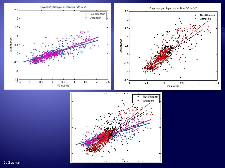
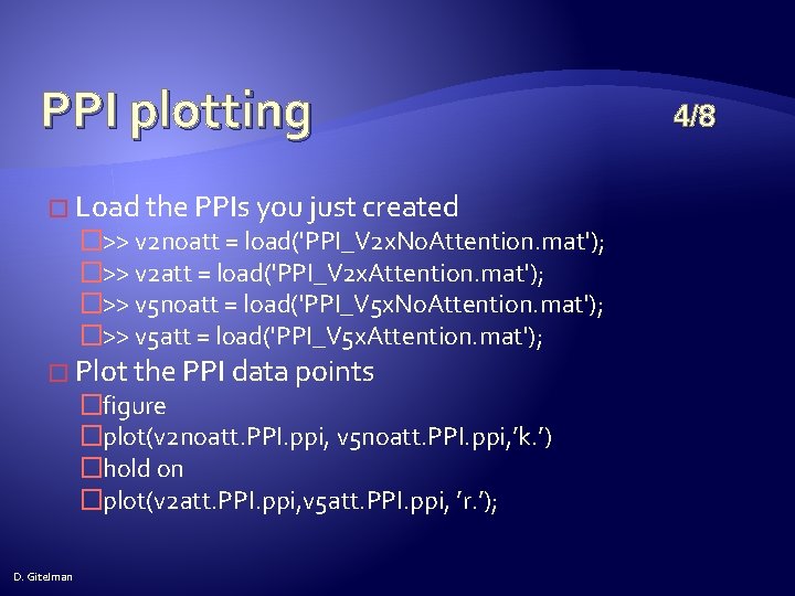
PPI plotting � Load the PPIs you just created �>> v 2 noatt = load('PPI_V 2 x. No. Attention. mat'); �>> v 2 att = load('PPI_V 2 x. Attention. mat'); �>> v 5 noatt = load('PPI_V 5 x. No. Attention. mat'); �>> v 5 att = load('PPI_V 5 x. Attention. mat'); � Plot the PPI data points �figure �plot(v 2 noatt. PPI. ppi, v 5 noatt. PPI. ppi, ’k. ’) �hold on �plot(v 2 att. PPI. ppi, v 5 att. PPI. ppi, ’r. ’); D. Gitelman 4/8
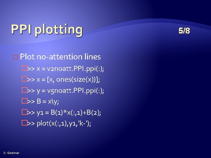
PPI plotting � Plot no-attention lines �>> x = v 2 noatt. PPI. ppi(: ); �>> x = [x, ones(size(x))]; �>> y = v 5 noatt. PPI. ppi(: ); �>> B = xy; �>> y 1 = B(1)*x(: , 1)+B(2); �>> plot(x(: , 1), y 1, 'k-'); D. Gitelman 5/8
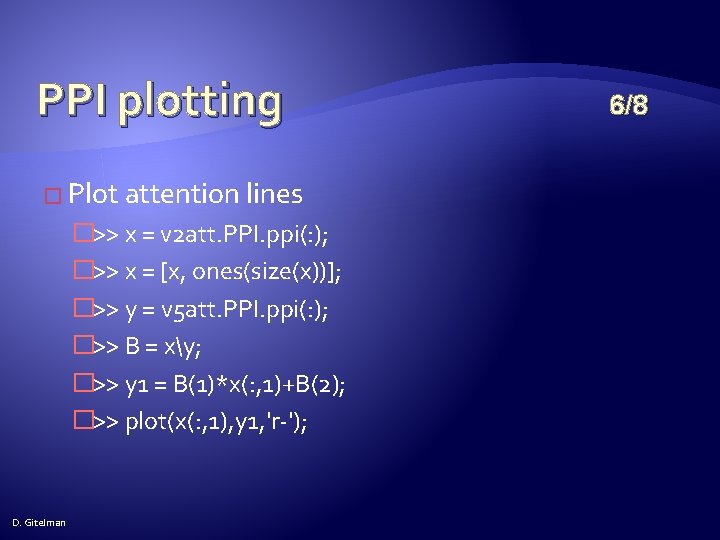
PPI plotting � Plot attention lines �>> x = v 2 att. PPI. ppi(: ); �>> x = [x, ones(size(x))]; �>> y = v 5 att. PPI. ppi(: ); �>> B = xy; �>> y 1 = B(1)*x(: , 1)+B(2); �>> plot(x(: , 1), y 1, 'r-'); D. Gitelman 6/8
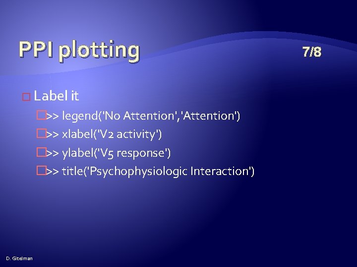
PPI plotting � Label it �>> legend('No Attention', 'Attention') �>> xlabel('V 2 activity') �>> ylabel('V 5 response') �>> title('Psychophysiologic Interaction') D. Gitelman 7/8
PPI plotting D. Gitelman
PPI Questions � Including main effects in my PPI analysis takes away all the interaction results. Do I have to include main effects? �Absolutely! Otherwise, the inference on interactions will be confounded by main effects. � Should I include movement regressors in my PPI analysis? �Yes, include any nuisance effects you would normally include. D. Gitelman
PPI Questions � What does it mean to adjust for different effects? �Adjusting for effects means keeping effects of interest and removing effects of no interest, e. g. , movement regressors, block effects, etc. �In order to adjust an effects of interest F-contrast should be set up before trying to extract the data. D. Gitelman
PPI Questions � Should VOIs be based on the exact locations of group effects or should they be subject specific? �Both. VOI s should be subject specific, but should be “close enough” to a maxima based on a group analysis, the literature , etc. “Close enough” depends on where the maxima is in the brain, the smoothness of the data, etc. Close enough in the caudate might be 5 mm, while in the parietal cortex, close enough might be 1 cm. D. Gitelman
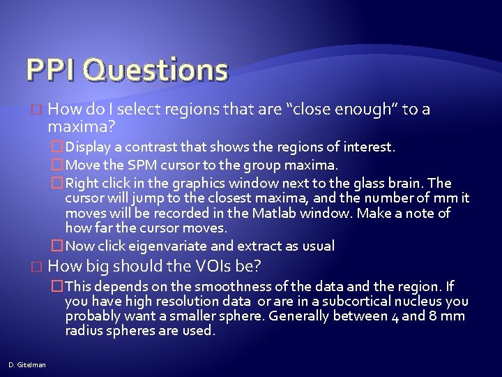
PPI Questions � How do I select regions that are “close enough” to a maxima? �Display a contrast that shows the regions of interest. �Move the SPM cursor to the group maxima. �Right click in the graphics window next to the glass brain. The cursor will jump to the closest maxima, and the number of mm it moves will be recorded in the Matlab window. Make a note of how far the cursor moves. �Now click eigenvariate and extract as usual � How big should the VOIs be? �This depends on the smoothness of the data and the region. If you have high resolution data or are in a subcortical nucleus you probably want a smaller sphere. Generally between 4 and 8 mm radius spheres are used. D. Gitelman
PPI Questions � How is a group PPI analysis done? �The con images from the interaction term can be brought to a standard second level analysis (onesample t-test within a group, two-sample t-test between groups, ANOVA’s, etc. ) D. Gitelman
THE END D. Gitelman