PATHOPHYSIOLOGY OF PAIN Prof J Hanek MD Ph


 localised in body")


")


 It occurs as a result of injury to or dysfunction of")

 a) Predominating peripheral")

 Neurophysiologic and neuroanatomic changes that may")
 Development atypical connections between nerve sprouts or demyelinated axons in the region of")


 Causalgia - severe burning pain")

")


 it is dull, deep, not well defined,")
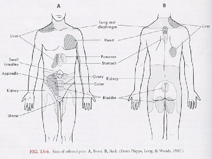
 Refered pain = when an algogenic process affecting a")


 ● Chest pain is only a late and inconstant marker")
 Congenital")


- Slides: 60
PATHOPHYSIOLOGY OF PAIN Prof. J. Hanáček, MD, Ph. D
● Alteration in sensory function dysfunctions of the general or special senses • Dysfunctions of the general senses chronic pain, abnormal temperature regulation, tactile dysfunction Definitions of pain • Pain is a complex unpleasant phenomenon composed of sensory experiences that include time, space, intensity, emotion, cognition, and motivation • Pain is an unpleasant or emotional experience originating in real or potential damaged tissue • Pain is an unpleasant phenomenon that is uniquely experienced by each individual; it cannot be adequately defined, identified, or measured by an observer
The experience of pain Three hierarchial levels interact usually to produce complex picture of pain: 1. sensory - discriminative 2. motivational - affective 3. cognitive - evaluative 1. Sensory - discriminative system (location, intensity, quality, and temporal and spatial aspects of pain) 2. Motivational - affective system determines the individual´s approach-avoidance behaviours (depression, anxiety) 3. Cognitive - evaluative system (thoughts concerning the cause and significance of pain). It may block or modulate the perception of pain
Pain categories 1. Somatogenic pain is pain with cause (usually known) localised in body tissue: a/ nociceptive pain – somatic, visceral b/ neuropatic pain 2. Psychogenic pain is pain for which there is no known physical cause but processing of sensitive information in CNS is disturbed In this type of pain the psychological evaluation yields evidence that the pain itself is predominantly sustained by psychological factors Acute and chronic pain Acute pain is a protective mechanism that alerts the individual to a condition or experience that is immediately harmful to the body Onset - usually sudden
Relief - after the chemical mediators that stimulate the nociceptors , are removed • This type of pain mobilises the individual to prompt action to relief it • Stimulation of autonomic nervous system can be observed during this type of pain (mydriasis, tachycardia, tachypnoe, sweating, vasoconstriction) Responses to acute pain - increased heart rate - diaphoresis - increased respiratory rate - elevated blood pressure - blood sugar - gastric acid secretion - pallor or flushing, dilated pupils - gastric motility - blood flow to the viscera, kidney and skin - nausea occasionally occurs
Psychological and behavioural response to acute pain - fear - general sense of unpleasantness or unease - anxiety Chronic pain is persistent or intermittent usually defined as lasting at least 6 months The cause is often unknown, often develops insidiously, very often is associated with a sense of hopelessness and helplessness. Depression often results
Psychological response to chronic pain Intermittent pain produces a physiologic response similar to acute pain Persistent pain allows for adaptation (functions of the body are normal but the pain is not reliefed) Chronic pain produces significant behavioural and psychological changes The main changes are: - depression - an attempt to keep pain - related behaviour to a minimum - sleeping disorders - preoccupation with the pain - tendency to deny pain
Pain threshold and pain tolerance The pain threshold is the point at which a stimulus is perceived as pain It does not vary significantly among healthy people or in the same person over time Perceptual dominance- intense pain at one location of the body may cause an increase in the pain threshold in another location • The pain tolerance is expressed as duration of time or the intensity of pain that an individual will endure before initiation overt pain responses. It is influenced by - person s cultural prescriptions - expectations - role behaviours - physical and mental health
• Pain tolerance is generally decreased: - with repeated exposure to pain, - by fatigue, anger, boredom, apprehension, - sleep deprivation • Tolerance to pain may be increased: - by alcohol consumption, - medication, hypnosis, - warmth, distracting activities, - strong beliefs or faith Pain tolerance varies greatly among people and in the same person over time A decrease in pain tolerance is also evident in children, teenagers and elderly
Age and perception of pain Children and the elderly may experience or express pain differently than adults Infants in the first 1 to 2 days of life are less sensitive to pain (or they simply lack the ability to verbalise the pain experience). A full behavioural response to pain is apparent at 3 to 12 month of life Older children, between the ages of 15 and 18 years, tend to have a lower pain threshold than do adults Pain threshold tends to increase with ageing This change is probably caused by peripheral neuropathies and changes in the thickness of the skin
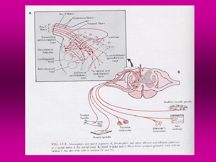
Neuroanatomy of pain The portions of the nervous system responsible for the sensation and perception of pain may be divided into three areas: 1. afferent pathways 2. CNS 3. efferent pathways The afferent portion is composed of: a) nociceptors (nerve endings of nociceptive nerve cells) b) afferent nerve fibres c) spinal cord network
Afferent pathways terminate in the dorsal horn of the spinal cord (1 st afferent neuron) ● 2 nd afferent neuron creates spinal part of afferent system The portion of CNS involved in the interpretation of the pain signals are the limbic system, reticular formation, thalamus, hypothalamus and cortex ● The efferent pathways, composed of the fibers connecting the reticular formation, midbrain, and substantia gelatinosa. are involved in different behavioral and psychological responses to pain, and thay are responsible for modulating pain sensation
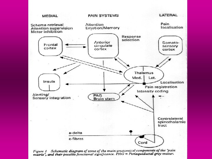
The brain first perceives the sensation of pain • The thalamus, sensitive cortex : perceiving describing localizing of pain • Parts of thalamus, brainstem and reticular formation: - identify dull longer-lasting, and diffuse pain • The reticular formation and limbic system: - control the emotional and affective response to pain Because the cortex, thalamus and brainstem are interconnected with the hypothalamus and autonomic nervous system, perception of pain is associated with an autonomic response
The role of afferent and efferent pathways in processing of pain information Nociceptive pain Nociceptors: Endings of small unmyelinated and lightly myelinated afferent neurons Stimulators: Chemical, mechanical and thermal noxae Mild stimulation positive, pleasurable sensation (e. g. tickling) Strong stimulation pain Location: In muscles, tendons, epidermis, subcutanous tissue, joints, visceral organs - they are not evenly distributed in the body (in skin more then in internal structures)
Nociceptive pain: - mechanisms involved in development Transduction is the process by which afferent nerve endings participate in translating noxious stimuli (e. g. , a pinprick) into nociceptive impulses
Afferent pathways • From nociceptors transmitted by small A-delta fibers and C- fibers to the spinal cord form synapses with neurons in the dorsal horn (DH) • From DH transmitted to higher parts of the spinal cord and to the rest of the CNS by spinothalamic tracts *The small unmyelinated C- neurons are responsible for the transmission of diffuse burning or aching sensations *Transmission through the larger, myelinated A- delta fibers occurs much more quickly. A delta - fibers carry well-localized, sharp pain sensations
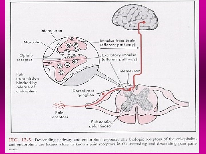
Efferent analgesic system Its role: - inhibition of afferent pain signals Mechanisms: - pain afferents on their way up to CNS send branches to periaqueductal gray (PAG) - gray matter surrounding the cerebral aqueduct in the midbrain, and stimulates the neurons there activation of efferent (descendent) anti-nociceptive pathways - from there the impulses are transmitted through the spinal cord to the dorsal horn - there thay inhibit or block transmission of nociceptive signals at the level of dorsal horn
Enk – enkefalinergic PAG – paraaqueductal gray EAA – excitatory amino acids RVM – rostral ventro-medial medulla Descendent antinociceptive systém
Antinociceptive placebo effect (Zubieta J-Ket al. , 2005)
The role of the spinal cord in pain processing • Most of afferent pain fibers terminate in the DH of the spinal segment that they enter. Some, however, extend toward the head or the foot for several segments before terminating • The A- fibers, some large A-delta fibers and small C- fibers terminate in the laminae of dorsal horn and in the substantia gelatinosa (SG) • The laminae than transmit specific information (about burned or crushed skin, about gentle pressure) to 2 nd afferent neuron
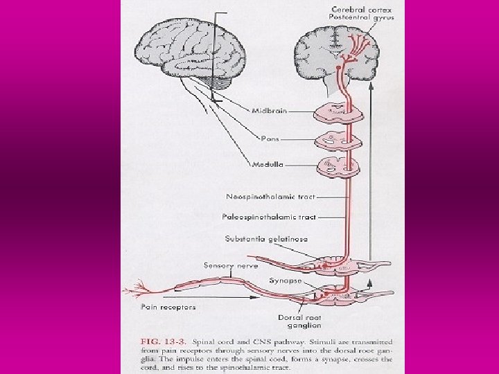
• 2 nd afferent neurons transmit the impulse from the SG and laminae through the ventral and lateral horn, crossing in the same or adjacent spinal segment, to the other side of the cord. From there the impulse is carried through the spinothalamic tract to the brain. The two divisions of spinothalamic tract are known: 1. the neospinothalamic tract - it carries information to the mid brain, thalamus and post central gyrus (where pain is perceived) 2. the paleospinothalamic tract - it carries information to the reticular formation, pons, limbic system, and mid brain (more synapses to different structures of brain)
PAG – periaqueductal gray PB – parabrachial nucleus in pons VMpo – ventromedial part of the posterior nuclear complex MDvc – ventrocaudal part of the medial dorsal nucleus VPL – ventroposterior lateral nucleus ACC – anterior cingulate cortex PCC – posterior cingulate cortex HT – hypothalamus S 1, S 2 – first and second somatosensory cortical areas PPC – posterior parietal complex SMA – supplementary cortical areas AMYG – amygdala PF – prefrontal cortex
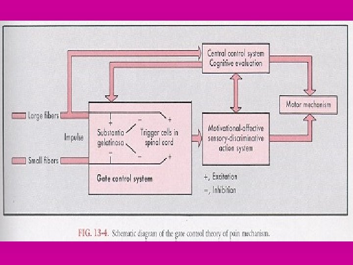
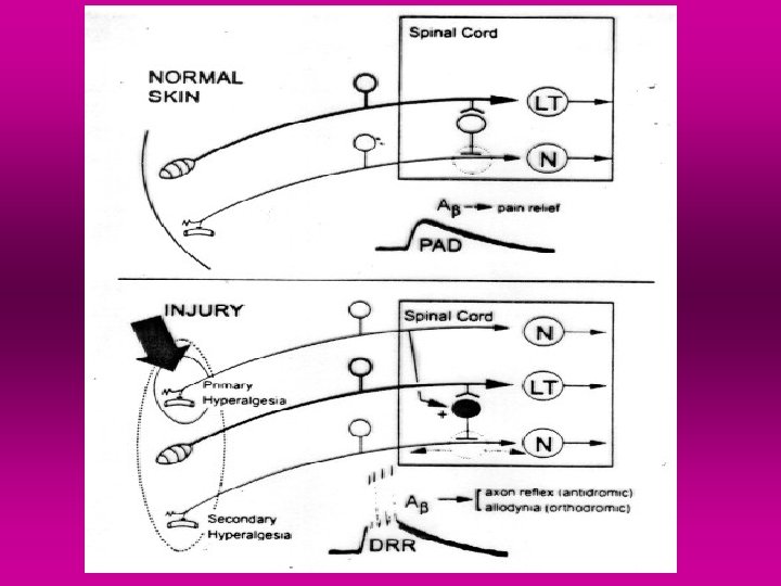
Theory of pain production and modulation • Most rational explanation of pain production and modulation is based on gate control theory (created by Melzack and Wall) • According to this theory, nociceptive impulses are transmitted to the spinal cord through large A- delta and small C- fibers • These fibers create synapses with neurons in the SG • The cells in this structure function as a gate, regulating transmission of impulses to CNS Stimulation of larger nerve fibers (A-alfa, A-beta) causes the cells in SG to "close the gate„ for transport of painful information centrally. • A closed gate leads to decreases stimulation of T-cells (the 2 nd afferent neuron), which decreases transmission of impulses, and diminishes pain perception
ØStimulation of small fibers input inhibits cells in SG and "open the gate". • An open gate increases the stimulation of T-cells transmission of impulses enhances pain perception • In addition to gate control through large and small fibers stimulation, the central nervous system, through efferent pathways, may close, partially close, or open gate , too. Cognitive functioning may thus modulate pain perception Action of endorphins(ED) All ED act by attaching to opiate receptors on the plasma membrane of the afferent neuron. The result than is inhibition of releasing of the neurotransmitter, thus blocking the transmission of the painful stimulus
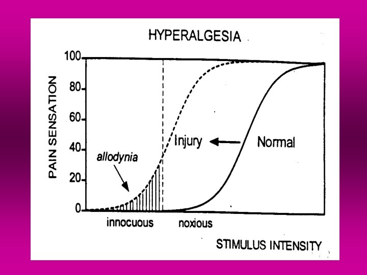
Neuropathic pain (NP) It occurs as a result of injury to or dysfunction of the nervous system itself, peripheral or central. The nerve injury may be induced by pathology in surrounding tissue. Characteristics of NP – it may mimic quality of somatic pain – it may have characteristic of „disesthetic“ pain (e. g. uncomfortable, unfamiliar sensation such as burning, shock-like, tingling – may be associated with reffered pain, allodynia, hyperalgesia, hyperpathia Hyperpathia – exaggerated pain responses following a stimulus often with aftersensations and intense emotional reaction
What causes neuropathic pain? Neuropathic pain often seems to have no obvious cause; but, some common causes of neuropathic pain include: - Hereditary disorders Metabolic disorders, Nerve ischemia, Nerve compression, - Traumatic nerve damage - Toxic nerve damage - Infection of nerve tissue - Immune mediated nerve tissue damage Example of some diseases leading to NP development Alcoholism, Amputation, Back, and Leg, and Hip problems, Chemotherapy, Diabetes mellitus, Facial nerve problems, HIV syndrome, Multiple sclerosis, Shingles (Herpes zoster), Spine surgery What are the symptoms of neuropathic pain? a) Stimulus indipendent pain b) Stimulus evoked pain
Neuropathic pain – subtypes (according a primary location of sustaining mechanism) a) Predominating peripheral generator: e. g. compression or entrapment neuropathies, plexopathies, radiculopathies, polyneuropathies b) Predominating central generator: e. g. spinal cord injury, post-stroke pain Deaferentation pain - form of neuropathic pain: a term implying that sensory deficit in the painful area is a prominent feature (anesthesia dolorosa) • Phantom pain- pain localizei into non-existing organ (tissue)
• Hypersensitivity – increased sensitivity of the system involved in the pain processing • Hyperalgesia – increased the pain sensitivity to noxious stimuli Allodynia - phenomenon characterised by painful sensations provoked by non-noxious stimuli, (e. g. touch), transmitted by fast- conducting nerve fibres Mechanism: changes of the response characteristics of second - order spinal neurons, so that normally inactive or weak synaptic contact mediating non-noxius stimuli acquire the capability to activate a neuron that normally responds only to impulses signaling pain
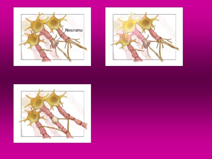
Pathomechanisms involved in genesis of neuropathic pain 1) Neurophysiologic and neuroanatomic changes that may occur in peripherally generated neuropathic pain a) Abnormal nerve morphology – grow multiple nerve sprouts, – some of these sprouts may form neuromas Nerve sprouts and neuromas can generate spontaneuos activity Areas of spontaneuos activity ( sensitivity) are associated with a change in Na+ receptors concentration – at sites of demyelination – are more sensitive to physical stimuli (manifested as tenderness) Neural axon injury Neural axon sprouting and neuroma
b) Development atypical connections between nerve sprouts or demyelinated axons in the region of nerve damage – permitting „cross–talk“ between somatic or sympathetic efferents and nociceptors c) Anterograde and retrograde transport of coumpounds stimulation of nerve cell body to production of specific genes
Common clinical forms of neuropathic pain Peripheral neuralgias after trauma or surgery lumbosacral and cervical rhizotomy, ● peripheral neuralgia Most peripheral neuralgias are the result of trauma or surgery. Such a conditions does not necessary occur as a result of damaging a major nerve trunk but may be caused by an incision involving only small nerve branches (incisional pain) Mechanism: the pain is due to neuroma formation in the scar tissue (? )
Deaferentation pain following spinal cord injury Incidence of severe pain due to spinal cord and cauda equina lesions ranges from 35 to 92 % of patients This pain is ascribed to 3 causes: 1. mechanically induced pain (fracture bones, myofascial pain) 2. radicular pain (compression of nerve root) 3. central pain (deaferentation mechanism)
Psychogenic pain – mechanism Dysfunction of central mechanisms responsible for processing of sensoric afferent informations - releasing of mediators decreasing pain threshold - prolonged muscle contraction due to psychogenic stress - incresed activity of SNS decreasing pain threhold - inhibition of activity of descending antinociceptive system
Clinical Manifestation of Pain Acute Pain We can distinguish two types of acute pain: 1. Somatic 2. Visceral – referred Somatic pain is superficial coming from the skin or close to the surface of the body. Visceral pain refers to pain in internal organs, the abdomen, or chest. Referred pain is pain that is present in an area removed or distant from its point of origin. The area of referred pain is supplied by the nerves from the same spinal segment as the actual site of pain
Different types of chronic somatic pain I. Nervous system intact 1. nociceptive pain 2. nociceptive - neurogenic pain (nerve trunk pain) II. Permanent functional and/or morphological abnormalities of the nervous system (preganglionic, spinal - supraspinal) 1. neurogenic pain 2. neuropathic pain 3. deafferentation pain
The most common chronic pain 1. Persistent low back pain – result of poor muscle tone, inactivity, muscle strain, sudden vigorous exercise 2. Chronic pain associated with cancer 3. Neuralgias 4. Myofascial pain syndromes 5. Hemiagnosia 6. Phantom limb pain
Neuralgias - results from damages of peripheral nerves a) Causalgia - severe burning pain appearing 1 to 2 weeks after the nerve injury associated with discoloration and changes in the texture of the skin in the affected area. b) Reflex sympathetic dystrophies - occur after peripheral nerve injury and is characterised by continuous severe burning pain. Vasomotor changes are present (vasodilatation vasoconstriction cool cyanotic and edematous extremities). Myofascial pain syndromes - second most common cause of chronic pain. These conditions include: myositis, fibrositis, myalgia, muscle strain, injury to the muscle and fascia The pain is a result of muscle spasm, tenderness and stiffness
Hemiagnosia – is a loss of ability to identify the sorce of pain on one side (the affected side) of the body. Application of painful stimuli to the affected side thus produces anxiety, moaning, agitation and distress but no attempt to withdrawal from or push aside the offending stimulus. Emotional and autonomic responses to the pain my be intensified. ● Hemiagnosia is associated with stroke that produces paralysis and hypersensitivity to painful stimuli in the affected side Phantom limb pain - is pain that an individual feels in amputated limb
Pathophysiology of muscle pain Muscle pain - a part of somatic deep pain, (MP) - it is common in rheumathology and sports medicine - is rather diffuse and difficult to locate MP is not a prominent feature of the serious progressive diseases affecting muscle, e. g. the muscular dystrophies, denervation , or metabolic myopathies, but it is a feature of rhabdomyolysis Muscles are relatively insensitive to pain when elicited by needle prick or knife cut, but overlying fascia is very sensitive to pain. Events, processes which may lead to muscular pain are: ● metabolic events: • metabolic depletion ( ATP muscular contracture) • accumulation of unwanted metabolities (K+, bradykinin)
Pathophysiology of visceral pain Visceral pain: Types - angina pectoris, myocardial infarction, acute pancreatitis, cephalic pain, prostatic pain, nephrlolythiatic pain Receptors: unmyelinated C - fibres For human pathophysiology the kinds of stimuli apt to induce pain in the viscera are important. It is well-known that the stimuli likely to induce cutaneous pain are not algogenic in the viscera. This explains why in the past the viscera were considered to be insensitive to pain
Adequate stimuli of inducing visceral pain: 1. abnormal distention and contraction of the hollow viscera muscle walls 2. rapid stretching of the capsule of such solid visceral organs as are the liver, spleen, pancreas. . . 3. abrupt anoxemia of visceral muscles 4. formation and accumulation of pain - producing substances 5. direct action of chemical stimuli (oesophagus, stomach) 6. traction or compression of ligaments and vessels 7. inflammatory processes 8. necrosis of some structures (myocardium, pancreas)
Characteristic feature of true visceral pain a) it is dull, deep, not well defined, and differently described by the patients b) sometimes it is difficult to locate this type of pain because it tends to irradiate c) it is often accompanied by a sense of malaise d) it induces strong autonomic reflex phenomena - diffuse sweating, vasomotor responses, changes of arterial pressure and heart rate, and an intense psychic alarm reaction -"angor animi" – e. g. in angina pectoris) e) when organ capsules or other structures, e. g. myocardium are involved, however, the pain is usually well localized and described as sharp , stubbing, or thobbing • There are many visceral sensation that are unpleasant but below the level of pain, e. g. feeling of disagreeable fullness or acidity of the stomach or undefined and unpleasant thoracic or abdominal sensation. These visceral sensation may precede the onset of visceral pain
Refered visceral pain (transferred pain) Refered pain = when an algogenic process affecting a viscus recurs frequently or becomes more intense and prolonged, the location becomes more exact and the painfull sensation is progressively felt in more superficial struftures ● Refered pain may be accompanied by allodynia and cutaneous and muscular hyperalgesia Mechanisms involved in refered pain creation: a) convergence of impulses from viscera and from the skin in the CNS: Sensory impulses from the viscera create an irritable focus in the segment at which they enter the spinal cord. Afferent impulses from the skin entering the same segment are thereby facilitated, giving rise to true cutaneous pain. b) senzitization of neurons in dorsal horn
Painful visceral afferent impulses activate anterior horn motor cells to produce rigidity of the muscle (viscero -motor reflexes) A similar activation of anterolateral autonomic cells induces pyloerection, vasoconstriction, and other sympathetic phenomena These mechanisms, which in modern terms can be defined as positive sympathetic and motor feedback loops, are fundamental in reffered pain It is clear that painful stimulation of visceral structures evokes a visceromuscular reflex, so that some muscles contract and become a new source of pain
It has been observed that the local anesthetic block of the sympathetic ganglia led to the disappearance, or at least to a marked decrease, of reffered pain, allodynia, hyperalgesia. In some conditions, reffered somatic pain is long-lasting, increases progressively, and is accompanied by dystrophy of somatic structures. Possible mechanisms: - onset of self-maintaining vicious circle impulses: peripheral tissue afferent fibers central nervous system peripheral tissue somatic and sympathetic efferent fibres
Intricate conditions - in some types of pain, e. g. chest pain, is difficult to distinguish the true cause of pain because such kind of pain may be related to cervical osteoarthrosis, esophageal hernia, cholecystitis, MI, other pathologic processes. It is diffcult to ascertain whether this pain is due to a simple addition of impulses from different sources in the CNS or to somatovisceral and viscerosomatic reflexes mechanisms. It has been demonstrated that the mnemonic process is facilitated if the experience to be retained is repeated many times or is accompanied by pleasant or unpleasant emotions. Pain is, at least in part, a learned experience - e. g. during the first renal colic, true somatic pain followed visceral pain after a variable interval. In subsequent episodes of renal colic pain, somatic pain developed promptly and was not preceded by true visceral pain. This is probably due to the activation of mnemonic traces.
Silent myocardial ischemia (SMI) ● Chest pain is only a late and inconstant marker of episodes of transient MI in vasospastic angina (30 %), in stable angina (50 %) • Mechanisms of SMI a) Lack of the pain is, in part, related to the duration and severity of MI. Episodes shorter than 3 min, and those accompanied by a modest impairment of left ventricle ( in end-diastolic pressure inferior to 6 mm Hg) are always painless. Longer and more severe episodes are acccompanied by chest pain in some instances but not in others. b) Pacients with predominantly SMI appear to have a generalized defective perception of pain ( threshold and tolerance). Mechanism: level of circulating -endorphin (? )
Disturbances in pain perception and nociception Most of the disturbances are congenital a) Congenital analgesia - nociceptive stimuli are not processed and/or integrated at a level of brain. Patient does not feel a pain b) Congenital sensoric neuropathy - nociceptive stimuli are not transmitted by peripheral nerves or by spinal afferent tracts. Acquired disturbances in pain perception and nociception They may occur at syringomyely, disturbances of parietal lobe of brain, in patients suffering from neuropathy (e. g. chronic diabetes mellitus)
Development of neuropathic chronic pain after spinal cord injury
Neural axon injury
Measurements of pain intensity