
Causes of proptosis and Thyroid Ophthalmopathy Dr Ajai














- Slides: 55
Causes of proptosis and Thyroid Ophthalmopathy Dr. Ajai Agrawal Additional Professor, Department of Ophthalmology, A. I. I. M. S. , Rishikesh 1
Acknowledgement • Upto. Date: Graves’ orbitopathy: Diagnosis and Treatment. • Cummings Otolaryngology. Girod, Douglas A. ; Wemer, Richard D. ; Larsen, Christopher G. . Published January 1, 2015. © 2015. • Endocrinology : Adult and Paediatric. Burch, Henry B. ; Bahn, Rebecca S. . Published January 1, 2016. © 2016. • Some of the images used were taken from eyetext. net 2
Learning Objectives • At the end of this class the students shall be able to : • Enumerate the common causes of proptosis • Understand the pathogenesis and clinical features of thyroid eye disease. • Enumerate and elicit the common eye signs of thyroid ophthalmopathy. • Have a basic understanding of principles of management of the disease. 3
Question • A 44 -year-old female patient presents with bilateral protrusion of the eyeball along with retraction of the eyelids and lid lag on downgaze. What is her most likely diagnosis? • a. Grave’s disease • b. Orbital pseudotumor • c. Orbital cellulitis • d. Orbital varices 4
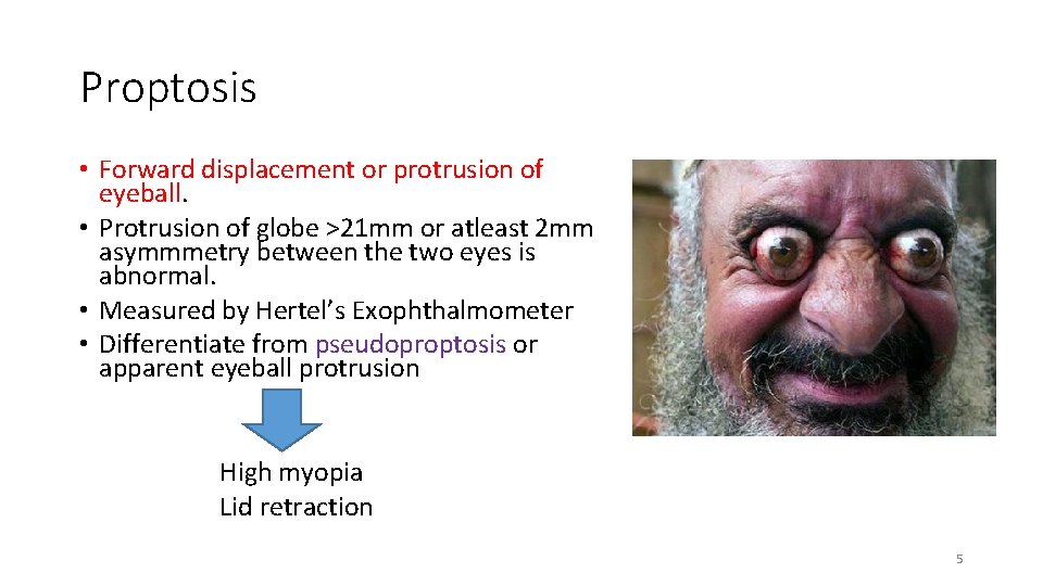
Proptosis • Forward displacement or protrusion of eyeball. • Protrusion of globe >21 mm or atleast 2 mm asymmmetry between the two eyes is abnormal. • Measured by Hertel’s Exophthalmometer • Differentiate from pseudoproptosis or apparent eyeball protrusion High myopia Lid retraction 5
Proptosis • Axial and non-axial(Abaxial) • Unilateral and Bilateral proptosis Congenital: Dermoid Cyst Traumatic: Orbital hemorrhage Inflammatory: Orbital cellulitis Orbital psudotumour Vascular: Orbital varix Cystic: Orbital cysticercosis Tumours of the orbit Inflammation: Thyroid orbitopathy Sarcoidosis Neoplasia: Lymphoma Leukaemia Vascular malformation: Arterio-venous shunts Lacrimal lesions: Sjogren’s syndrome 6
Introduction of Thyroid eye disease • Thyroid eye disease is an autoimmune disease producing symptoms related to inflammation, accumulation of fluid in the orbit and also to adipogenesis raising intra-orbital pressure. • Synonyms • Graves’ ophthalmopathy/orbitopathy (GO) • Thyroid eye disease (TED) • Thyroid associated ophthalmopathy (TAO) • Dysthyroid ophthalmopathy 7
Epidemiology • Prevalence of thyroid ophthalmopathy = 0. 4% Women > Men • But severity greater in men • Bimodal age distribution – Peak incidence in fourth and sixth decades of life • May be exacerbated by stress and smoking • Most common cause of exophthalmos • >50% of cases with Graves’ disease have eye involvement 8
Etiology • Graves’ hyperthyroidism (90%) • Hypothyroid Hashimoto’s thyroiditis • Euthyroid subjects with no current or past evidence of thyroid hyper or hypofunction (so called euthyroid Graves’ disease). • In patients with Grave’s disease, eye signs may precede, coincide with or follow the hyperthyroidism 9
Risk factors • Smoking (strongest modifiable risk factor) • Family history • Monozygotic twins 10
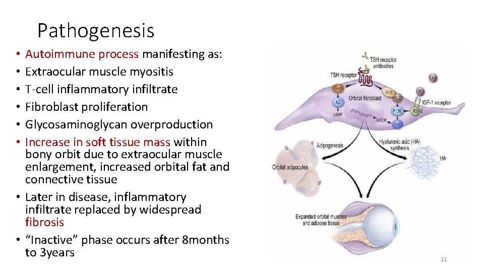
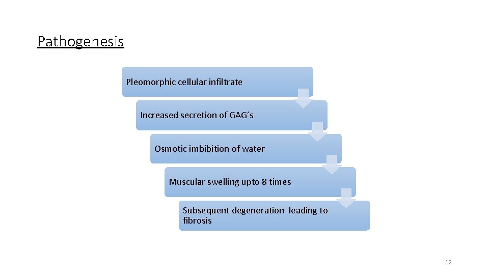
Pathogenesis Autoimmune process manifesting as: Extraocular muscle myositis T-cell inflammatory infiltrate Fibroblast proliferation Glycosaminoglycan overproduction Increase in soft tissue mass within bony orbit due to extraocular muscle enlargement, increased orbital fat and connective tissue • Later in disease, inflammatory infiltrate replaced by widespread fibrosis • “Inactive” phase occurs after 8 months to 3 years • • • 11
Pathogenesis Pleomorphic cellular infiltrate Increased secretion of GAG’s Osmotic imbibition of water Muscular swelling upto 8 times Subsequent degeneration leading to fibrosis 12
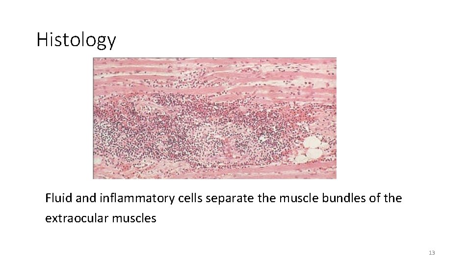
Histology Fluid and inflammatory cells separate the muscle bundles of the extraocular muscles 13
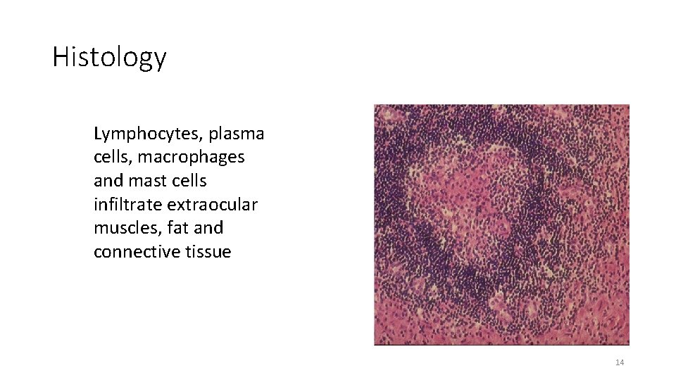
Histology Lymphocytes, plasma cells, macrophages and mast cells infiltrate extraocular muscles, fat and connective tissue 14
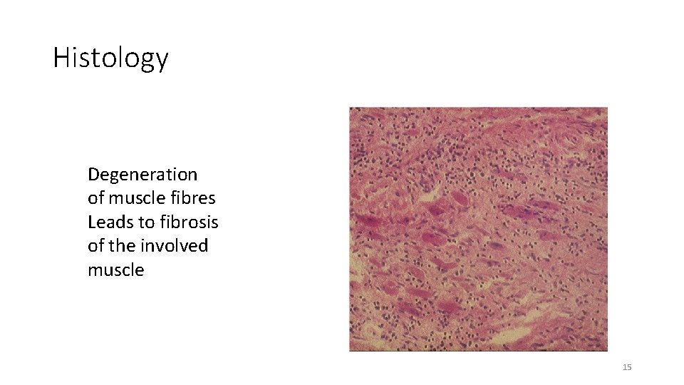
Histology Degeneration of muscle fibres Leads to fibrosis of the involved muscle 15
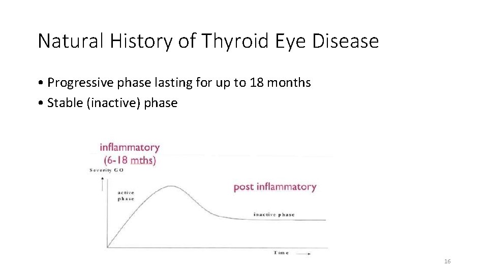
Natural History of Thyroid Eye Disease • Progressive phase lasting for up to 18 months • Stable (inactive) phase 16
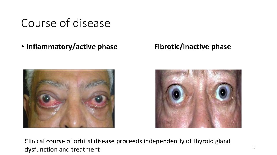
Course of disease • Inflammatory/active phase Fibrotic/inactive phase Clinical course of orbital disease proceeds independently of thyroid gland dysfunction and treatment 17
Symptoms • Foreign body sensation • Epiphora (tearing) • Photophobia • Bulging of eyes • Puffiness of eye lids • Diplopia • Visual loss 18
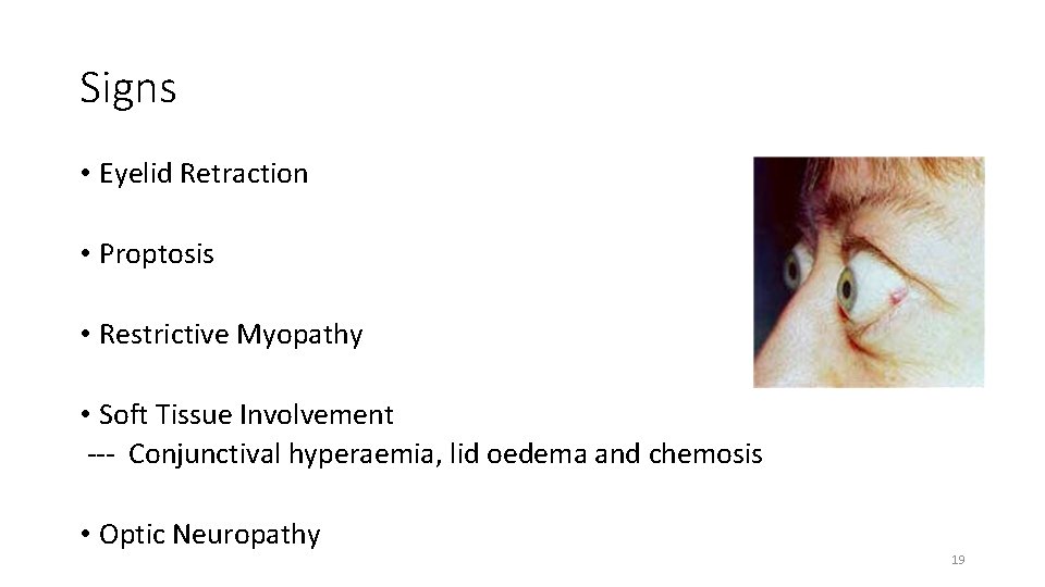
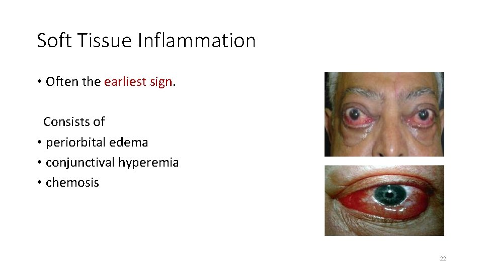
Signs • Eyelid Retraction • Proptosis • Restrictive Myopathy • Soft Tissue Involvement --- Conjunctival hyperaemia, lid oedema and chemosis • Optic Neuropathy 19
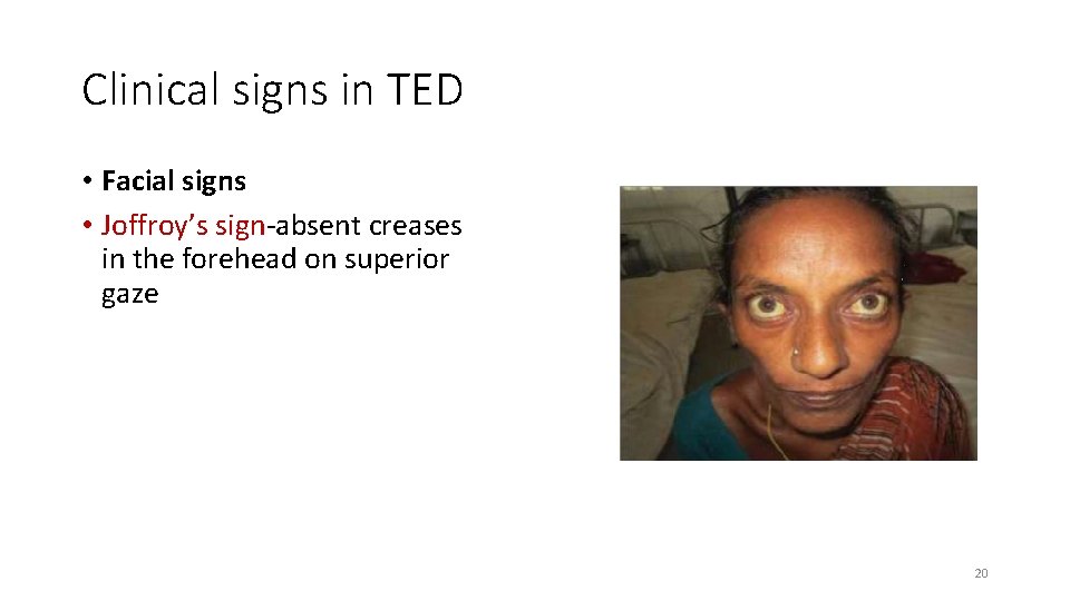
Clinical signs in TED • Facial signs • Joffroy’s sign-absent creases in the forehead on superior gaze 20
Clinical eye lid signs in TED • Kocher’s sign- staring appearance • Von Graefe’s sign- lid lag on downgaze • Dalrymple’s sign- lid retraction • Stellwag’s sign- incomplete & infrequent blinking • Enroth ’s sign- edema of lower lid • Griffith’s sign- lower lid lag on upgaze 21
Soft Tissue Inflammation • Often the earliest sign. Consists of • periorbital edema • conjunctival hyperemia • chemosis 22
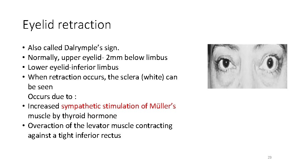
Eyelid retraction • Also called Dalrymple’s sign. • Normally, upper eyelid- 2 mm below limbus • Lower eyelid-inferior limbus • When retraction occurs, the sclera (white) can be seen Occurs due to : • Increased sympathetic stimulation of Müller’s muscle by thyroid hormone • Overaction of the levator muscle contracting against a tight inferior rectus 23
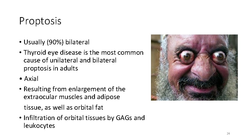
Proptosis • Usually (90%) bilateral • Thyroid eye disease is the most common cause of unilateral and bilateral proptosis in adults • Axial • Resulting from enlargement of the extraocular muscles and adipose tissue, as well as orbital fat • Infiltration of orbital tissues by GAGs and leukocytes 24
Proptosis • It does not respond to hyperthyroidism treatment • Is permanent in 70% of cases. • Severe proptosis prevents adequate lid closure • May lead to severe exposure keratopathy and corneal ulceration. 25
Restrictive Myopathy • Eye movements are restricted due to oedema in extraocular muscles during infiltrative stage and subsequent fibrosis. • Despite expansion of the extraocular muscles , the muscle fibres themselves are normal. • IR>MR>SR>LR • Pressure exerted by a fibrotic inferior rectus muscle on the globe may cause a spike in intraocular pressure during upgaze. 26
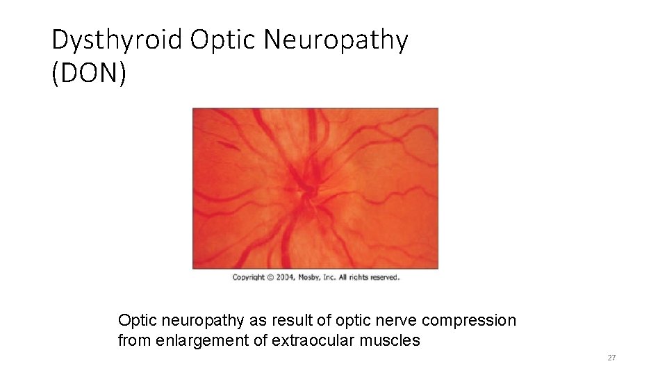
Dysthyroid Optic Neuropathy (DON) Optic neuropathy as result of optic nerve compression from enlargement of extraocular muscles 27
WERNER´S CLASSIFICATION - NOSPECS • Class 0: No signs or symptoms • Class 1: Only signs (lid retraction, stare ± lid lag) • Class 2: Soft tissue involvement • Class 3: Proptosis • Class 4: Extraocular muscle involvement • Class 5: Corneal involvement • Class 6: Sight loss (optic nerve involvement) 28
EUGOGO classification • Mild : eyelid swelling , lid retraction, proptosis • Moderate-Severe : Active disease (EOM dysfunction, diplopia , proptosis >25 mm) • Very severe : Compressive Optic Neuropathy , Corneal exposure (needs emergent surgery) 29
VISA classification • V (Vision) , I (inflammation), S (Strabismus) , A (Appearance) • Vision/CON • Inflammation/Congestion : based on documented change of inflammation rather than absolute value • Strabismus/Motility : measuring ductions and alignments • Appearance/Exposure • Score of 5 or more —> Active disease or progression (Consider Steroids) 30
Differential Diagnosis • Orbital tumors (primary or metastatic) • Orbital pseudotumor • Wegener’s granulomatosis • Orbital infection • Carotid-cavernous sinus fistula 31
Diagnosis • Characteristic eye findings • Thyroid dysfunction • Imaging 32
Blood investigations • • Highly sensitive & specific -- T 4(thyroxine) + TSH or serum TSH If eye findings associates with euthyroid Graves’ disease – Thyroid peroxidase antibody Antibody to thyroglobulin • • • Others Free T 4 index Thyroid-stimulating immunoglobulin Antithyroid antibodies Serum T 3 33
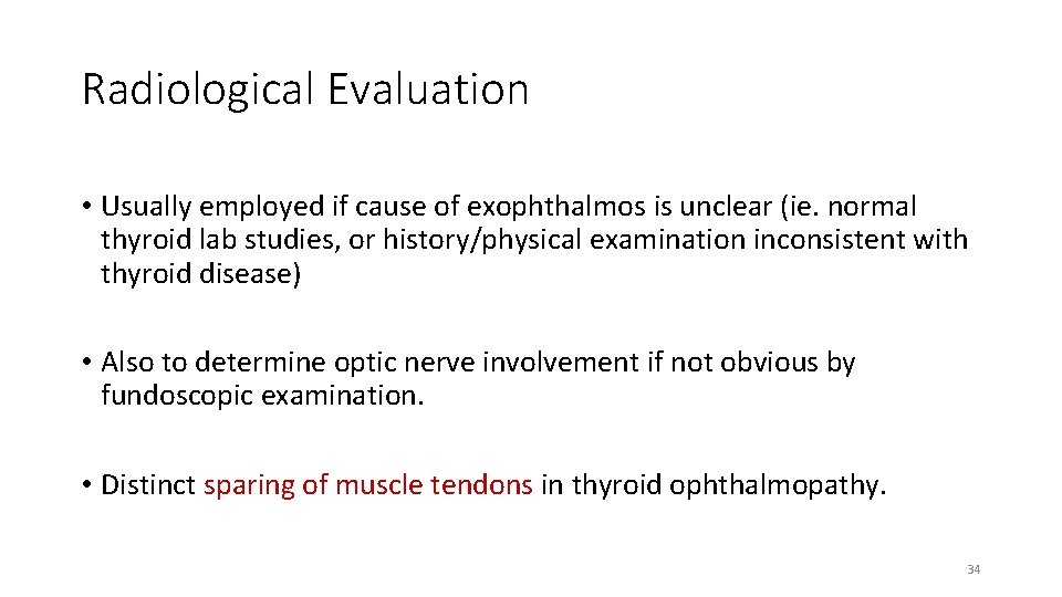
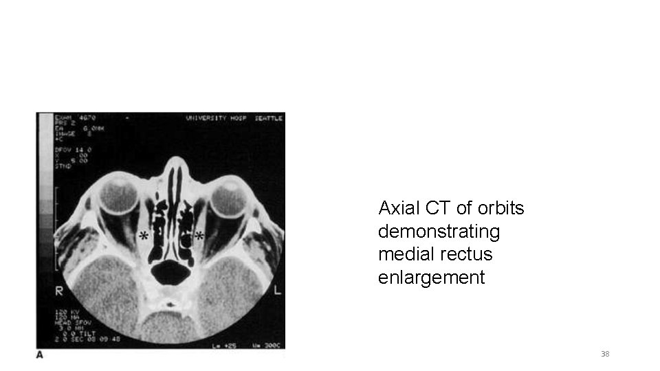
Radiological Evaluation • Usually employed if cause of exophthalmos is unclear (ie. normal thyroid lab studies, or history/physical examination inconsistent with thyroid disease) • Also to determine optic nerve involvement if not obvious by fundoscopic examination. • Distinct sparing of muscle tendons in thyroid ophthalmopathy. 34
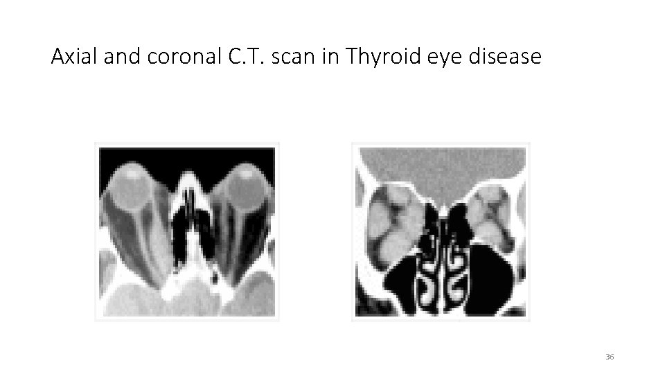
Radiological Evaluation • CT scan is currently the imaging study of choice. • MRI is sensitive for showing compression of the optic nerve. • Neuroimaging usually reveals • Thick muscle belly with tendon sparing • Usually IR & MR • Bilateral muscle enlargement is the norm • Unilateral cases usually represent asymmetric involvement rather than normality of the less involved side 35
Axial and coronal C. T. scan in Thyroid eye disease 36
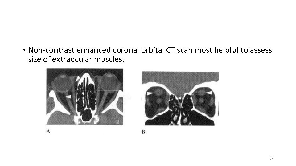
• Non-contrast enhanced coronal orbital CT scan most helpful to assess size of extraocular muscles. 37
Axial CT of orbits demonstrating medial rectus enlargement 38
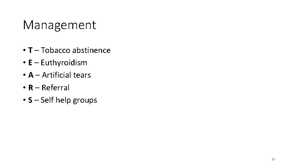
Management • T – Tobacco abstinence • E – Euthyroidism • A – Artificial tears • R – Referral • S – Self help groups 39
Medical Management of Hyperthyroidism • Anti-thyroid drugs : thinoamides (PTU) , carbimazole , methimazole. • Thionamides inhibit synthesis of thyroid hormones. • Need 6 -8 weeks to achieve euthyroid state • Side effects of anti-thyroid drugs Skin rash , urticarial , arthralgia , fever 40
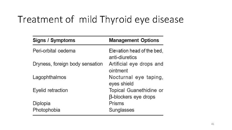
Treatment of mild Thyroid eye disease 41
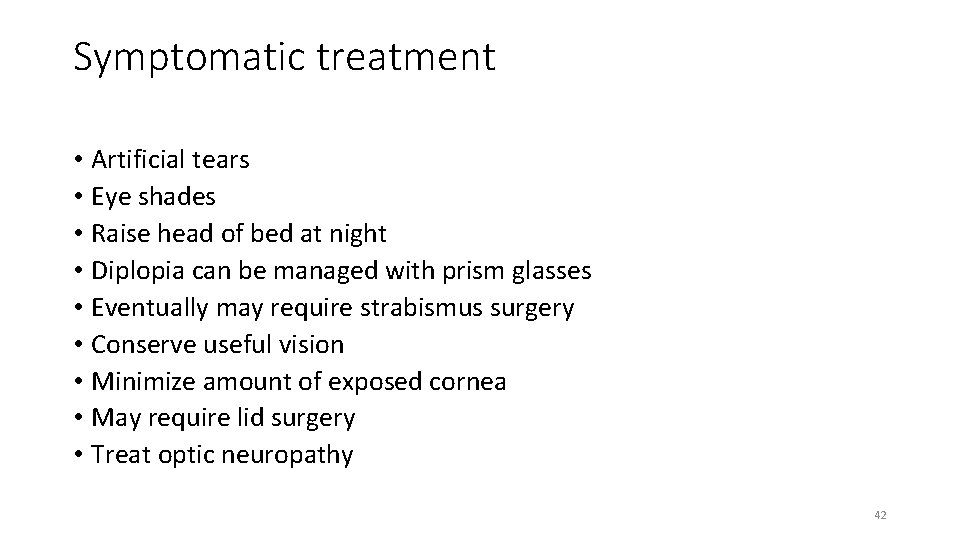
Symptomatic treatment • Artificial tears • Eye shades • Raise head of bed at night • Diplopia can be managed with prism glasses • Eventually may require strabismus surgery • Conserve useful vision • Minimize amount of exposed cornea • May require lid surgery • Treat optic neuropathy 42
Selenium • 200 microgram/day for 6 months • For Mild disease • Antioxidant effect • Immunomodulatory effect : reduce thyroid autoantibodies • Reduces severity of disease and improve quality of life 43
Corticosteroids • Intravenous , Oral • IV pulses are more effective and have less side effects • IV dose (max 8 grams) : 500 mg weekly for 6 weeks and then 250 mg weekly for 6 weeks • Relapse is common (20%) • Steroid response is evident usually 2 -4 weeks later • Moderate to severe TED : 71% respond to IV steroid vs 51% with oral • IV steroids for compressive Optic Neuropathy 44
Rituximab • Chimeric mono-clonal antibody targets CD 20 • CD 20 is expressed on more than 95% of B cells and plasma cells • RTX depletes 95% of mature B cells , blocks Ab production , and decreases inflammatory cytokine release • For steroid-refractory disease • Side effects : Allergic reaction (mild) PML (severe) 45
Orbital Radiation • Mechanism : lymphocyte sterilization, destruction of tissue monocytes • 20 Gy in 10 divided sessions over 2 weeks • May have a role in patients with TED who have restricted ocular motility or active disease • Some studies have shown benefit (controversial) • More suited for patients > 35 years of age • Contra-indicated in pre-existing retinopathy (diabetes , hypertensive) 46
Botulinum Toxin • Neurotoxin , inhibits acetylcholine release • For upper lid retraction (transconjunctival , transcutaneous route) • Effect on Muller’s muscle and LPS • Side effects of Botox : bruising , ptosis and diplopia 47
Orbital Decompression for TED • Decompression usually in stable phase of disease. • Indications • compressive optic neuropathy • severe exposure keratopathy • Post-operative complications (diplopia, vision loss) • Outcome is variable : degree of fibrosis , fat expansion , bone available, duration of optic neuropathy. • Decompression —> Muscle Surgery —> Lid surgery 48
Strabismus Surgery for TED • In the stable phase with stable alignments for 6 months • Aim is single binocular vision in primary and reading position • Typically involves release of the restricted muscle by recession rather than resection • Conjunctival dissection is challenging • Use of adjustable sutures is strongly recommended due to the variability in fibrosis, resulting in unpredictable results. • Oblique surgery can increase area of single binocular vision 49
Eye lid surgery • The most common indication for lid surgery is upper lid retraction. • Graded Muller’s and levator aponeurosis weakening. • Lower lid lengthening is indicated in lower lid retraction. 50
Psychological Impact of TED • Disfigurement/altered facial appearance • Misinterpretation as hostile or angry • Almost 50% of TED suffer depression and/or anxiety • 90% of TED have appearance concerns (young females) • 44% have self-confidence issues • Multidisciplinary approach (psychiatric included) • Support groups 51
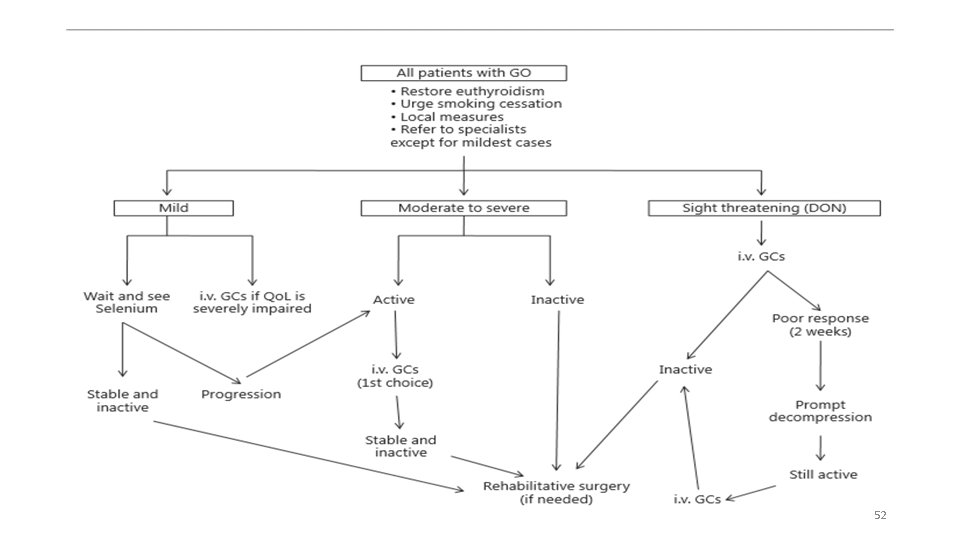
52
Conclusion • Activation of thyrotropin receptor on orbital fibroblast by circulating autoantibodies plays a primary role in development of thyroid ophthalmopathy. • Management is based on accurate assessment of both severity and activity of disease. • Immunosuppressive therapy is reserved for patients with clinically active moderate to severe disease 53
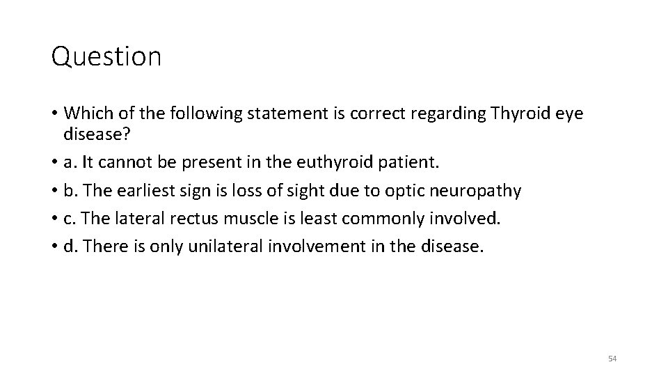
Question • Which of the following statement is correct regarding Thyroid eye disease? • a. It cannot be present in the euthyroid patient. • b. The earliest sign is loss of sight due to optic neuropathy • c. The lateral rectus muscle is least commonly involved. • d. There is only unilateral involvement in the disease. 54
Thank You 55