
TUBERCULOSIS the disease in children and implications for
TUBERCULOSIS. . . the disease in children and implications for the care of today’s child. . . from a clinician’s view. H. S. Patterson, M. D.
TB IN CHILDREN OBJECTIVES • Understand, in simple terms, the pathophysiology and clinical course of TB in children. • Understand the difference between TB infection and TB disease • Understand how to make the diagnosis of TB in a child.
TB IN CHILDREN OUTLINE • • • historical and epidemiologic perspectives diagnostic considerations natural course mortality/complications why prophylaxis? look alike
TUBERCULOSIS. . . The greatest killer of all time. . . The captain of all these men of death. . . during this century and the last, one billion people have died from tuberculosis

TUBERCULOSIS. . . “The White Plague” Oliver Wendell Holmes Sr, 1861
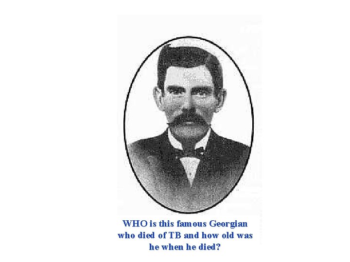
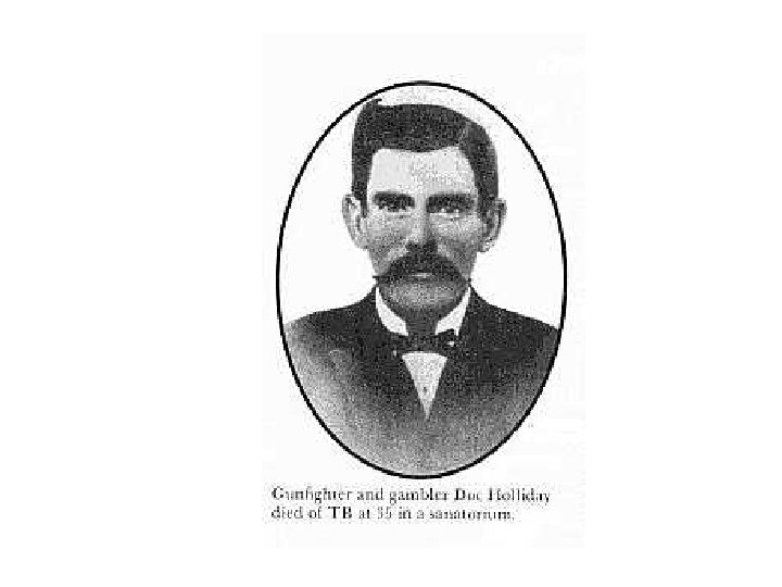
WHO is this famous Georgian who died of TB and how old was he when he died?
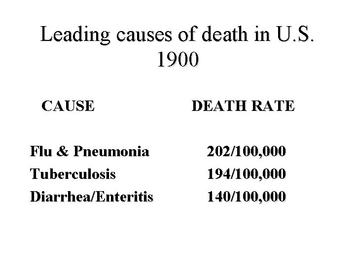
Leading causes of death in U. S. 1900 CAUSE Flu & Pneumonia Tuberculosis Diarrhea/Enteritis DEATH RATE 202/100, 000 194/100, 000 140/100, 000
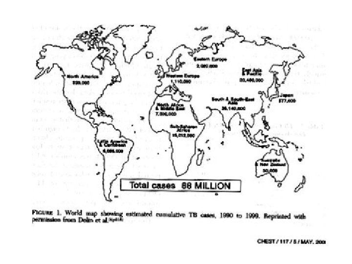
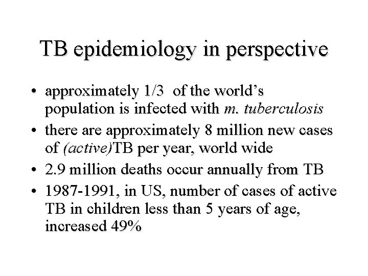
TB epidemiology in perspective • approximately 1/3 of the world’s population is infected with m. tuberculosis • there approximately 8 million new cases of (active)TB per year, world wide • 2. 9 million deaths occur annually from TB • 1987 -1991, in US, number of cases of active TB in children less than 5 years of age, increased 49%
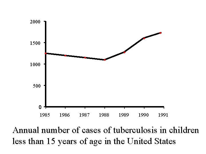
2000 1500 1000 500 0 1985 1986 1987 1988 1989 1990 1991 Annual number of cases of tuberculosis in children less than 15 years of age in the United States
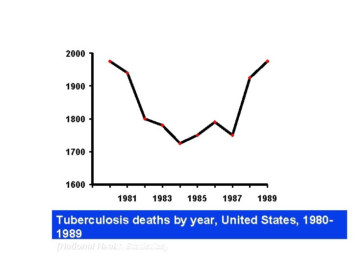
2000 1900 1800 1700 1600 1981 1983 1985 1987 1989 Tuberculosis deaths by year, United States, 19801989 (National Health Statistics)
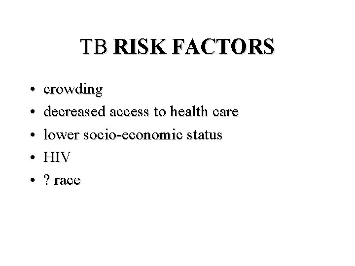
TB RISK FACTORS • • • crowding decreased access to health care lower socio-economic status HIV ? race

Historical TB
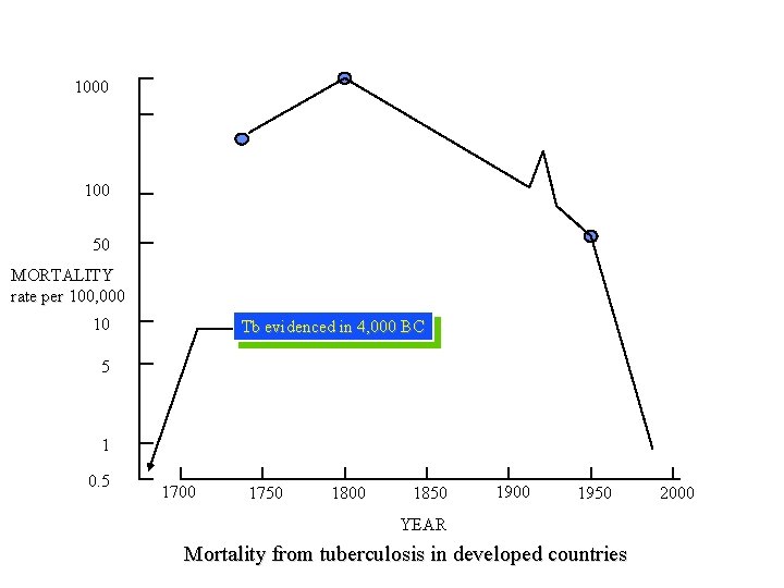
1000 100 50 MORTALITY rate per 100, 000 10 Tb evidenced in 4, 000 BC 5 1 0. 5 1700 1750 1800 1850 1900 1950 YEAR Mortality from tuberculosis in developed countries 2000
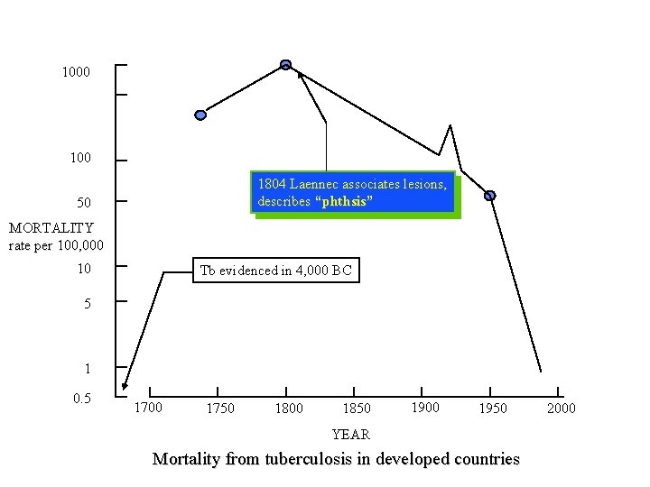
1000 1804 Laennec associates lesions, describes “phthsis” 50 MORTALITY rate per 100, 000 10 Tb evidenced in 4, 000 BC 5 1 0. 5 1700 1750 1800 1850 1900 1950 YEAR Mortality from tuberculosis in developed countries 2000
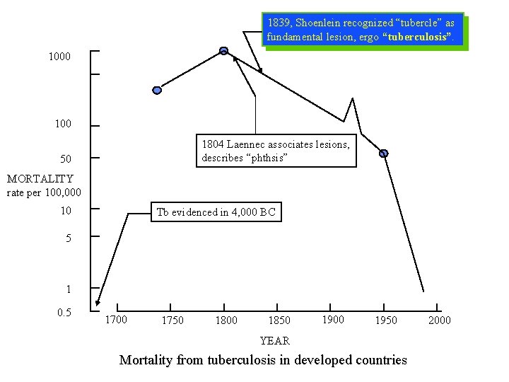
1839, Shoenlein recognized “tubercle” as fundamental lesion, ergo “tuberculosis”. 1000 1804 Laennec associates lesions, describes “phthsis” 50 MORTALITY rate per 100, 000 10 Tb evidenced in 4, 000 BC 5 1 0. 5 1700 1750 1800 1850 1900 1950 YEAR Mortality from tuberculosis in developed countries 2000
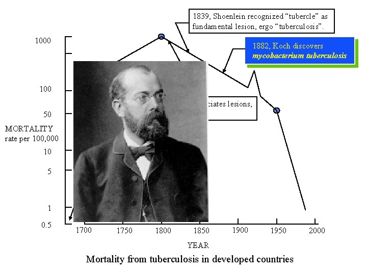
1839, Shoenlein recognized “tubercle” as fundamental lesion, ergo “tuberculosis”. 1000 1882, Koch discovers mycobacterium tuberculosis 100 1804 Laennec associates lesions, describes “phthsis” 50 MORTALITY rate per 100, 000 10 Tb evidenced in 4, 000 BC 5 1 0. 5 1700 1750 1800 1850 1900 1950 YEAR Mortality from tuberculosis in developed countries 2000
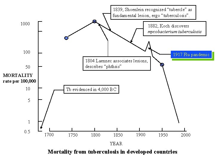
1839, Shoenlein recognized “tubercle” as fundamental lesion, ergo “tuberculosis”. 1000 1882, Koch discovers mycobacterium tuberculosis 100 1917 Flu pandemic 1804 Laennec associates lesions, describes “phthsis” 50 MORTALITY rate per 100, 000 10 Tb evidenced in 4, 000 BC 5 1 0. 5 1700 1750 1800 1850 1900 1950 YEAR Mortality from tuberculosis in developed countries 2000
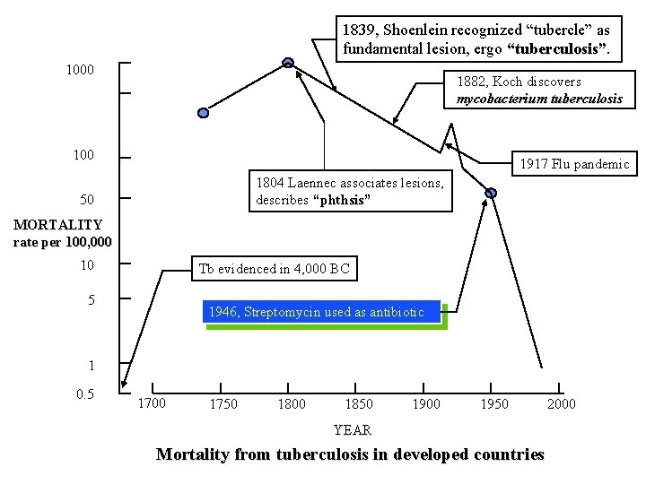
1839, Shoenlein recognized “tubercle” as fundamental lesion, ergo “tuberculosis” 1000 1882, Koch discovers mycobacterium tuberculosis 100 1917 Flu pandemic 1804 Laennec associates lesions, describes “phthsis” 50 MORTALITY rate per 100, 000 10 Tb evidenced in 4, 000 BC 5 1946, Streptomycin used as antibiotic 1 0. 5 1700 1750 1800 1850 1900 1950 YEAR Mortality from tuberculosis in developed countries 2000
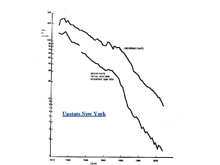
Upstate New York
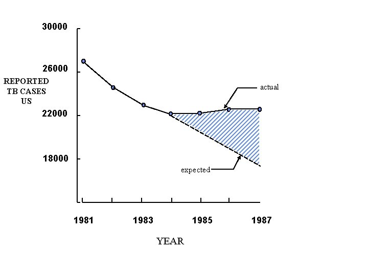
30000 26000 REPORTED TB CASES US actual 22000 18000 expected 1981 1983 1985 YEAR 1987
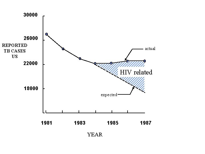
30000 26000 REPORTED TB CASES US actual 22000 HIV related 18000 expected 1981 1983 1985 YEAR 1987
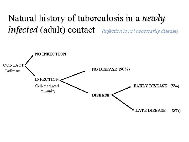
Natural history of tuberculosis in a newly infected (adult) contact (infection is not necessarily disease) NO INFECTION CONTACT Defenses NO DISEASE (90%) INFECTION Cell-mediated immunity EARLY DISEASE (5%) DISEASE LATE DISEASE (5%)
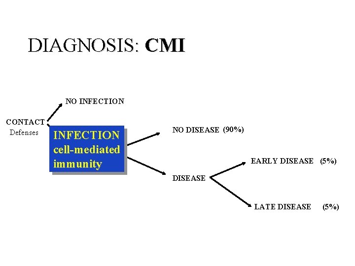
DIAGNOSIS: CMI NO INFECTION CONTACT Defenses INFECTION cell-mediated immunity NO DISEASE (90%) EARLY DISEASE (5%) DISEASE LATE DISEASE (5%)
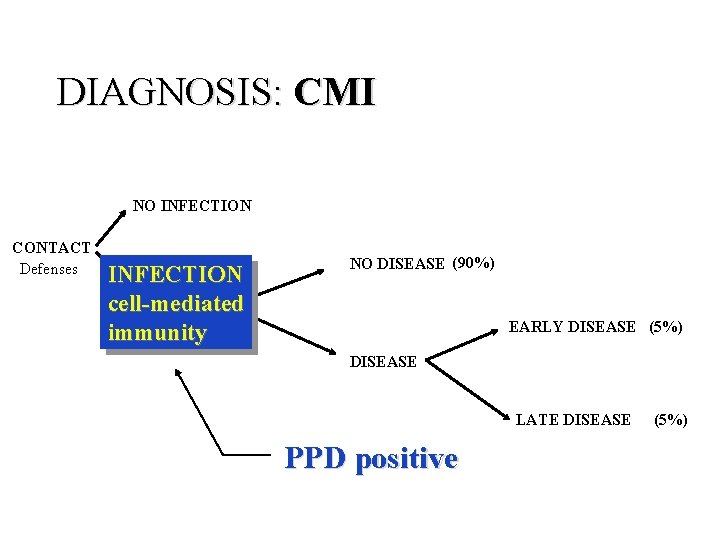
DIAGNOSIS: CMI NO INFECTION CONTACT Defenses INFECTION cell-mediated immunity NO DISEASE (90%) EARLY DISEASE (5%) DISEASE LATE DISEASE PPD positive (5%)
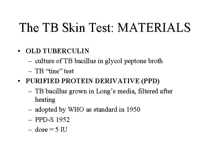
The TB Skin Test: MATERIALS • OLD TUBERCULIN – culture of TB bacillus in glycol peptone broth – TB “tine” test • PURIFIED PROTEIN DERIVATIVE (PPD) – TB bacillus grown in Long’s media, filtered after heating – adopted by WHO as standard in 1950 – PPD-S 1952 – dose = 5 IU
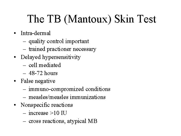
The TB (Mantoux) Skin Test • Intra-dermal – quality control important – trained practioner necessary • Delayed hypersensitivity – cell mediated – 48 -72 hours • False negative – immuno-compromized conditions – measles/measles immunizations • Nonspecific reactions – increase >10 IU – cross reactions, atypical MB
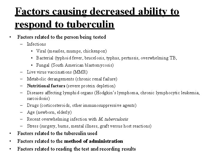
Factors causing decreased ability to respond to tuberculin • Factors related to the person being tested – Infections • Viral (measles, mumps, chickenpox) • Bacterial (typhoid fever, brucelosis, typhus, pertussis, overwhelming TB, • Fungal (South American blastomycosis) – Live virus vaccinations (MMR) – Metabolic derangements (chronic renal failure) – Nutritional factors (severe protein depletion) – Diseases affecting lymphid organs (Hodgkin’s lymphoma, chronic lymphocytic leukemia, sarcoidosis) – Drugs (corticosteroids, other immunosuppressive agents) – Age (newborn, elderly) – Recent overwhelming infection with M. tuberculosis – Stress (surgery, burns, mental illness, graft versus host reactions) • • • Factors related to the tuberculin used Factors related to the method of administration Factors related to reading the test and recording results
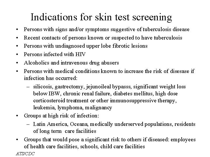
Indications for skin test screening • • • Persons with signs and/or symptoms suggestive of tuberculosis disease Recent contacts of persons known or suspected to have tuberculosis Persons with undiagnosed upper lobe fibrotic lesions Persons infected with HIV Alcoholics and intravenous drug abusers Persons with medical conditions known to increase the risk of disesase if infection has occurred: – silicosis, gastrectomy, jejunoileal bypasss, significant weight loss below IBW, chronic renal failure, diabetes mellitus, high dose corticosteroid treatment or other immunosuppressive therapy, leukemia, lymphoma, malignancy • Groups at high risk of infection: – Latin America, Oceana, medically underserved populations, residents of long term care facilities • Groups that would pose a significant risk to others if diseased: employees of health care facilities, schools, child care facilities ATS/CDC
TB PATHOPHYSIOLOGY • • • systemic infection primary infection/disease progressive primary disease miliary disease meningitis chronic TB (re-activation)
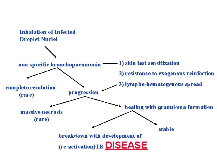
Inhalation of Infected Droplet Nuclei non-specific bronchopneumonia 1) skin test sensitization 2) resistance to exogenous reinfection 3) lympho-hematogenous spread complete resolution (rare) progression massive necrosis (rare) healing with granuloma formation breakdown with development of (re-activation)TB DISEASE stable
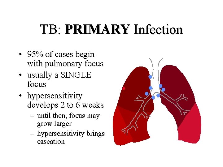
TB: PRIMARY Infection • 95% of cases begin with pulmonary focus • usually a SINGLE focus • hypersensitivity develops 2 to 6 weeks – until then, focus may grow larger – hypersensitivity brings caseation
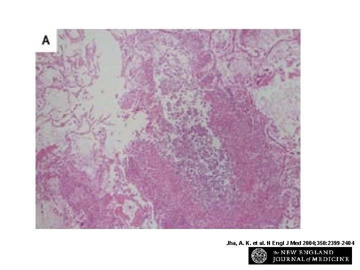
Tissue Specimens Obtained at Autopsy from the Patient Jha, A. K. et al. N Engl J Med 2004; 350: 2399 -2404
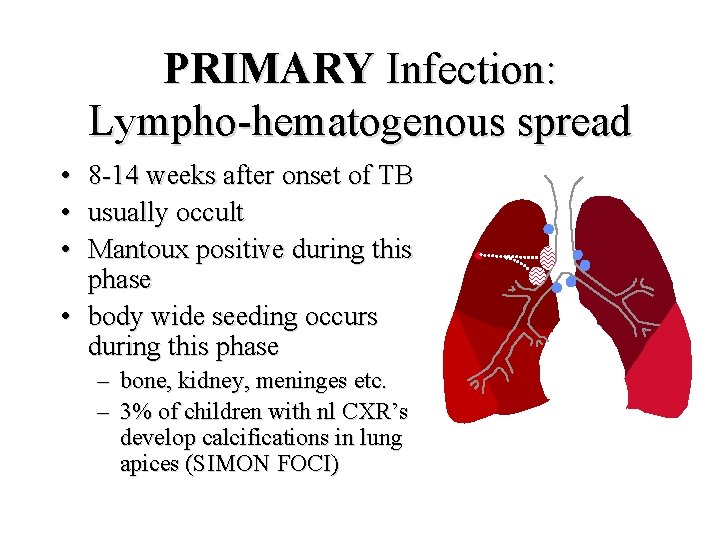
PRIMARY Infection: Lympho-hematogenous spread • • • 8 -14 weeks after onset of TB usually occult Mantoux positive during this phase • body wide seeding occurs during this phase – – bone, kidney, meninges etc. 3% of children with nl CXR’s develop calcifications in lung apices (SIMON FOCI)
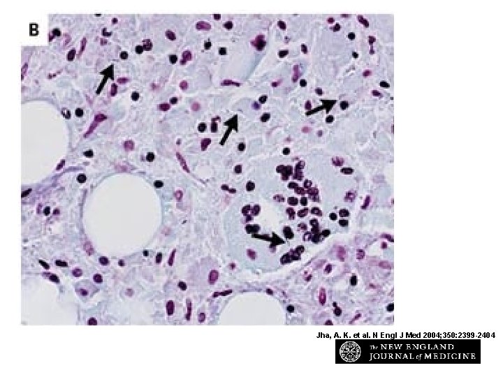
Jha, A. K. et al. N Engl J Med 2004; 350: 2399 -2404
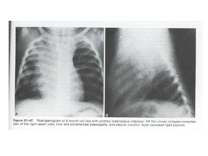
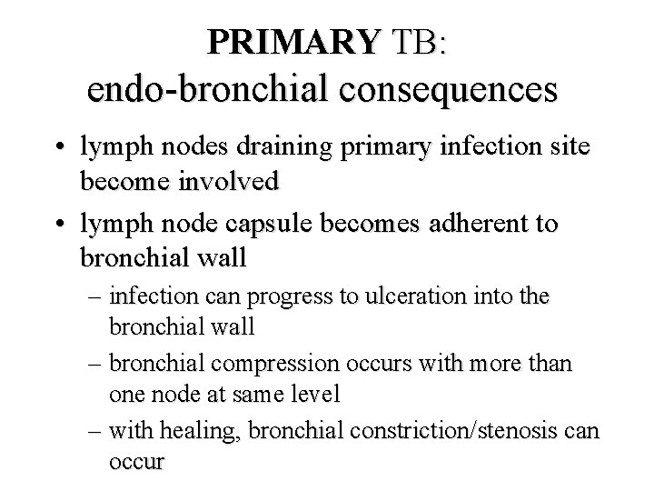
PRIMARY TB: endo-bronchial consequences • lymph nodes draining primary infection site become involved • lymph node capsule becomes adherent to bronchial wall – infection can progress to ulceration into the bronchial wall – bronchial compression occurs with more than one node at same level – with healing, bronchial constriction/stenosis can occur
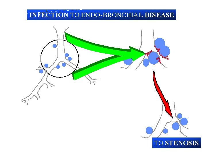
INFECTION TO ENDO-BRONCHIAL DISEASE TO STENOSIS
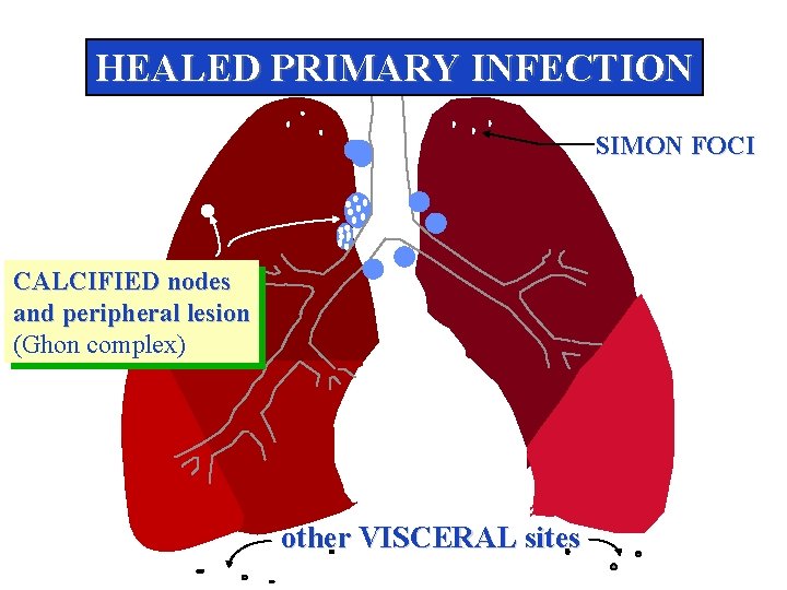
HEALED PRIMARY INFECTION SIMON FOCI CALCIFIED nodes and peripheral lesion (Ghon complex) other VISCERAL sites
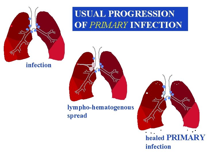
USUAL PROGRESSION OF PRIMARY INFECTION infection lympho-hematogenous spread healed PRIMARY infection
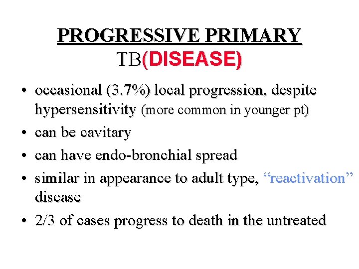
PROGRESSIVE PRIMARY TB(DISEASE) • occasional (3. 7%) local progression, despite hypersensitivity (more common in younger pt) • can be cavitary • can have endo-bronchial spread • similar in appearance to adult type, “reactivation” disease • 2/3 of cases progress to death in the untreated
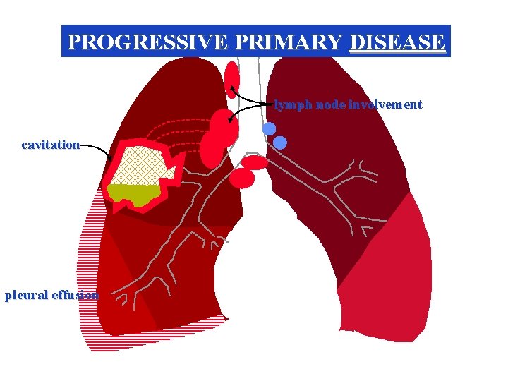
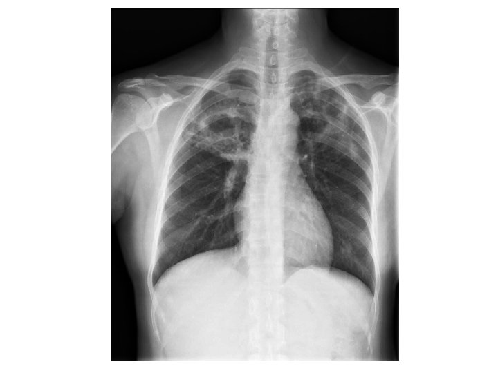
PROGRESSIVE PRIMARY DISEASE lymph node involvement cavitation pleural effusion
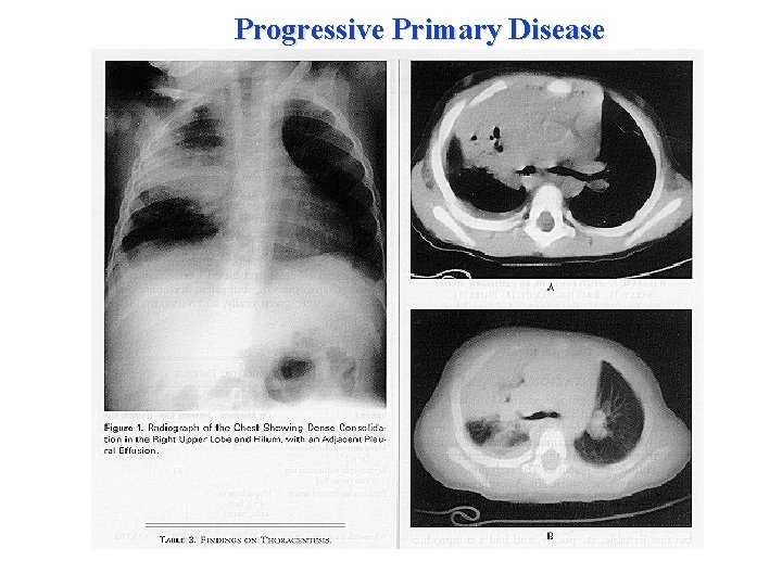
Progressive Primary Disease
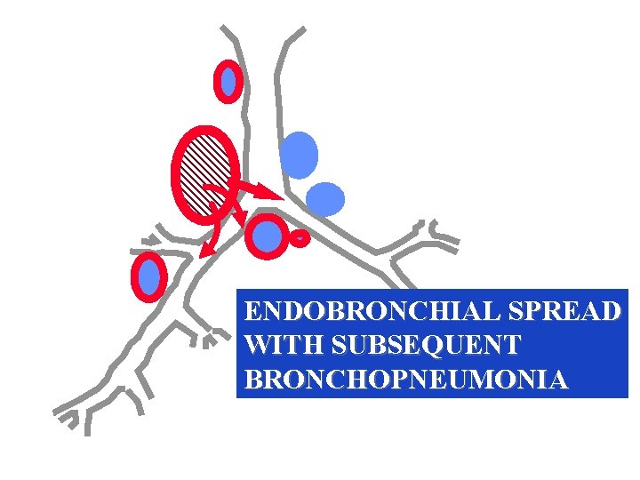
ENDOBRONCHIAL SPREAD WITH SUBSEQUENT BRONCHOPNEUMONIA
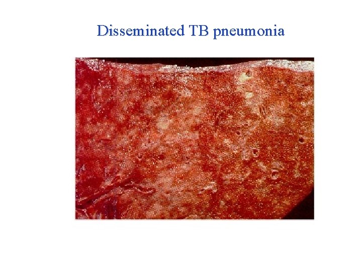
Disseminated TB pneumonia
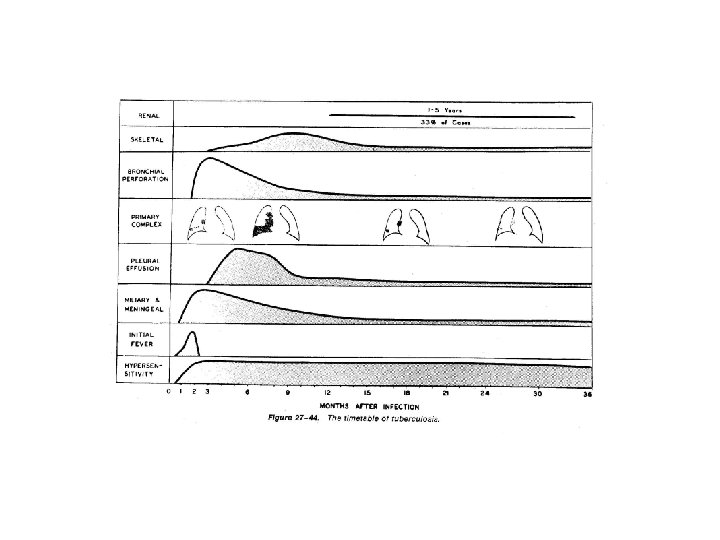
MILIARY Disease Generalized Hematogenous Tuberculosis • generalized dissemination through bloodstream – caseous focus ruptures into blood vessel – growth of tubercle within the blood vessel – may be acute, occult or chronic • • • uniformly fatal if not treated rare usually occurs in the first 4 months after primary infection
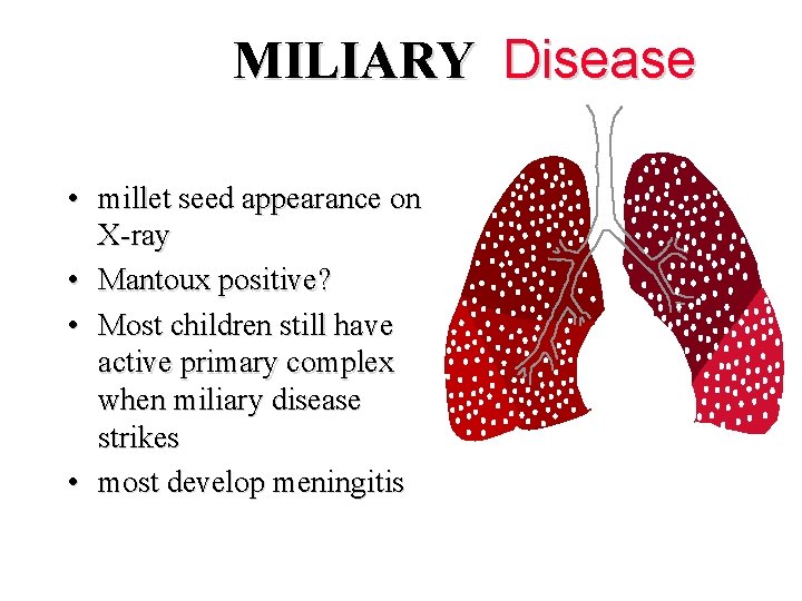
MILIARY Disease • millet seed appearance on X-ray • Mantoux positive? • Most children still have active primary complex when miliary disease strikes • most develop meningitis
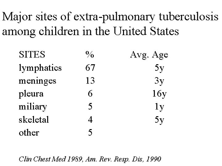
Major sites of extra-pulmonary tuberculosis among children in the United States SITES lymphatics meninges pleura miliary skeletal other % 67 13 6 5 4 5 Avg. Age 5 y 3 y 16 y 1 y 5 y Clin Chest Med 1989, Am. Rev. Resp. Dis, 1990
TB MENINGITIS • • • primary seeding caseous lesion adjacent meninges growth/discharge usually insidious prophylaxis is preventive
TB MENINGITIS • 40% of cases ocur within 3 months of estimated onset of TB – rarely < 1 month • 90% with TB menningitis PPD positive – 94% of these have positive CXR’s • average duration from meningitis diagnosis to death (untreated) 19. 5 days
CHRONIC PULMONARY TB (re-activation/adult type) • occurs only after primary infection – 0. 5 -10 % incidence (7% by Lincoln) – usually not less than 1 year of age – most cases occur when primary infection aquired after 7 years of age • minute areas of bronchopneumonia • tissue hypersensitivity - cavity formation • few symptoms early on in disease
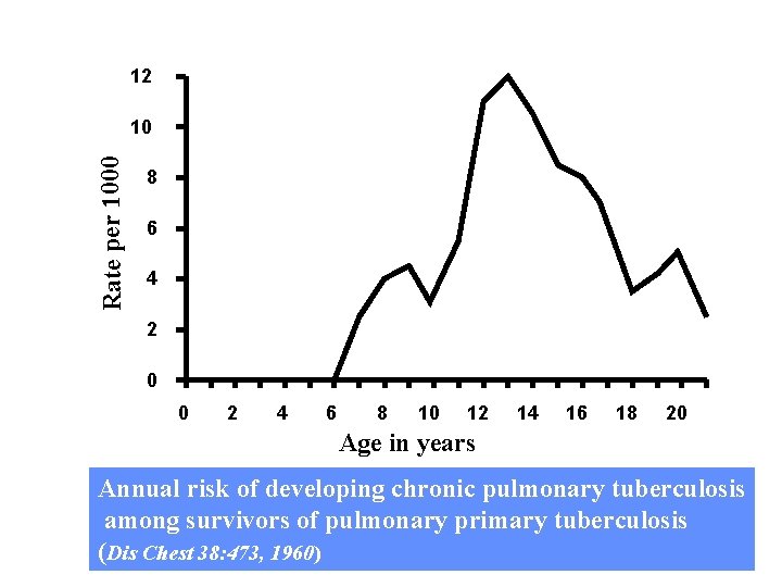
12 Rate per 1000 10 8 6 4 2 0 0 2 4 6 8 10 12 14 16 18 20 Age in years Annual risk of developing chronic pulmonary tuberculosis among survivors of pulmonary primary tuberculosis (Dis Chest 38: 473, 1960)
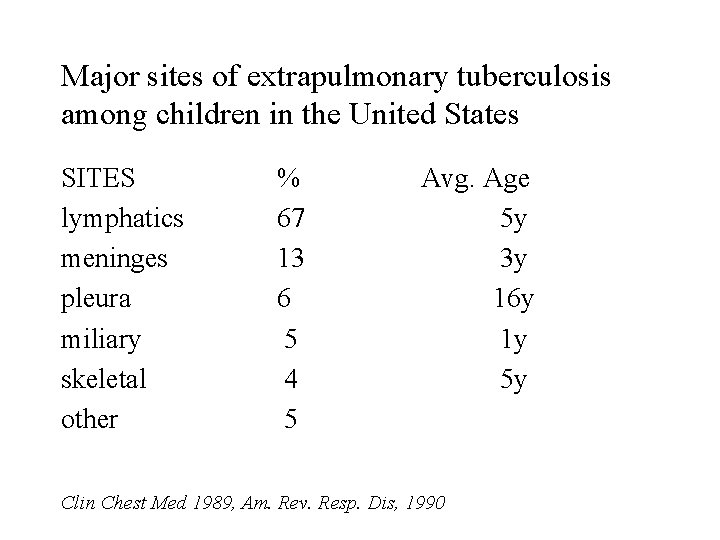
Major sites of extrapulmonary tuberculosis among children in the United States SITES lymphatics meninges pleura miliary skeletal other % 67 13 6 5 4 5 Avg. Age 5 y 3 y 16 y 1 y 5 y Clin Chest Med 1989, Am. Rev. Resp. Dis, 1990
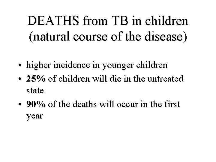
DEATHS from TB in children (natural course of the disease) • higher incidence in younger children • 25% of children will die in the untreated state • 90% of the deaths will occur in the first year
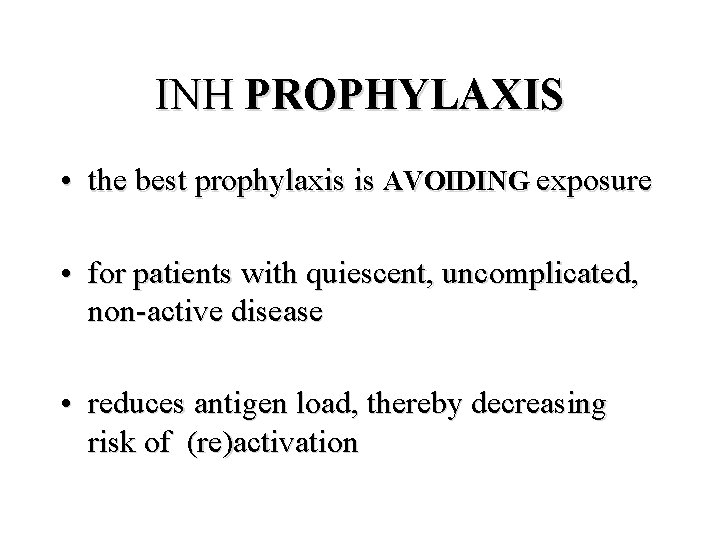
INH PROPHYLAXIS • the best prophylaxis is AVOIDING exposure • for patients with quiescent, uncomplicated, non-active disease • reduces antigen load, thereby decreasing risk of (re)activation
PULMONARY TUBERCULOSIS LOOK ALIKE. . .
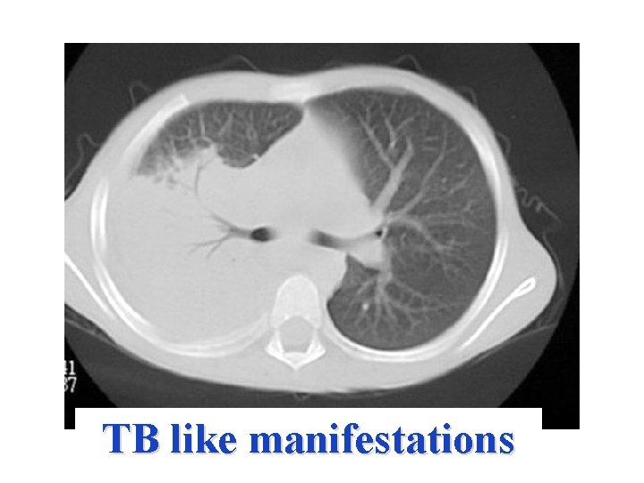
TB like manifestations
TB IN CHILDREN SUMMARY • There is no such thing as a positive PPD from just “being exposed” to the disease • primary vs reactivation disease • children with primary TB are rarely “communicable” • untreated, there is high morbidity and mortality • children with quiescent disease deserve prophylaxis • prophylaxis is “essentially” curative
- Slides: 62