PNEUMONIA AND LUNG DISEASES BDS 2017 BATCH DR
 DR. WALEEM AHMAD ASSISTANT PROFESSOR")
PNEUMONIA AND LUNG DISEASES (BDS 2017 BATCH) DR. WALEEM AHMAD ASSISTANT PROFESSOR

LUNG DISEASES • Respiratory diseases, or lung diseases, are pathological conditions affecting the organs and tissues that make gas exchange difficult in airbreathing. • They include conditions of the respiratory tract including the trachea, bronchioles, alveoli, pleurae, pleural cavity, and the nerves and muscles of respiration.

• Respiratory diseases range from mild and selflimiting, such as the common cold, to life-threatening diseases such as bacterial pneumonia, pulmonary embolism, acute asthma, lung cancer, and severe acute respiratory syndromes.

Classification Respiratory diseases can be classified in many different ways, including by the organ or tissue involved, by the type and pattern of associated signs and symptoms, or by the cause of the disease.
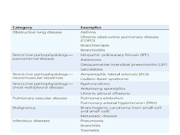

I. OBSTRUCTIVE LUNG DISEASE • Asthma, chronic bronchitis, bronchiectasis and chronic obstructive pulmonary disease (COPD) are all obstructive lung diseases characterised by airway obstruction.

II. RESTRICTIVE LUNG DISEASES • Restrictive lung diseases are characterized by a loss of lung compliance, causing incomplete lung expansion and increased lung stiffness. • Restrictive lung diseases may be due to specific causes which can be intrinsic to the parenchyma of the lung, or extrinsic to it.

• Intrinsic lung diseases includes Pneumoconiosis caused by long-term exposure to dusts, Radiation fibrosis, as a consequence of another disease such as rheumatoid arthritis, Hypersensitivity pneumonitis , Acute respiratory distress syndrome (ARDS), a severe lung condition occurring in response to a critical illness or injury, Infant respiratory distress syndrome due to a deficiency of surfactant in the lungs of a baby born prematurely and Tuberculosis.

• Extrinsic Diseases includes Nonmuscular diseases of the upper thorax such as kyphosis, pectus carinatum and pectus excavatum, Diseases restricting lower thoracic/abdominal volume (e. g. obesity, diaphragmatic hernia, or the presence of ascites) and Pleural thickening.

III. CHRONIC RESPIRATORY DISEASES • Chronic respiratory diseases are long-term diseases of the airways and other structures of the lung. They are characterized by a high inflammatory cell recruitment (neutrophil) and/or destructive cycle of infection.

IV. RESPIRATORY TRACT INFECTIONS • Infections can affect any part of the respiratory system(URTI, LRTI). A. Upper respiratory tract infection: • The most common upper respiratory tract infection is the common cold. • Sinusitis, tonsillitis, otitis media, pharyngitis and laryngitis are also considered upper respiratory tract infections.

B. Lower respiratory tract infection: • The most common lower respiratory tract infection is pneumonia, an infection of the lungs which is usually caused by bacteria, particularly Streptococcus pneumoniae in Western countries. • Worldwide, tuberculosis is an important cause of pneumonia. • Other pathogens such as viruses and fungi can cause pneumonia for example severe acute respiratory syndrome and pneumocystis pneumonia

V. TUMORS A. Malignant tumors: • Malignant tumors of the respiratory system, particularly primary carcinomas of the lung, are a major health problem responsible for 15% of all cancer diagnoses and 30% of all cancer deaths. • The major histological types of respiratory system cancer are:

1. Small cell lung cancer 2. Non-small cell lung cancer – Adenocarcinoma of the lung – Squamous cell carcinoma of the lung – Large cell lung carcinoma
 • Lymphoma • Head and")
• Other lung cancers (carcinoid, Kaposi’s sarcoma, melanoma) • Lymphoma • Head and neck cancer • Pleural mesothelioma, almost always caused by exposure to asbestos dust.

B. Benign tumors • Benign tumors are relatively rare causes of respiratory disease. Examples of benign tumors are: 1. Pulmonary hamartoma 2. Congenital malformations such as pulmonary sequestration and congenital cystic adenomatoid malformation (CCAM).

VI. PLEURAL CAVITY DISEASES 1. Pleural effusion- A collection of fluid in the pleural cavity is known as a pleural effusion 2. Pneumothorax- A pneumothorax is a hole in the pleura covering the lung allowing air in the lung to escape into the pleural cavity. The affected lung "collapses" like a deflated balloon.

VII. PULMONARY VASCULAR DISEASE Pulmonary vascular diseases are conditions that affect the pulmonary circulation. • Pulmonary embolism, a blood clot that forms in a vein, breaks free, travels through the heart and lodges in the lungs (thromboembolism). Large pulmonary emboli are fatal, causing sudden death

• Pulmonary arterial hypertension, elevated pressure in the pulmonary arteries. Most commonly it is idiopathic (i. e. of unknown cause) but it can be due to the effects of another disease, particularly COPD.

• Pulmonary hemorrhage, inflammation and damage to capillaries in the lung resulting in blood leaking into the alveoli. This may cause blood to be coughed up. Pulmonary hemorrhage can be due to autoimmune disorders such as granulomatosis with polyangiitis and Goodpasture's syndrome.

PNEUMONIA • Pneumonia is defined as an acute respiratory illness associated with recently developed radiological pulmonary shadowing which may be segmental, lobar or multilobar.

CLASSIFICATION

Pneumonia: Classification I Based on anatomical involvement Lobar Pneumonia Bronchopneumonia Interstitial Pneumonia

Pneumonia: Classification II Classification By Aetiology • Primary Pneumonia • Secondary Pneumonia (including Aspiration Pneumonia) • Suppurative Pneumonia (necrotizing pneumonia) Common Organisms Less Common Organisms Streptococcous pneumoniae Klebsiella pneumoniae Haemophilus influenza Streptococcus pneumonia Moraxella catarrhalis Pseudomonas aeruginosa Staphylococcus aureus Coxiella burnetii Legionella pneumophilia Chlamydia pneumoniae Mycoplasma pneumoniae Chlamydia psittaci Actinomyces israeli Viruses (including SARS) Table 1: Organisms responsible for primary pneumonia

Pneumonia: Classification III Classification by mode of acquiring pneumonia • Community-acquired Pneumonia • Hospital acquired pneumonia, or Ventilator associated pneumonia) • Health Care Associated Pneumonia • Pneumonia in immuno-compromised host

Pneumonia: Pathophysiology • Pneumonia results from the proliferation of microbial pathogens at the alveolar level and the host's response to those pathogens. • Microorganisms gain access to the lower respiratory tract in several ways. • Aspiration from the oropharynx; (most common) • Inhalation of contaminated droplets; • Hematogenous spread • Contiguous extension.
 • Defense Mechanisms against Pneumonia • Hairs and turbinates of")
Pneumonia: Pathophysiology (contd. ) • Defense Mechanisms against Pneumonia • Hairs and turbinates of the nares • Branching architecture of the tracheobronchial tree • Muco-ciliary clearance and local antibacterial factors • Gag reflex and the cough mechanism • the normal flora adhering to mucosal cells of the oropharynx • Alveolar Macrophages, local proteins (e. g. , surfactant proteins A and D)
 • Only when the capacity of the alveolar macrophages to")
Pneumonia: Pathophysiology (contd. ) • Only when the capacity of the alveolar macrophages to ingest or kill the microorganisms is exceeded does clinical pneumonia become manifest. • The alveolar macrophages initiate the inflammatory response to bolster lower respiratory tract defenses. Mediators Responsible Effects IL-1 and TNF Fever IL – 8 , GCSF stimulates release & attraction of neutrophils to the lungs Causes peripheral leucocytosis, increased purulent secretions Inflammatory mediators (macrophages) & neutrophils Localised alveolar capillary leak Table 2: Mediators responsible in patho-physiology of Pneumonia
 Mechanism Clinical Features Hemoptysis Leaky alveolar capillaries Radiologic infiltrates Rales")
Pneumonia: Pathophysiology (contd. ) Mechanism Clinical Features Hemoptysis Leaky alveolar capillaries Radiologic infiltrates Rales on auscultation Alveolar filling and action of bacterial pathogens Hypoxemia Decreased compliance of lungs Dyspnoea Reduction in lung volume and compliance Intra-pulmonary shunting of blood Death

Pneumonia: Pathology Stages/Phases of Pneumonia Characteristic Features Oedematous Phase Alveoli filled with proteinaceous exudate and bacteria Rapidly followed by Red hepatization phase Stage of Red hepatization Presence of erythrocytes in the cellular intraalveolar exudate Neutrophil influx Bacteria can be seen in specimens Stage of Gray hepatization (successful containment of the infection & improvement in gas exchange) Erythrocytes extravasation ceases, extravasated ones get lysed and degraded Neutrophils predominant Abundant fibrin deposition Disappearance of bacteria Macrophages reappear Debris (PMN, Bacteria, fibrin) cleared Inflammatory response cleared Table 3: Different Pathologic Phases of Pneumonia Stage of Resolution *Has been described best for lobar pneumococcal pneumonia.

CAP & HAP: A Comparison Community Acquired Definition It indicates pneumonia occuring in a person in a community (outside hospital) Predisposing Cigarette smoking Factors Upper respiratory tract infections Alcohol Corticosteroid therapy Old age Recent influenza infection Indoor air pollution Mode of Spread Droplet Infection Infecting Agent S. Pneumoniae, S. aureus, H. influenza Viruses (influenza, parainfluenza, measles, Herpes simplex, Varicella, CMV) Hospital Acquired Refers to a new episode of pneumonia occurring at least 2 days after admission to hospital. Reduced host defences against bacteria Aspiration of nasopharyngeal/gastric secretions Bacteria introduced into lower respiratory tract Bacteraemia Early-Onset: similar to CAP Late-Onset: Escherichia, Pseudomonas, Klebsiella, MRSA, anaerobes
 Community Acquired Hospital Acquired Can vary from indolent to")
CAP & HAP: Presentation(contd. ) Community Acquired Hospital Acquired Can vary from indolent to fulminating Universally agreed diagnostic presentation criteria lacking Pulmonary Symptoms : breathlessness, Should be considered in any cough, -non-productive or productive hospitalised /ventilated patient (mucoid, purulent , blood stained) who develops: haemoptysis, plueritic chest pain, may able purulent sputum (or to speak sentences/ short of breath endotracheal secretions), Systemic Features : fever a/w chills n new radiological infiltrates, rigors, tachycardia, vomiting, decreased an otherwise unexplained appetite, headache, fatigue, myalgias, increase in oxygen arthralgia requirement, Elderly: New-onset/ progressive confusion a core temperature > 38. 3°C, Severely ill: septic shock or organ-failure and Tachypnoea, use of accessory muscles, leucocytosis or leucopenia. increased/decreased vocal fremitus, Percussion note: dull to flat Bronchial Breathing, Crackles Whispering pectoriloquy, pleural friction rub

CAP & HAP: Investigation Community Acquired Chest Radiology: to confirm the diagnosis and to exclude complications Pulse oximetry to monitor response to oxygen therapy, if Sa. O 2 < 93%, features of severe pneumonia, identify ventilatory failure or acidosis Cell count: neutrophil leucocytosis Hospital Acquired Circulating biomarkers may assist with the diagnosis but are currently non-specific. Appropriate investigations are similar to CAP, microbiological confirmation preferrable. In mechanically ventilated patients, bronchoscopy-directed protected Microbiologic Studies: for severe CAP brush specimens or bronchoalveolar and for those that do not respond to initial lavage (BAL) may be performed. therapy (Gram stain, sputum culture, blood culture, Polymerase Chain Endotracheal aspirates are easy to Reaction, Serology, Antigen Detection) obtain but less reliable. Renal Function Tests: Urea & electrolytes Liver Function Tests Elevated C-Reactive Protein

Chest Radiology In Pneumonia Right Upper Lobar Pneumonia Right Upper Lobe Pneumonia with air bronchograms Right Middle Lobar Pneumonia Left Lobar Pneumonia with pleural effusion

Hospital Care Associated Pneumonia • HCAP represents a transition between classic CAP and typical HAP • Refers to the development of pneumonia in a person who has spent at least 2 days in hospital within the last 90 days, attended a haemodialysis unit, received intravenous antibiotics, or been resident in a nursing home or other long-term care facility. (Davidson-21 st Ed. ) • MRSA in particular is more common in HCAP than in traditional HAP/VAP. • Patients at greatest risk for HCAP are not well defined. • Patients from nursing homes are not always at elevated risk for infection with MDR pathogens. • Low risk of MDR infection if they have not recently received antibiotics and are independent in most activities of daily living. • Patients receiving home infusion therapy or undergoing chronic dialysis are probably at particular risk for MRSA pneumonia • In general, management of HCAP due to MDR pathogens is similar to that of MDR HAP/VAP.

Pneumonia: Severity Assessment • There are currently two sets of criteria: • Pneumonia Severity Index (PSI), a prognostic model used to identify patients at low risk of dying; and • CURB-65 criteria, a severity-of-illness score. Assessment for need for ICU care provided by severity criteria proposed by the Infectious Diseases Society of America (IDSA) and the American Thoracic Society (ATS) ≥ ≥ ≥ ≤ ≤

CAP TREATMENT: GENERAL CONSIDERATION • Adequate hydration, oxygen therapy for hypoxemia, and assisted ventilation • Patients with severe CAP who remain hypotensive despite fluid resuscitation may have adrenal insufficiency and may respond to glucocorticoid treatment. • Immunomodulatory therapy in the form of drotrecogin alfa (activated) should be considered for CAP patients with persistent septic shock and APACHE II scores of 25, particularly if the infection is caused by S. pneumoniae. • Once the etiologic agent(s) and susceptibilities are known, therapy may be altered to target the specific pathogen(s). • Switch to oral treatment is appropriate as long as the patient can ingest and absorb the drugs, is hemodynamically stable, and is showing clinical improvement. • The duration of treatment for CAP has generated considerable interest

CAP: Empirical Treatment for Out-Patients Modality Regimen Previously healthy and no antibiotics in past 3 months: A macrolide [clarithromycin (500 mg PO bid) or azithromycin (500 mg PO once, then 250 mg qd)] or Doxycycline (100 mg PO bid) Comorbidities or antibiotics in past 3 months: select an alternative from a different class A respiratory fluoroquinolone [moxifloxacin (400 mg PO qd), gemifloxacin (320 mg PO qd), levofloxacin (750 mg PO qd)] or A β-lactam [preferred: high-dose amoxicillin (1 g tid) or amoxicillin/clavulanate (2 g bid); alternatives: ceftriaxone (1– 2 g IV qd), cefpodoxime (200 mg PO bid), cefuroxime (500 mg PO bid)] plus a macrolide In regions with a high rate of "high-level" pneumococcal macrolide resistance, consider alternatives listed above for patients with comorbidities.

CAP: Empirical Treatment for In-Patients Modality Regimen Non ICU Patients A respiratory fluoroquinolone [moxifloxacin (400 mg PO or IV qd), gemifloxacin (320 mg PO qd), levofloxacin (750 mg PO or IV qd)] A β-lactam [cefotaxime (1– 2 g IV q 8 h), ceftriaxone (1– 2 g IV qd), ampicillin (1– 2 g IV q 4– 6 h), ertapenem (1 g IV qd in selected patients)] plus a macrolide [oral clarithromycin or azithromycin or IV azithromycin (1 g once, then 500 mg qd)] ICU Patients A β-lactam [cefotaxime (1– 2 g IV q 8 h), ceftriaxone (2 g IV qd), ampicillin-sulbactam (2 g IV q 8 h)] plus Azithromycin or a fluoroquinolone

CAP: Empirical Treatment Special Concerns Organism being considered Treatment Regimen Pseudomonas An antipneumococcal, antipseudomonal β-lactam [piperacillin/tazobactam (4. 5 g IV q 6 h), cefepime (1– 2 g IV q 12 h), imipenem (500 mg IV q 6 h), meropenem (1 g IV q 8 h)] plus either ciprofloxacin (400 mg IV q 12 h) or levofloxacin (750 mg IV qd) The above β-lactams plus an aminoglycoside [amikacin (15 mg/kg qd) or tobramycin (1. 7 mg/kg qd) and azithromycin] The above β–lactams plus an aminoglycoside plus an antipneumococcal fluoroquinolone CA-MRSA Add linezolid (600 mg IV q 12 h) or vancomycin (1 g IV q 12 h).

• • • CAP TREATMENT: FAILURE TO RESPOND Patients who are slow to respond to therapyshould be reevaluated at about day 3 (sooner if their condition is worsening rather than simply not improving) Check the drug, dose of the correct drug Pathogen resistant to the drug selected, or a sequestered focus (e. g. , a lung abscess or empyema). Consider possibility of an unsuspected pathogen (e. g. , CAMRSA, M. tuberculosis, or a fungus). Re-consider your Diagnosis: tuberculosis, pulmonary edema, pulmonary embolism, lung carcinoma, radiation and hypersensitivity pneumonitis, and connective tissue disease involving the lungs. Nosocomial superinfections—both pulmonary and extrapulmonary

HAP: Emperical Treatment Categories Patients without Risk Factors for MDR Pathogens Patients with Risk Factors for MDR Pathogens Treatment Ceftriaxone (2 g IV q 24 h) or Moxifloxacin (400 mg IV q 24 h), ciprofloxacin (400 mg IV q 8 h), or levofloxacin (750 mg IV q 24 h) or Ampicillin/sulbactam (3 g IV q 6 h) or Ertapenem (1 g IV q 24 h) 1. A β-lactam: Ceftazidime (2 g IV q 8 h) or cefepime (2 g IV q 8– 12 h) or Piperacillin/tazobactam (4. 5 g IV q 6 h), imipenem (500 mg IV q 6 h or 1 g IV q 8 h), or meropenem (1 g IV q 8 h) plus 2. A second agent active against gram-negative bacterial pathogens: Gentamicin or tobramycin (7 mg/kg IV q 24 h) or amikacin (20 mg/kg IV q 24 h) or Ciprofloxacin (400 mg IV q 8 h) or levofloxacin (750 mg IV q 24 h) plus 3. An agent active against gram-positive bacterial pathogens: Linezolid (600 mg IV q 12 h) or Vancomycin (15 mg/kg, up to 1 g IV, q 12 h)

PNEUMONIA: COMPLICATIONS • • • • Para-pneumonic effusion Empyema Retention of sputum causing lobar collapse Pulmonary embolism and DVT Pneumothorax (S. aureus) Suppurative pneumonia/ lung abscess ARDS, renal failure, multi-organ failure Ectopic abscess formation (S. aureus), Metastatic infection Pericarditis, hepatitits, myocarditis, meningoencephalitis Pyrexia due to drug hypersensitivity Exacerbation of comorbid illnesses. Complicated pleural effusion: diagnostic/therapeutic tapping required If the fluid has a p. H of <7, a glucose level of <2. 2 mmol/L, and a lactate dehydrogenase concentration of >1000 U/L or if bacteria are seen or cultured, then the fluid should be drained; a chest tube is usually required.

PNEUMONIA: FOLLOW UP • Fever and leukocytosis usually resolve within 2– 4 days in otherwise healthy patients with CAP, but physical findings may persist longer. • Chest radiographic abnormalities are slowest to resolve and may require 4– 12 weeks to clear • Patients may be discharged from the hospital once their clinical conditions are stable, with no active medical problems requiring hospital care. • For a patient whose condition is improving and who (if hospitalized) has been discharged, a follow-up radiograph can be done ~4– 6 weeks later. • If relapse or recurrence is documented, particularly in the same lung segment, the possibility of an underlying neoplasm must be considered.

PNEUMONIA: PROGNOSIS • Depends on • Patient's age, • Comorbidities, and • Site of treatment (inpatient or outpatient). • Young patients without comorbidity do well and usually • recover fully after ~2 weeks. • Older patients and those with comorbid conditions can take several weeks longer to recover fully. • Overall mortality rate for the outpatient group is <1%. • Overall mortality rate for Inpatient group is estimated at 10%, with ~50% of deaths directly attributable to pneumonia.

PNEUMONIA: PREVENTION • The main preventive measure is vaccination. • In the event of an influenza outbreak, unprotected patients at risk from complications should be vaccinated immediately and given chemoprophylaxis with either oseltamivir or zanamivir for 2 weeks—i. e. , until vaccine-induced antibody levels are sufficiently high. • Because of an increased risk of pneumococcal infection, even among patients without obstructive lung disease, smokers should be strongly encouraged to stop smoking. • 7 -valent pneumococcal conjugate vaccine: produces T cell– dependent antigens that result in long-term immunologic memory.

THANKS
- Slides: 47