Kharkov National Medical University Department of Histology Cytology


 2 – lipids (hydrophobic tails)")
 Cholesterol (non-polar)")
 Glycerol Fatty acids (hydrophobic tails)")


 Organelles")


 n")
 endoplasmic reticulum n n is a membranous network of sac-like structures cisternae.")

 endoplasmic reticulum, SER")


 n Ribosomes n n")


- Slides: 77
Kharkov National Medical University Department of Histology, Cytology and Embryology Lecture 1. Introduction. Cytology
Histology – is the science that studies microscopic structure and function of the human organism, the organization of the tissues and organs. Cytology – is the science that study the structure and functions of the cell. Embryology – is the science that research embryonic development (formation) of human body
Cytology
n n - the The cell is: smallest structural and functional unit, - the elementary level of organization of the multicellular organisms. - a self-regulating, self-regenerating and self-governing biological system n cell possesses all features of the whole organism, such as metabolism, growth, irritability, movement, and others.
Types of cells in human body
Cells produce matrix:
Light and electron microscopy are 2 mane methods in histology
Levels of biological systems n Biomolecules n membranes n organelles CELL
Membrane contents: n n 1 – lipids (hydrophilic head) 2 – lipids (hydrophobic tails) 3 – proteins 4 – carbohydrates (only outer cell membrane)
Lipids may be: n n Phospholipids – triglycerides (polar) Cholesterol (non-polar)
Phospholipids : n n n Phosphate group (hydrophilic heads) Glycerol Fatty acids (hydrophobic tails)
Proteins may constitute close to 50% of membrane content n n n function : 1 - channels, in the membranes 2 - pumps, 3 - receptors, 4 - enzymes, 5 - integrative, 6 - structural
Membranes form: n n Outer cell membrane – cytolemma or plasmalemma Organelles Vesicles Nucleus - nuclear envelop
Structure of a typical cell 1. Cell membrane 2. Nucleus 3. Cytoplasm organelles Cytosol = matryx, inclusions hialoplasm
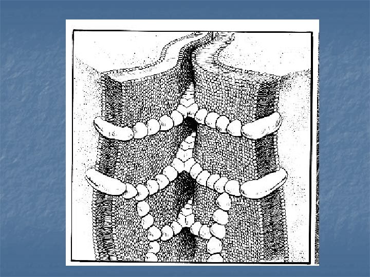
Cell junction n - consist of multiprotein complexes that provide contact between neighbouring cells or between a cell and the extracellular matrix.
Where Cells contact -- Cell junction G 1 2
Cell junction n Communicating or Gap junctions junction Tight junction Desmosomes
Tight junction n present in different types of epithelia two layers of glycocalyx are fused. act as a barrier, that prevents the movement of molecules into the intercellular spaces
G
Gap junction n n allow for direct chemical communication between adjacent cellular cytoplasm through diffusion without contact of the extracellular fluid numerous in muscle tissue
Gap junction n n Consists of six connexin proteins, interacting to form a cylinder with a pore in the centre connexon. This protrudes across the cell membrane, and when two adjacent cell connexons interact, they
Desmosome n n is the most common type of junction Provides cell attachment
Inside the cell : n Cytoplasm and nucleus
Inside the cell … n n n Cytoplasm consists of: Matrix (hialoplasm, cytozol) Organelles Inclusions
Inclusions n n Inclusions are "nonliving" components of the cell. They include granules with secretions, pigment granules, lipid droplets, and glycogen.
Organelles: classification by structure Membranous or "membrane-bound" n n Non-membranous
Organelles: classification by function n General (present in every cell, perform general function) n Special (in specialised cell, perform special function)
Rough (rough-surfaced) endoplasmic reticulum n n is a membranous network of sac-like structures cisternae. the cisternal space (or lumen) is continuous with the perinuclear space but separate from the cytosol.
n n The surface of the RER is studded with ribosomes giving it a "rough" appearance (hence its name). Function - synthesis of proteins
Smooth(-surfaced) endoplasmic reticulum, SER
SER consists of tubules that are located near the cell periphery. Function: n It synthesizes lipids - phospholipids and steroids. n It also carries out the metabolism of carbohydrates (synthesis of glycogen, gluconeogenesis ), n drug detoxification, and steroid metabolism. n n Storage of Ca-ions (only in muscle cell)
n Golgi apparatus n n n also known as the Golgi complex, Golgi body, or simply the Golgi, is an organelle found in most eukaryotic cells. It was identified in 1897 by the Italian physician Camillo Golgi and named after him in 1898. Loks as a pack of sacs.
Golgi apparatus n Golgi complex is connected with endoplasmic reticulum
Golgi apparatus Functions. 1. synthesis of substances, which has begun in endoplasmic reticulum and is accomplished in the Golgi complex. n
Golgi apparatus Functions. n 2. formation of compound molecules – glycoproteins, lipoproteins.
Golgi apparatus Functions. n 3. production of lysosomes and secretory vesicles.
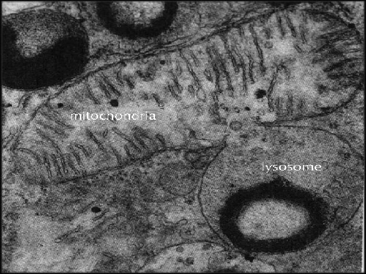
Mitochondrion n is a membraneenclosed organelle found in most eukaryotic cells.
Mitochondrion n A mitochondrion contains outer and inner membranes composed of phospholipid bilayers and proteins. Folds of inner membrane – cristae Inside M. lie matryx
Mitochondrion n Mitochondria provide energy for various cellular functions , Produce ATP molecules by Krebs cycle
Lysosome n n n Lysosomes are round vesicles that contain acid hydrolase enzymes that break down waste materials and cellular debris. Lysosomal enzymes help in digesting the materials within phagosomes. They can be described as the stomach of the cell.
Lysosome Cycle n n lysosomes are formed from Golgi complex The nearly produced lysosome is primary lysosome
Lysosome Cycle n n primary lysosome fuses with the phagosome -secondary lysosome or phagolysosome part of undigesting material may remain within the cell as residual bodies.
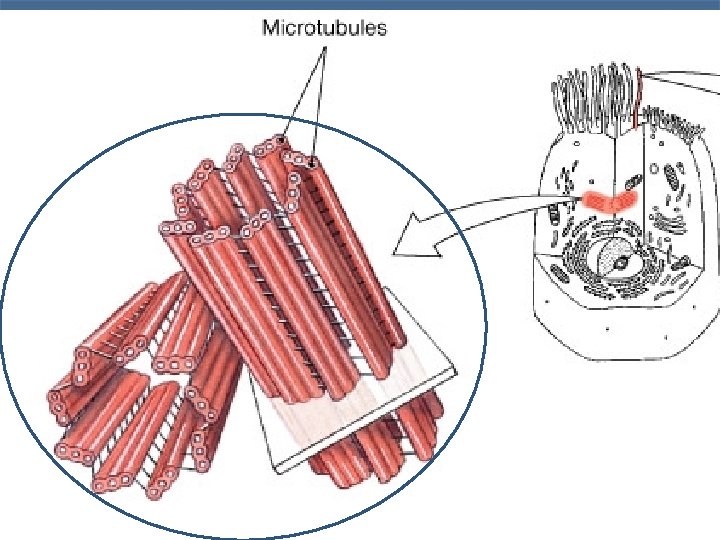
Non-membranous organelles: n Microfilaments Microtubules Centrioles (Cell Center) n Ribosomes n n
The cytoskeleton n n - is made up of three kinds of protein filaments: Actin filaments (also called microfilaments) Intermediate filaments Microtubules
Microfilaments, Microtubules form “Skeleton” of the cell
Cell center Centriole = 9 x 3 microtubules; 2 centrioles = cell center = Β-tubulin function -formation of mitotic spindle (mitosis, meiosis), flagella, basal bodies and cilia
Cell center
Nucleus n Eukaryotes have nucleus
Nucleus is a membrane-limited structure: n n Nucleolemma nuclear envelope Nucleoplasm Nucleolus Chromatin, chromosomes
Nuclear envelope n n n - Consists of two membranes. The outer layer is continuous with endoplasmic reticulum. The inner layer provides attachment to the ends of the chromosomes.
Nuclear envelope n There are gaps, called nuclear pores n The nuclear pore transports some substances from nucleus into cytoplasm
Nucleolus n n Nucleolus is the site very amplificated molecule of DNA. It is the site of active synthesis of ribosomal RNA.
n In the nucleolus RNA binds with protein and forms ribosomal subunits, which leave the nucleus via nuclear pores to the cytoplasm as ribosomes.
Chromosome - DNA molecules which contain genetic information n n DNA molecule is coiled around the histone core, which consists of eight gistone molecules. Such particles, consisting of gistone core and DNA, are called nucleosomes.
Chromatin n n is the combination of DNA and proteins that make up the contents of the nucleus of a cell. The primary functions of chromatin are 1) to package DNA into a smaller volume to fit in the cell, 2) to strengthen the DNA to allow mitosis, 3) to prevent DNA damage, and 4) to control gene expression and DNA replication
n n Sites where chromatin fibrils are packed very tightly together are heterochromatin sites – non-active. there are less condensed chromatin fibrils loops euchromatin sites - active.
n Euchromatin predominates in metabolically active nuclei, n n Heterochromatin predominates in metabolically inactive nuclei
Chromosome is an organized structure of DNA and protein found in cells.
Cell Cycle n n n The life of a somatic cell is a cyclic process It is called cell cycles consists of two periods: interphase and mitosis.
Interphase is a period between two divisions of the cell. Consists of 3 phases G 1 , S , G 2
In G 1 phase: n cell grows, performs its routine functions.
In G 1 phase n n Nondividing cells complete period G 1 by growing old and death of the cell Dividing cell – prepare for next phase
S- phase = synthesis phase n n DNA molecules are duplicated each chromosome now consists of two DNA molecules or two chromatids. At the beginning of this phase, the chromosome number is 2 N and at the end, the chromosome number is 4 N.
G 2 phase n n In this phase synthesis of proteins, which are required for cell division, takes place. After phase G 2 mitosis always begins
G 0 phase n cell can leave the cycle in any phase, except G 2, and enter to so-called G 0 phase (outside the cycle). The cell that leaves the cycle is considered as reserve stem cell.
Mitosis is the process of somatic cells division. Mitosis consists of four phase: prophase, metaphase, anaphase, telophase.
Prophase n n Chromosomes becomes more and more coiled become recognisable. the nuclear membrane breaks down and the nucleoli disappear
n n Two centrioles separate and move to opposite poles of the cell. Сentrioles produce a number of microtubules which pass from one centriole to other and form a spindle of division.
Metaphase n - chromosomes move to a position midway between the two centrioles at the equator of the cell and form the equatorial plate
anaphase n n - the chromatids separate and move to opposite poles of the cell At the end of anaphase chromatids are called chromosomes.
Telophase n n n - two daughter nuclei are formed by appearance of nuclear membranes around them. The chromosomes gradually elongate and become indistinct. Nucleoli reappear.