Cytologic features Background Proteinaceous fluid with many hemosiderin
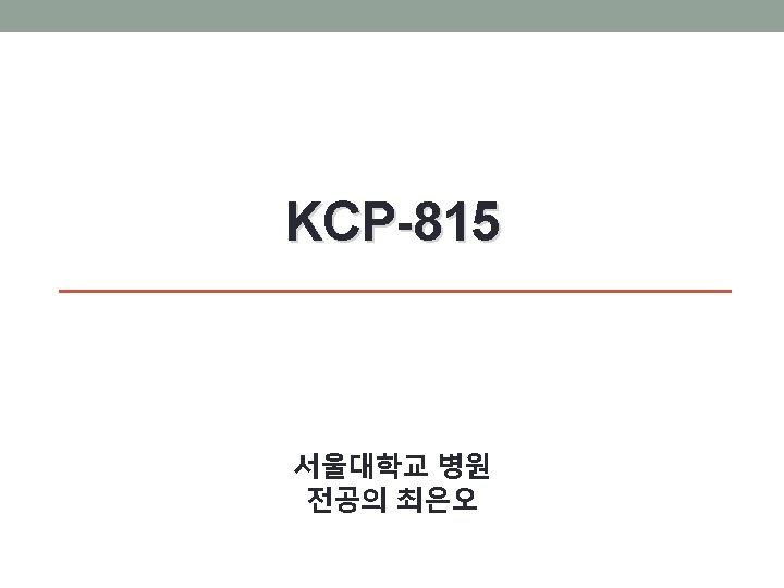


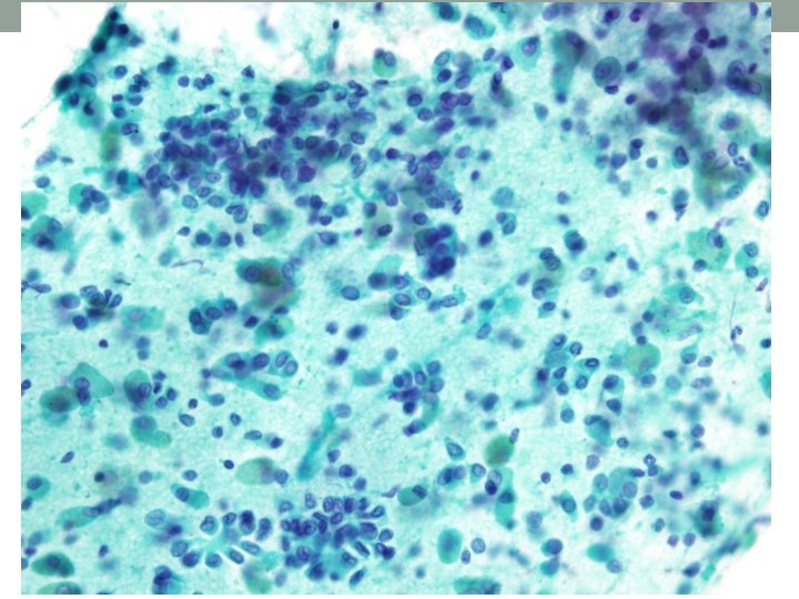
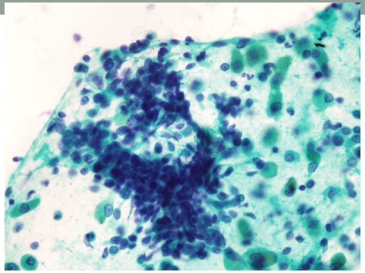
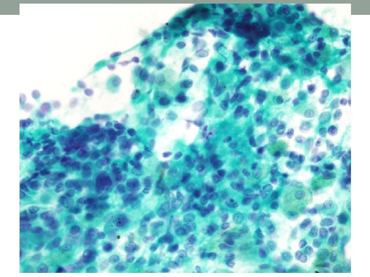
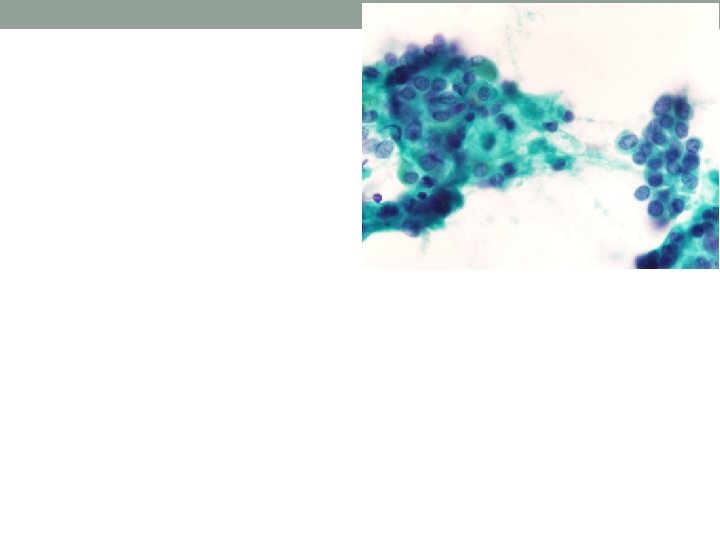


Cytologic features Background Proteinaceous fluid with many hemosiderin or anthracotic pigment-laden macrophages Cellularity Moderate Architecture Scattered epithelial cell clusters exhibiting papillary, sometimes ill-formed acini, rosette-like pattern Cell Small to medium in size Round or columnar cells Relatively bland-looking, but occasionally large cells with mild to moderate nuclear pleomorphism and nuclear hyperchromasia Cytoplasm Small to moderate amount of cytoplasm Nucleus Mostly, uniform round to oval nuclei Smooth to irregular nuclear contour Fine, stippled chromatin pattern Inconspicuous nucleoli Intranuclear inclusion

Differential diagnosis • Adenocarcinoma with lepidic pattern • Carcinoid tumor • Papillary adenoma • Sclerosing hemangioma

Clinical and radiologic features ADC with lepidic pattern Carcinoid tumor Sclerosing hemangioma Papillary adenoma • Less commonly • 1 -2% of all lung • Marked female • Rare incidence associated with smoking than other types of NSCLC • Less than 4% of NSCLCs • May present as single or multiple nodules tumors • From childhood to the ninth decades (mean age: 47 years) • Similar frequencies in men and women • Mostly, central masses causing bronchial obstruction predilection (80%) • Peak incidence in fifth decade • Mostly, solitary lesion • Multifocal in 4% of cases • Males appear to predominate • Between 7 and 60 years • Peripheral parenchymal lesion

ADC with lepidic pattern Carcinoid tumor Sclerosing Hemangioma • Highly cellular • Cells occur singly • Papillary clusters of • Epithelial cell aspirates with and in clusters bland cuboidal to • Stripped nuclei columnar cells in cells and/or or flat sheets spindled cells • Round to oval nuclei of uniform size • Nuclear membrane folds and grooves • Intranuclear pseudoinclusions bland-looking clusters of sheet mononuclear cells or papillary • Small plasmacytoid • Hyalinized stromal papillary clusters • Scan to moderate cytoplasm • Stippled granular chromatin Papillary adenoma tissue fragments • Some scattered epithelial cells with nuclear pleomorphism and pattern • Round to oval nuclei • Mild nuclear atypia • Fine chromatin intranuclear pattern, inclusions sometimes small • Hemosiderin-laden macrophages in the background nucleoli • Scanty cytoplasm, high N/C ratio
 Lung, aspiration cytology: Adenocarcinoma with lepidic pattern")
Cytologic diagnosis MICRO (2 pap) Lung, aspiration cytology: Adenocarcinoma with lepidic pattern
- Slides: 14