Primary and secondary CNSLymphoma Prof Nossrat Firusian Recklinghausen

- Slides: 34
Primary and secondary CNS-Lymphoma Prof. Nossrat Firusian, Recklinghausen, Germany
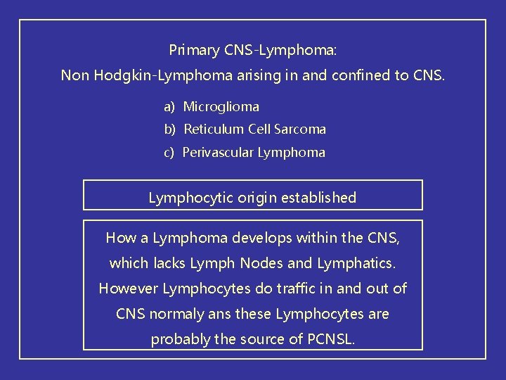
Primary CNS-Lymphoma: Non Hodgkin-Lymphoma arising in and confined to CNS. a) Microglioma b) Reticulum Cell Sarcoma c) Perivascular Lymphoma Lymphocytic origin established How a Lymphoma develops within the CNS, which lacks Lymph Nodes and Lymphatics. However Lymphocytes do traffic in and out of CNS normaly ans these Lymphocytes are probably the source of PCNSL.
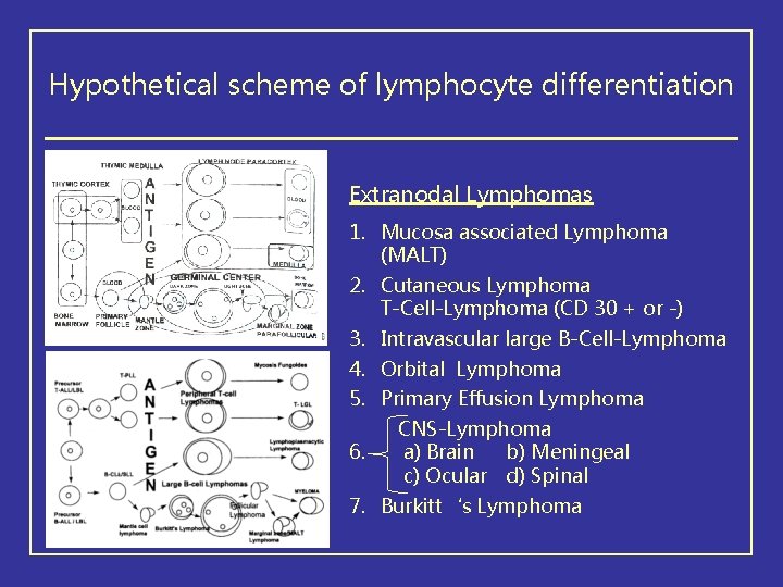
Hypothetical scheme of lymphocyte differentiation Extranodal Lymphomas 1. Mucosa associated Lymphoma (MALT) 2. Cutaneous Lymphoma T-Cell-Lymphoma (CD 30 + or -) 3. Intravascular large B-Cell-Lymphoma 4. Orbital Lymphoma 5. Primary Effusion Lymphoma 6. CNS-Lymphoma a) Brain b) Meningeal c) Ocular d) Spinal 7. Burkitt‘s Lymphoma
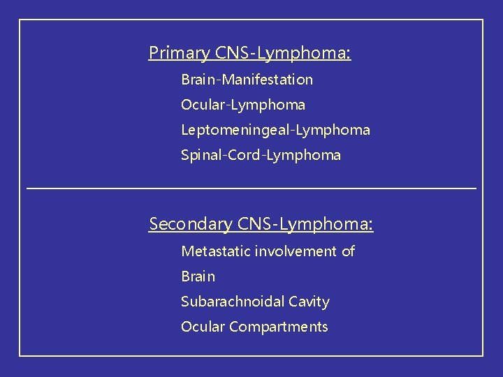
Primary CNS-Lymphoma: Brain-Manifestation Ocular-Lymphoma Leptomeningeal-Lymphoma Spinal-Cord-Lymphoma Secondary CNS-Lymphoma: Metastatic involvement of Brain Subarachnoidal Cavity Ocular Compartments
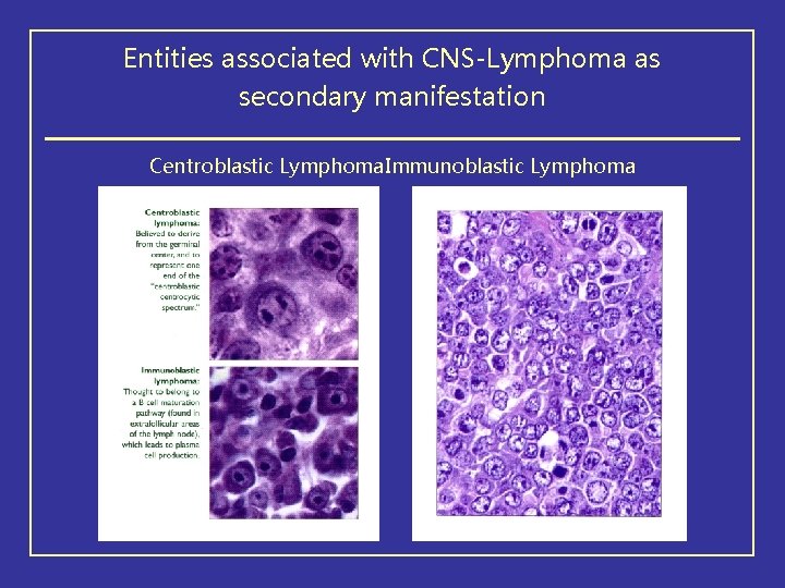
Entities associated with CNS-Lymphoma as secondary manifestation Centroblastic Lymphoma. Immunoblastic Lymphoma
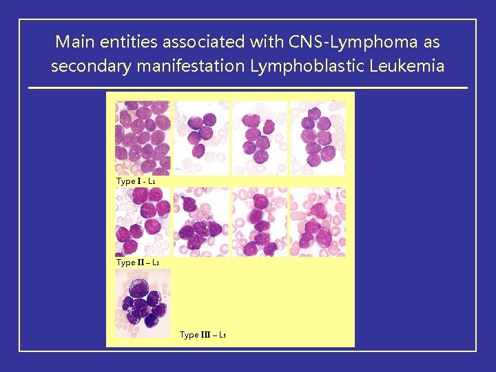
Main entities associated with CNS-Lymphoma as secondary manifestation Lymphoblastic Leukemia Type I - L 1 Type II – L 2 Type III – L 3
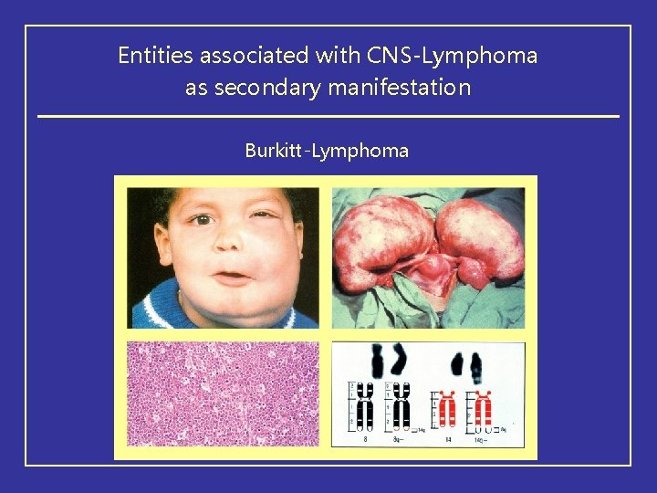
Entities associated with CNS-Lymphoma as secondary manifestation Burkitt-Lymphoma
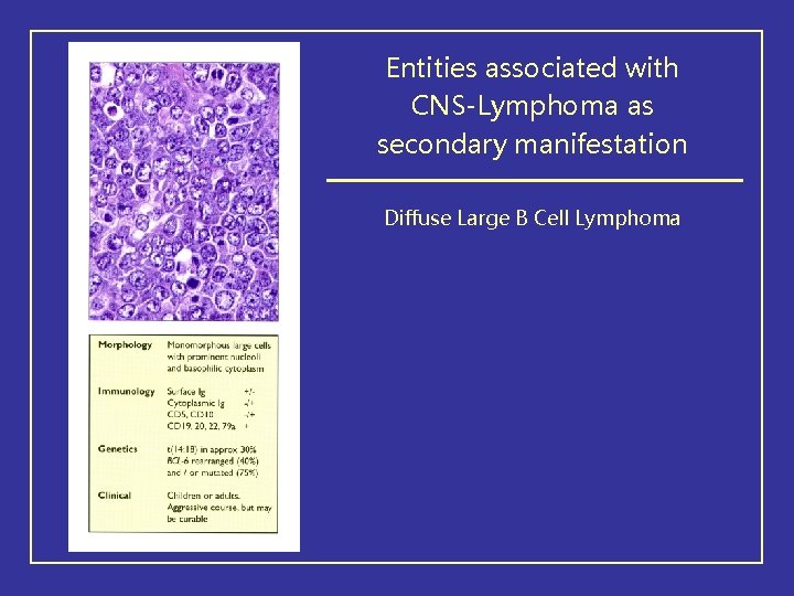
Entities associated with CNS-Lymphoma as secondary manifestation Diffuse Large B Cell Lymphoma
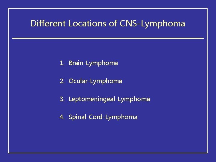
Different Locations of CNS-Lymphoma 1. Brain-Lymphoma 2. Ocular-Lymphoma 3. Leptomeningeal-Lymphoma 4. Spinal-Cord-Lymphoma
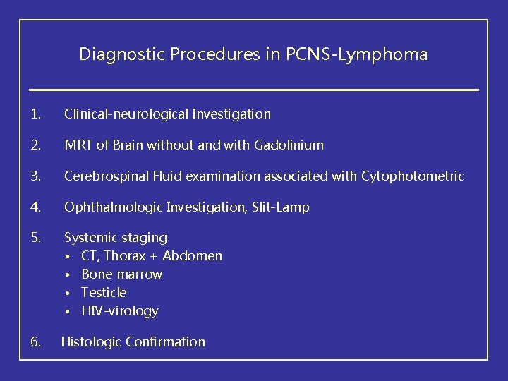
Diagnostic Procedures in PCNS-Lymphoma 1. Clinical-neurological Investigation 2. MRT of Brain without and with Gadolinium 3. Cerebrospinal Fluid examination associated with Cytophotometric 4. Ophthalmologic Investigation, Slit-Lamp 5. Systemic staging • CT, Thorax + Abdomen • Bone marrow • Testicle • HIV-virology 6. Histologic Confirmation
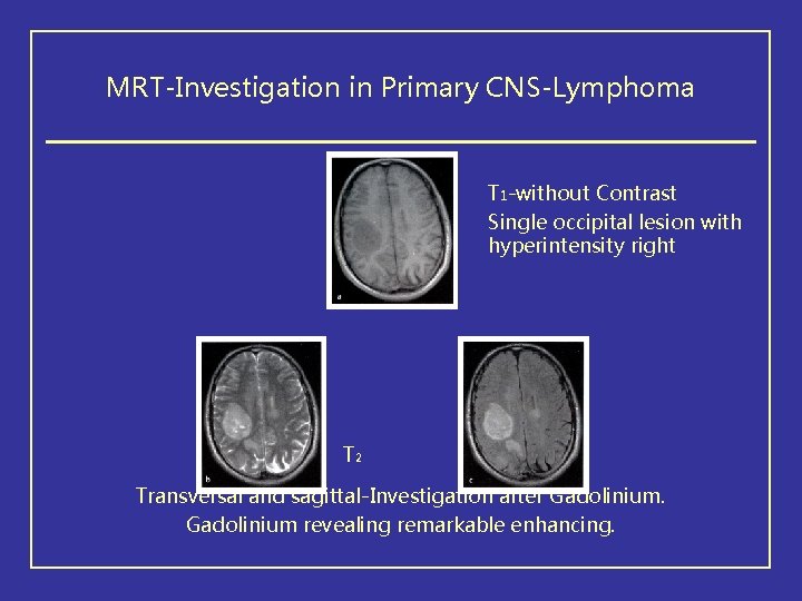
MRT-Investigation in Primary CNS-Lymphoma T 1 -without Contrast Single occipital lesion with hyperintensity right T 2 FLAIR Transversal and sagittal-Investigation after Gadolinium revealing remarkable enhancing.
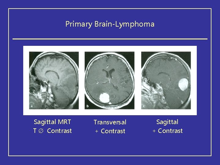
Primary Brain-Lymphoma Sagittal MRT T Contrast Transversal + Contrast Sagittal + Contrast
MRT in Primary CNS-Lymphoma a – c: T 2 -MRT-Investigation revealing multiple lesions within both hemisphere e: FLAIR-Sequence revealing remarkable enhancement f – g: Remarkable decreased enhancement after Administration of Gadolinium
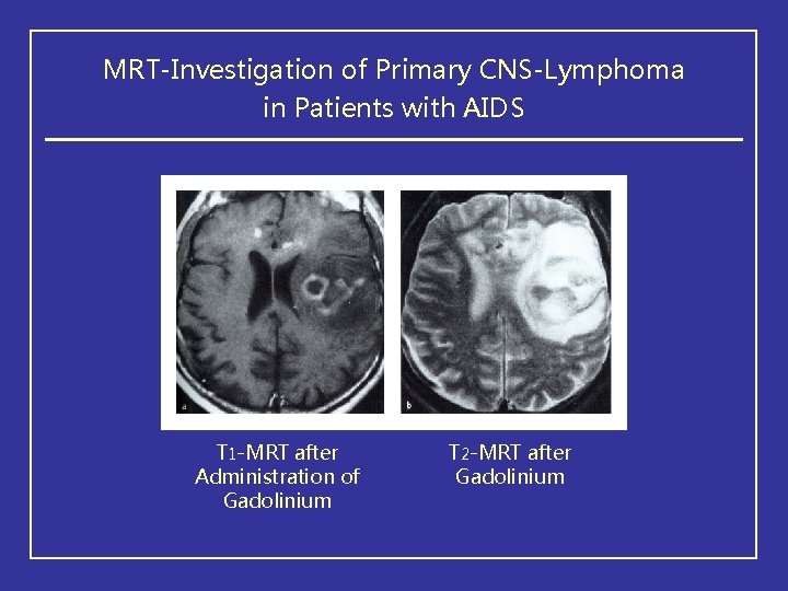
MRT-Investigation of Primary CNS-Lymphoma in Patients with AIDS T 1 -MRT after Administration of Gadolinium T 2 -MRT after Gadolinium
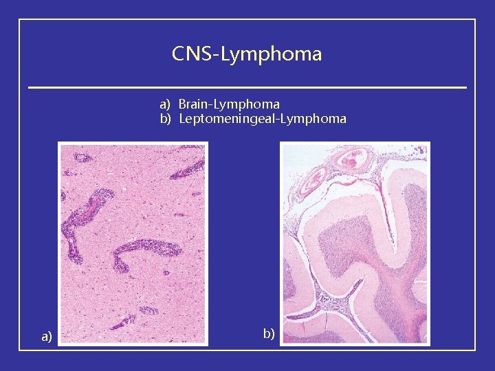
CNS-Lymphoma a) Brain-Lymphoma b) Leptomeningeal-Lymphoma a) b)
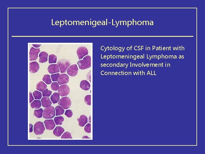
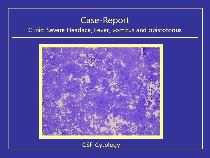
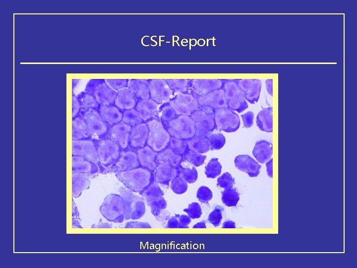
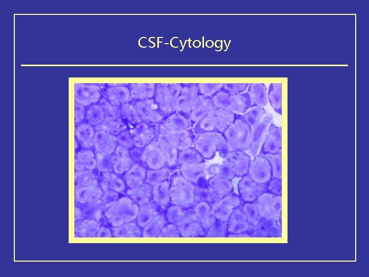
Leptomenigeal-Lymphoma Cytology of CSF in Patient with Leptomeningeal Lymphoma as secondary Involvement in Connection with ALL
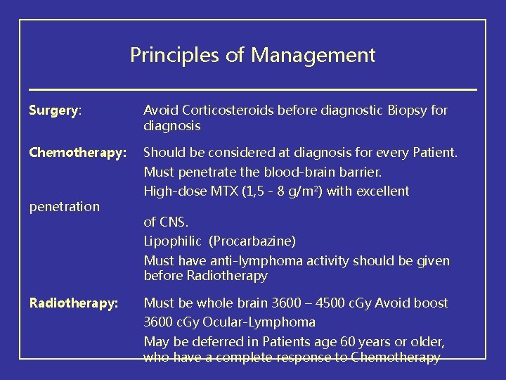
Principles of Management Surgery: Avoid Corticosteroids before diagnostic Biopsy for diagnosis Chemotherapy: Should be considered at diagnosis for every Patient. Must penetrate the blood-brain barrier. penetration High-dose MTX (1, 5 - 8 g/m²) with excellent of CNS. Lipophilic (Procarbazine) Must have anti-lymphoma activity should be given before Radiotherapy: Must be whole brain 3600 – 4500 c. Gy Avoid boost 3600 c. Gy Ocular-Lymphoma May be deferred in Patients age 60 years or older, who have a complete response to Chemotherapy
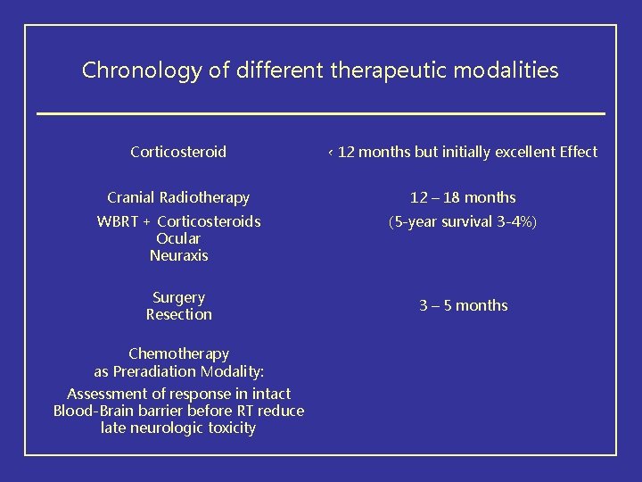
Chronology of different therapeutic modalities Corticosteroid ‹ 12 months but initially excellent Effect Cranial Radiotherapy 12 – 18 months WBRT + Corticosteroids Ocular Neuraxis (5 -year survival 3 -4%) Surgery Resection Chemotherapy as Preradiation Modality: Assessment of response in intact Blood-Brain barrier before RT reduce late neurologic toxicity 3 – 5 months
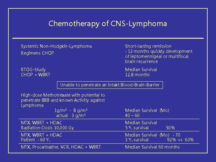
Chemotherapy of CNS-Lymphoma Systemic Non-Hodgkin-Lymphoma Regimens CHOP Short-lasting remission ‹ 12 months quickly development of leptomennigeal or multifocal brain recurrence RTOG-Study CHOP + WBRT Median Survival 12, 8 months Unable to penetrate an intakt Blood-Brain-Barrier High-dose Methotrexate with potential to penetrate BBB and known Activity against Lymphoma 1 g/m² - 8 g/m² actual 3 g/m² Median Survival (Mo) 40 – 60 MTX, WBRT + HDAC Radiation-Dosis 30. 000 Gy Median Survival 5 Y. survival MTX, WBRT + HDAC Patient ‹ 60 Y. Median Survival (Mo) › 70 3 Y. survival 92% vs 60% MTX, Procarbazine, VCR, HDAC + WBRT Median Survival 60 months 50%
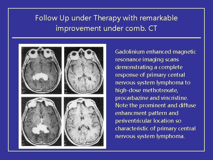
Follow Up under Therapy with remarkable improvement under comb. CT Gadolinium enhanced magnetic resonance imaging scans demonstrating a complete response of primary central nervous system lymphoma to high-dose methotrexate, procarbazine and vincristine. Note the prominent and diffuse enhancment pattern and periventricular location so characteristic of primary central nervous system lymphoma.
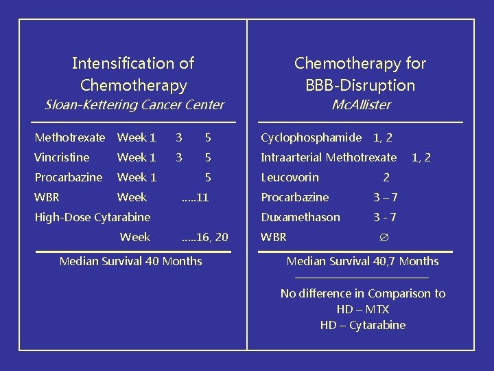
Intensification of Chemotherapy for BBB-Disruption Sloan-Kettering Cancer Center Mc. Allister Methotrexate Week 1 3 5 Cyclophosphamide Vincristine Week 1 3 5 Intraarterial Methotrexate Procarbazine Week 1 5 Leucovorin WBR Week …. . 11 High-Dose Cytarabine Week …. . 16, 20 Median Survival 40 Months 1, 2 2 Procarbazine 3– 7 Duxamethason 3 -7 WBR 1, 2 Median Survival 40, 7 Months No difference in Comparison to HD – MTX HD – Cytarabine
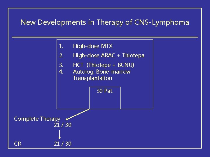
New Developments in Therapy of CNS-Lymphoma 1. High-dose MTX 2. High-dose ARAC + Thiotepa 3. 4. HCT (Thiotepe + BCNU) Autolog. Bone-marrow Transplantation 30 Pat. Complete Therapy 21 / 30 CR 21 / 30
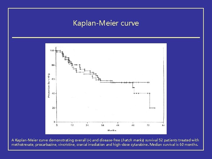
Kaplan-Meier curve A Kaplan-Meier curve demonstrating overall ( • ) and disease-free (hatch marks) survival 52 patients treated with methotrexate, procarbazine, vincristine, cranial irradiation and high-dose cytarabine. Median survival is 60 months.
Recurrent Disease of PCNS • No established second line Therapy • WBRT If Patients did not receive as part of initial Therapy • Ocular Radiotherapy in Patient with ocular Relapse (Bilateral) • HD MTX In Patient with long disease-free interval HD Cytarabine Tenrozolomide High-dose with Thiotepa, Busulfan and Cyclophosphamide followed by autologous Stem-Cell-Rescue - 3 year Survival • Leptomeningeal Relapse: Intrathecal or intraventricular MTX + Radiotherapy
Therapy of PCNSL in Immunocompromised Patients 1. Systemic Therapy more toxic in immunodeficient Patients 2. EBV-DNA-Identification in CSF by PCR 3. AIDS-related PCNSL occurs in Patients with CD 4 25 x 106 cell/L 4. Most important Component of PCNSL-Therapy in immunodeficient Patient: Reduction of Immunosupressiva, HAART 5. HAART + Ganciclovir 6. HAART + Corticosteroid + cranial Radiotherapy 7. HD MTX 3 g/m² 8. Monitoring of CSF-EBV-Level
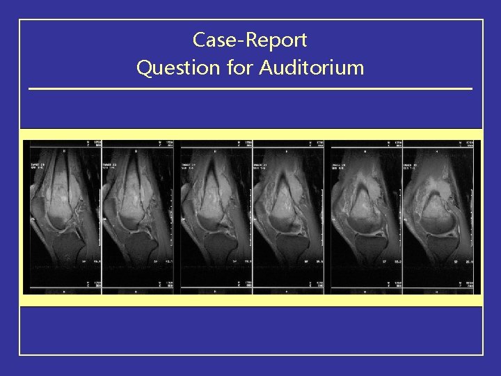
Case-Report Question for Auditorium
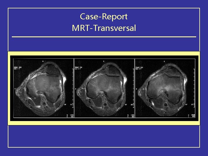
Case-Report MRT-Transversal
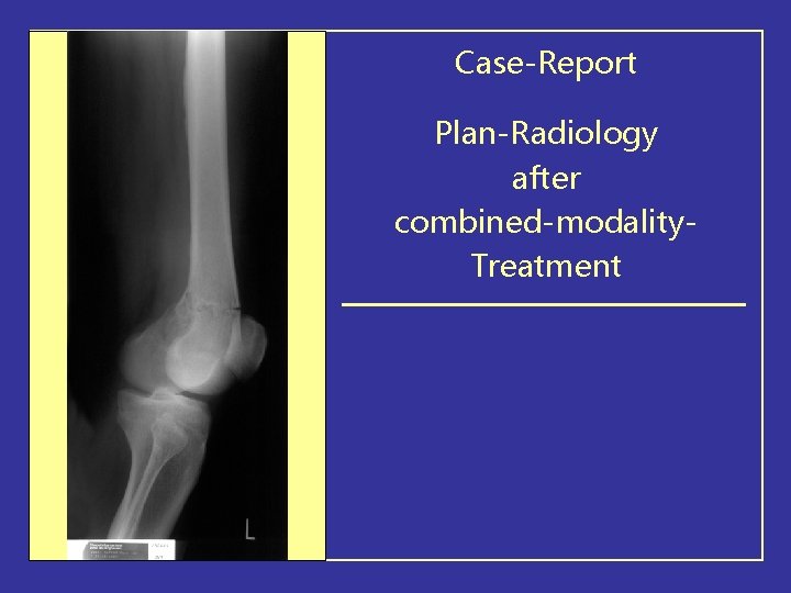
Case-Report Plan-Radiology after combined-modality. Treatment
Case-Report Clinic: Severe Headace, Fever, vomitus and opistotonus CSF-Cytology
CSF-Report Magnification
CSF-Cytology
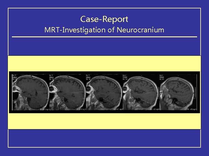
Case-Report MRT-Investigation of Neurocranium
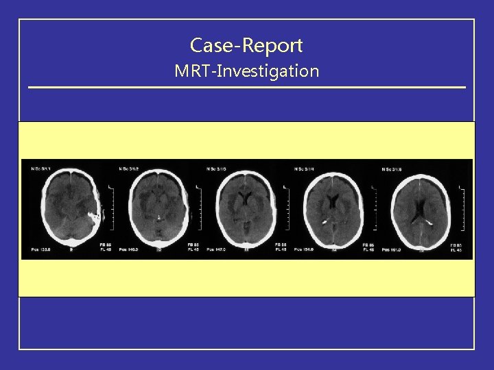
Case-Report MRT-Investigation
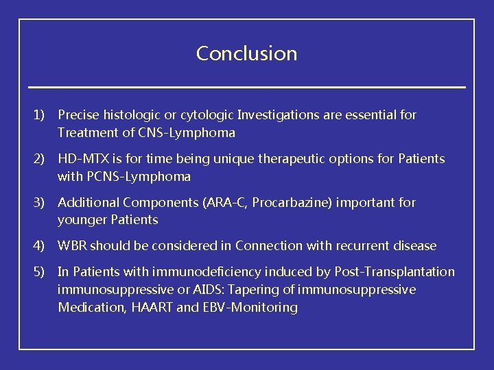
Conclusion 1) Precise histologic or cytologic Investigations are essential for Treatment of CNS-Lymphoma 2) HD-MTX is for time being unique therapeutic options for Patients with PCNS-Lymphoma 3) Additional Components (ARA-C, Procarbazine) important for younger Patients 4) WBR should be considered in Connection with recurrent disease 5) In Patients with immunodeficiency induced by Post-Transplantation immunosuppressive or AIDS: Tapering of immunosuppressive Medication, HAART and EBV-Monitoring