Membrane Dynamics Cell membrane structures and functions Membranes

Membrane Dynamics Cell membrane structures and functions – – – Membranes form fluid body compartments Membranes as barriers and gatekeepers How products move across membranes • i. e. , methods of transport – – – Distribution of water and solutes in cells & the body Chemical and electrical imbalances Membrane permeability and changes

The Cell

Plasma Membrane • Lipid bilayer and proteins in constantly changing fluid mosaic • Plays dynamic role in cellular activity • Separates intracellular fluid (ICF) from extracellular fluid (ECF) – Interstitial fluid (IF) = ECF that surrounds cells © 2013 Pearson Education, Inc.

The Cell Membrane • Fluid Mosaic Model – – Phospholipids Integral Proteins Peripheral Proteins Glycocalyx • Glycoproteins – MHC • Glycolipids – Cholesterol

Figure 3. 16 G proteins act as middlemen or relays between extracellular first messengers and intracellular second messengers that cause responses within the cell. Slide 2 Ligand (1 st Receptor G protein Enzyme messenger) 2 nd messenger 1 Ligand* (1 st messenger) binds to the receptor. The receptor changes shape and activates. Extracellular fluid Ligand Receptor * Ligands include hormones and neurotransmitters. © 2013 Pearson Education, Inc. Intracellular fluid

Figure 3. 16 G proteins act as middlemen or relays between extracellular first messengers and intracellular second messengers that cause responses within the cell. Slide 3 Ligand (1 st Receptor G protein Enzyme messenger) 1 Ligand* (1 st messenger) binds to the receptor. The receptor changes shape and activates. Ligand 2 The activated receptor binds to a G protein and activates it. The G protein changes shape (turns “on”), causing it to release GDP and bind GTP (an energy source). 2 nd messenger Extracellular fluid Receptor G protein GDP * Ligands include hormones and neurotransmitters. © 2013 Pearson Education, Inc. Intracellular fluid

Figure 3. 16 G proteins act as middlemen or relays between extracellular first messengers and intracellular second messengers that cause responses within the cell. Slide 4 Ligand (1 st Receptor G protein Enzyme messenger) 1 Ligand* (1 st messenger) binds to the receptor. The receptor changes shape and activates. 2 The activated receptor binds to a G protein and activates it. The G protein changes shape (turns “on”), causing it to release GDP and bind GTP (an energy source). 2 nd messenger 3 Activated G protein activates (or inactivates) an effector protein by causing its shape to change. Extracellular fluid Effector protein (e. g. , an enzyme) Ligand Receptor G protein GDP * Ligands include hormones and neurotransmitters. © 2013 Pearson Education, Inc. Intracellular fluid

Figure 3. 16 G proteins act as middlemen or relays between extracellular first messengers and intracellular second messengers that cause responses within the cell. Slide 5 Ligand (1 st Receptor G protein Enzyme messenger) 1 Ligand* (1 st messenger) binds to the receptor. The receptor changes shape and activates. 2 The activated receptor binds to a G protein and activates it. The G protein changes shape (turns “on”), causing it to release GDP and bind GTP (an energy source). 2 nd messenger 3 Activated G protein activates (or inactivates) an effector protein by causing its shape to change. Extracellular fluid Effector protein (e. g. , an enzyme) Ligand Receptor G protein GDP * Ligands include hormones and neurotransmitters. © 2013 Pearson Education, Inc. Inactive 2 nd messenger Active 2 nd messenger 4 Activated effector enzymes catalyze reactions that produce 2 nd messengers in the cell. (Common 2 nd messengers include cyclic AMP and Ca 2+. ) Intracellular fluid

Figure 3. 16 G proteins act as middlemen or relays between extracellular first messengers and intracellular second messengers that cause responses within the cell. Slide 6 Ligand (1 st Receptor G protein Enzyme messenger) 1 Ligand* (1 st messenger) binds to the receptor. The receptor changes shape and activates. 2 The activated receptor binds to a G protein and activates it. The G protein changes shape (turns “on”), causing it to release GDP and bind GTP (an energy source). 2 nd messenger 3 Activated G protein activates (or inactivates) an effector protein by causing its shape to change. Extracellular fluid Effector protein (e. g. , an enzyme) Ligand Receptor G protein GDP Inactive 2 nd messenger Activated kinase enzymes * Ligands include hormones and neurotransmitters. © 2013 Pearson Education, Inc. 4 Activated effector enzymes catalyze reactions that produce 2 nd messengers in the cell. (Common 2 nd messengers include cyclic AMP and Ca 2+. ) 5 Second messengers activate other enzymes or ion channels. Cyclic AMP typically activates protein kinase enzymes. Intracellular fluid

Figure 3. 16 G proteins act as middlemen or relays between extracellular first messengers and intracellular second messengers that cause responses within the cell. Slide 7 Ligand (1 st Receptor G protein Enzyme messenger) 1 Ligand* (1 st messenger) binds to the receptor. The receptor changes shape and activates. 2 The activated receptor binds to a G protein and activates it. The G protein changes shape (turns “on”), causing it to release GDP and bind GTP (an energy source). 2 nd messenger 3 Activated G protein activates (or inactivates) an effector protein by causing its shape to change. Extracellular fluid Effector protein (e. g. , an enzyme) Ligand Receptor G protein GDP Inactive 2 nd messenger Activated kinase enzymes * Ligands include hormones and neurotransmitters. © 2013 Pearson Education, Inc. 4 Activated effector enzymes catalyze reactions that produce 2 nd messengers in the cell. (Common 2 nd messengers include cyclic AMP and Ca 2+. ) 5 Second messengers activate other enzymes or ion channels. Cyclic AMP typically activates protein kinase enzymes. 6 Kinase enzymes activate other enzymes. Kinase enzymes transfer phosphate groups from ATP to specific proteins and activate a Cascade of cellular responses series of other enzymes that trigger (The amplification effect is various metabolic and structural tremendous. Each enzyme changes in the cell. catalyzes hundreds of reactions. ) Intracellular fluid

The Glycocalyx • "Sugar covering" at cell surface – Lipids and proteins with attached carbohydrates (sugar groups) • Every cell type has different pattern of sugars – Specific biological markers for cell to cell recognition – Allows immune system to recognize "self" and "non self" © 2013 Pearson Education, Inc. cells change it continuously – Cancerous

Thickness ~ 8 nm Cell Membrane Structure: Fluid Mosaic Model PLs Cholesterol Proteins: peripheral (associated) or integral

http: //www. youtube. com/watch ? v=Qqsf_UJcf. Bc

Passive Transport = Diffusion 1. Simple diffusion 2. Osmosis 3. Facilitated diffusion (= mediated transport) Active Transport Always protein-mediated Primary Secondary Receptor mediated transport

Movement across Membrane permeability varies for different molecules & cell types Two movement categories: • Passive and • Active depends on? ?

Passive Processes: Diffusion • Collisions cause molecules to move down or with their concentration gradient – Difference in concentration between two areas • Speed influenced by molecule size and temperature © 2013 Pearson Education, Inc.

Passive Processes • Molecule will passively diffuse through membrane if – It is lipid soluble, or – Small enough to pass through membrane channels, or – Assisted by carrier molecule PLAY Animation: Membrane Permeability © 2013 Pearson Education, Inc.
 substances diffuse directly through phospholipid bilayer")
Passive Processes: Simple Diffusion • Nonpolar lipid-soluble (hydrophobic) substances diffuse directly through phospholipid bilayer – E. g. , oxygen, carbon dioxide, fat-soluble vitamins PLAY Animation: Diffusion © 2013 Pearson Education, Inc.

Simple Diffusion

http: //www. youtube. com/watch ? v=s 0 p 1 ztrb. XPY&feature=rela ted

Facilitated Diffusion • Some molecules are too polar or too large to pass through the lipid bilayer. • Carrier proteins change shape after the molecules bind then envelopes the molecule and releases it • The binding site is moved from one side of the membrane to the other by a change in the confirmation of the carrier protein.

Carrier-Mediated Facilitated Diffusion • Transmembrane integral proteins are carriers • Transport specific polar molecules (e. g. , sugars and amino acids) too large for channels • Binding of substrate causes shape change in carrier then passage across membrane • Pearson Limited by Inc. number of carriers present © 2013 Education,

http: //highered. mcgrawhill. com/sites/0072495855/stu dent_view 0/chapter 2/animatio n__how_facilitated_diffusion_ works. html

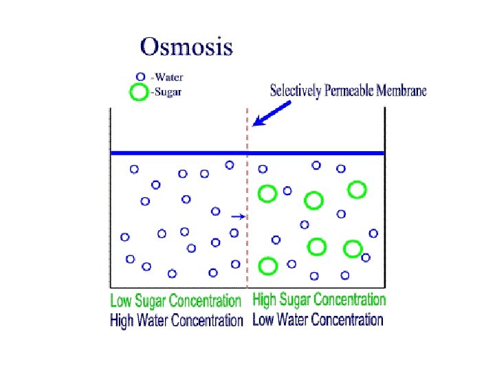
Passive Processes: Osmosis • Water concentration varies with number of solute particles because solute particles displace water molecules • Osmolarity - Measure of total concentration of solute particles • Water moves by osmosis until hydrostatic pressure (back pressure of water on membrane) and osmotic pressure (tendency of water to move into cell by osmosis) equalize © 2013 Pearson Education, Inc.

Tonicity • Tonicity: Ability of solution to alter cell's water volume – Isotonic: Solution with same nonpenetrating solute concentration as cytosol – Hypertonic: Solution with higher nonpenetrating solute concentration than cytosol – Hypotonic: Solution with lower nonpenetrating solute concentration than cytosol © 2013 Pearson Education, Inc.

Tonicity • Physiological term describing how cell volume changes if cell placed in the solution • Always comparative. Has no units. – Isotonic sol’n = No change in cell – Hypertonic sol’n = cell shrinks – Hypotonic = cell expands • Tonicity = Osmolarity • Number of particles in solution

Figure 3. 9 The effect of solutions of varying tonicities on living red blood cells. Hypertonic solutions Isotonic solutions Cells retain their normal size and shape in isotonic solutions (same solute/water concentration as inside cells; water moves in and out). Cells lose water by osmosis and shrink in a hypertonic solution (contains a higher concentration of solutes than are present inside the cells). © 2013 Pearson Education, Inc. Hypotonic solutions Cells take on water by osmosis until they become bloated and burst (lyse) in a hypotonic solution (contains a lower concentration of solutes than are present inside cells).

Membrane Transport: Active Processes • Two types of active processes – Active transport – Vesicular transport • Both require ATP to move solutes across a living plasma membrane because – Solute too large for channels – Solute not lipid soluble – Solute not able to move down © 2013 Pearson Education, Inc.
 – Bind specifically and reversibly with")
Active Transport • Requires carrier proteins (solute pumps) – Bind specifically and reversibly with substance • Moves solutes against concentration gradient – Requires energy © 2013 Pearson Education, Inc.

Active Transport: Two Types • Primary active transport – Required energy directly from ATP hydrolysis • Secondary active transport – Required energy indirectly from ionic gradients created by primary active transport © 2013 Pearson Education, Inc.

Active Transport • Movement from low conc. to high conc. • ATP needed • Creates state of disequilibrium • 1 o (direct) active transport – ATPases or “pumps” – Uniport and Antiport 2 o (indirect) active transport – Symport and antiport

Figure 3. 10 Primary active transport is the process in which solutes are moved across cell membranes against electrochemical gradients using energy supplied directly by ATP. Extracellular fluid Na+–K+ pump ATP-binding site K+ Cytoplasm 1 Three cytoplasmic Na + bind to pump protein. © 2013 Pearson Education, Inc. Slide 2

Figure 3. 10 Primary active transport is the process in which solutes are moved across cell membranes against electrochemical gradients using energy supplied directly by ATP. Extracellular fluid Na+–K+ pump ATP-binding site K+ Na+ bound Cytoplasm 1 Three cytoplasmic Na + bind to pump protein. P 2 Na+ binding promotes hydrolysis of ATP. The energy released during this reaction phosphorylates the pump. © 2013 Pearson Education, Inc. Slide 3

Figure 3. 10 Primary active transport is the process in which solutes are moved across cell membranes against electrochemical gradients using energy supplied directly by ATP. Extracellular fluid Na+–K+ pump ATP-binding site K+ Na+ bound Cytoplasm 1 Three cytoplasmic Na + bind to pump protein. P 2 Na+ binding promotes hydrolysis of ATP. The energy released during this reaction phosphorylates the pump. Na+ released P 3 Phosphorylation causes the pump to change shape, expelling Na+ to the outside. © 2013 Pearson Education, Inc. Slide 4

Figure 3. 10 Primary active transport is the process in which solutes are moved across cell membranes against electrochemical gradients using energy supplied directly by ATP. Extracellular fluid Na+–K+ pump K+ ATP-binding site Na+ bound Cytoplasm 1 Three cytoplasmic Na + bind to pump protein. P 2 Na+ binding promotes hydrolysis of ATP. The energy released during this reaction phosphorylates the pump. Na+ released P K+ 3 Phosphorylation causes the pump to change shape, expelling Na+ to the outside. P 4 Two extracellular K + bind to pump. © 2013 Pearson Education, Inc. Slide 5

Figure 3. 10 Primary active transport is the process in which solutes are moved across cell membranes against electrochemical gradients using energy supplied directly by ATP. Extracellular fluid Na+–K+ pump K+ ATP-binding site Na+ bound Cytoplasm 1 Three cytoplasmic Na + bind to pump protein. P 2 Na+ binding promotes hydrolysis of ATP. The energy released during this reaction phosphorylates the pump. Na+ released K+ bound P Pi K+ 5 K+ binding triggers release of the phosphate. The dephosphorylated pump resumes its original conformation. 3 Phosphorylation causes the pump to change shape, expelling Na+ to the outside. P 4 Two extracellular K + bind to pump. © 2013 Pearson Education, Inc. Slide 6

Figure 3. 10 Primary active transport is the process in which solutes are moved across cell membranes against electrochemical gradients using energy supplied directly by ATP. Extracellular fluid Na+–K+ pump K+ ATP-binding site Na+ bound Cytoplasm 1 Three cytoplasmic Na + bind to pump protein. P K+ released 6 Pump protein binds ATP; releases K + to the inside, and Na + sites are ready to bind Na+ again. The cycle repeats. 2 Na+ binding promotes hydrolysis of ATP. The energy released during this reaction phosphorylates the pump. Na+ released K+ bound P Pi K+ 5 K+ binding triggers release of the phosphate. The dephosphorylated pump resumes its original conformation. 3 Phosphorylation causes the pump to change shape, expelling Na+ to the outside. P 4 Two extracellular K + bind to pump. © 2013 Pearson Education, Inc. Slide 7

Figure 3. 10 Primary active transport is the process in which solutes are moved across cell membranes against electrochemical gradients using energy supplied directly by ATP. Extracellular fluid Na+–K+ pump K+ ATP-binding site Na+ bound Cytoplasm 1 Three cytoplasmic Na + bind to pump protein. P K+ released 6 Pump protein binds ATP; releases K + to the inside, and Na + sites are ready to bind Na+ again. The cycle repeats. 2 Na+ binding promotes hydrolysis of ATP. The energy released during this reaction phosphorylates the pump. Na+ released K+ bound P Pi K+ 5 K+ binding triggers release of the phosphate. The dephosphorylated pump resumes its original conformation. 3 Phosphorylation causes the pump to change shape, expelling Na+ to the outside. P 4 Two extracellular K + bind to pump. PLAY A&P © 2013 Pearson Education, Inc. Flix™: Resting Membrane Potential

• http: //www. youtube. com/watch? v=9 CBo Bewd. S 3 U&feature=related

• http: //www. youtube. com/watch? v=STz. O i. Rqzz. L 4&NR=1

Secondary Active Transport • Depends on ion gradient created by primary active transport • Energy stored in ionic gradients used indirectly to drive transport of other solutes © 2013 Pearson Education, Inc.

Cotransport Symport • Molecules are carried in same direction • Examples: Glucose and Na+ Antiport • Molecules are carried in opposite direction • Examples: Na+/K+ pump

Figure 3. 11 Secondary active transport is driven by the concentration gradient created by primary active transport. Extracellular fluid Na+-K+ pump Cytoplasm 1 Primary active transport The ATP-driven Na+-K+ pump stores energy by creating a steep concentration gradient for Na+ entry into the cell. © 2013 Pearson Education, Inc. Slide 2

Figure 3. 11 Secondary active transport is driven by the concentration gradient created by primary active transport. Extracellular fluid Slide 3 Glucose Na+-K+ pump Na+-glucose symport transporter loads glucose from extracellular fluid Na+-glucose symport transporter releases glucose into the cytoplasm Cytoplasm 1 Primary active transport The ATP-driven Na+-K+ pump stores energy by creating a steep concentration gradient for Na+ entry into the cell. © 2013 Pearson Education, Inc. 2 Secondary active transport As Na+ diffuses back across the membrane through a membrane cotransporter protein, it drives glucose against its concentration gradient into the cell.

Vesicular Transport • Transport of large particles, macromolecules, and fluids across membrane in membranous sacs called vesicles • Requires cellular energy (e. g. , ATP) © 2013 Pearson Education, Inc.

Vesicular Transport • Functions: – Endocytosis—transport into cell • Phagocytosis, pinocytosis, receptormediated endocytosis – Exocytosis—transport out of cell © 2013 Pearson Education, Inc.
 Macrophage")
Vesicular Transport Movement of macromolecules across cell membrane: 1. Phagocytosis (specialized cells only) Macrophage or Phagocytes 2. Pinocytosis “Cell drinking” 3. Receptor mediated endocytosis Down Regulation 4. Exocytosis

Vesicular Transport

Endocytosis • Phagocytosis – Pseudopods engulf solids and bring them into cell's interior – Form vesicle called phagosome • Used by macrophages and some white blood cells – Move by amoeboid motion • Cytoplasm flows into temporary extensions • Allows creeping © 2013 Pearson Education, Inc.

• http: //www. youtube. com/watch? v=Z_m XDv. ZQ 6 d. U

Endocytosis • Nonselective: • Pinocytosis for fluids & dissolved substances • Selective: • Receptor Mediated Endocytosis via clathrin- coated pits - Example: LDL cholesterol and Familial Hypercholesterolemia
 – Plasma membrane infolds, bringing extracellular fluid and dissolved")
Endocytosis • Pinocytosis (fluid-phase endocytosis) – Plasma membrane infolds, bringing extracellular fluid and dissolved solutes inside cell • Fuses with endosome – Most cells utilize to "sample" environment – Nutrient absorption in the small intestine – Membrane components recycled back to membrane © 2013 Pearson Education, Inc.

Pinocytosis

Endocytosis • Receptor-mediated endocytosis – Allows specific endocytosis and transcytosis • Cells use to concentrate materials in limited supply – Clathrin-coated pits provide main route for endocytosis and transcytosis • Uptake of enzymes, low-density lipoproteins, iron, insulin, and, unfortunately, viruses, diphtheria, and cholera toxins © 2013 Pearson Education, Inc.

Receptor Mediated Endocytosis • No. 1 uptake method in most cells • Receptors and substance is internalized into a coated pit-clathrin • Down Regulation

http: //www. youtube. com/watch ? v=Ki. LJl 3 Nwmp. U • http: //www. youtube. com/watch? v=4 g. Ltk 8 Yc 1 Zc&feature=related

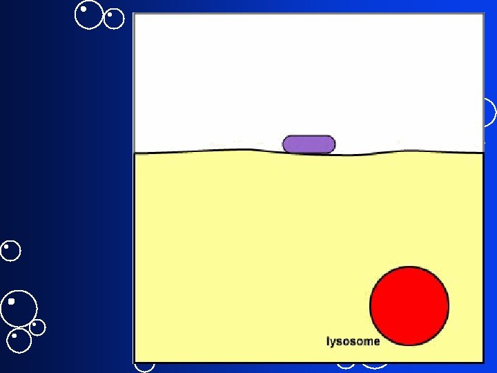
Figure 3. 14 b Exocytosis. Photomicrograph of a secretory vesicle releasing its contents by exocytosis (100, 000 x) © 2013 Pearson Education, Inc.

- Slides: 60