ERUPTIVE DISEASES PROF DR AYA VTRNEL MEASLES Measles


Incubation period: 10 - 12")

 Final stage: Rash starts as faint macules on the upper parts")


")


 ¨")


 in")

 ¨ Parvovirus B 19 ( discovered in 1975) member of")


 ¨ Human Herpes Virus (HHV) type 6 was discovered in")


 exotoxins")


 ¨ Primary infection of VARICELLA ZOSTER VIRUS (VZV). HUMAN HERPES VIRIDAE ¨")


 ¨ Virus replicated")


")


- Slides: 78
ERUPTIVE DISEASES PROF. DR. AYÇA VİTRİNEL
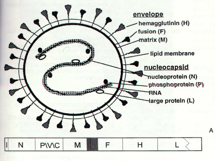
MEASLES ¨ Measles virus is a RNA virus of the genus ; MORBILLIVIRUS in the family PARAMYXOVIRIDAE ¨ During the prodromal period , short time after the rash appears=» found in nasopharenx, blood, urine ¨ Endemic throughout the world ¨ Maximal dissemination; Occurs by droplet spray during the prodromal period
MEASLES ¨ Contagious from 1 -2 days before onset of symptoms to 4 days after apperance of the rash. ¨ Infants acquire immunity transplacentally from mothers who have had measles or measles immunization. This immunity is usually complete for the first 4 -6 months of life.
Koplik spots
MEASLES ¨ Essential lesion is found in the skin, in the mucous membranes of the nasopharynx, bronchi, intestinal tract, conjunctivae ¨ Serous exudate and proliferation of mononuclear and a few polymorphonuclear cells occur around the capillaries. ¨ Hyperplasia of lymphoid tissue usually occurs particulary in the appendix.
MEASLES ¨ CLINICAL MANIFESTATIONS : 3 clinical stages ¨ 1)Incubation period: 10 - 12 days ¨ 2)Prodromal stage: 3 -5 days(av: 4 days) fever; subside rapidly within 2 days of rash. Dry cough. Coryza. Conjuctivitis. Koplik spots, patognomonic sign of measles
MEASLES ¨ Consist of serous exudate, proliferation of endothelial cells, grayish white dots have slight reddish areola opposite to the molars (rarely found within the mid partion of lowerlip, on the palate and on the lacrimal caruncle.
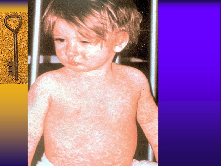
MEASLES ¨ 3) Final stage: Rash starts as faint macules on the upper parts of the neck, behind the ears, along the hairline , become maculopapular as the rash spreads rapidly over the entire face, neck, upper arms, upper part of the chest in the first 24 hours. ¨ Second day; spreads over the back, abdomen, arm and thigh It finally reaches the feet on the 2 nd. -3 rd. days. It begins to fade on the face.
MEASLES ¨ In the severe cases the rash is confluent, the skin is completely covered including the palms and soles. ¨ Often slightly hemorrhagic ¨ As the rash fades BROWNY DESQUAMATION and BROWNISH DISCOLORATION OCCUR. ¨ Hemorrahagic type: BLACK MEASLES bleeding may occur (mouth, nose, bowel) ¨ Atypical measles : occurs in recipient of killed measles virus vaccine
MEASLES ¨ Rash appears firstly ; palms, wrists, soles, ankles maculopapuler; vesicules, purpurical hemorrhagic. ¨ Koplic spots rarely occurs ¨ Headache, severe abdominal pain, vomiting myalgia, respiratory symptoms, pneumoniae can occur.
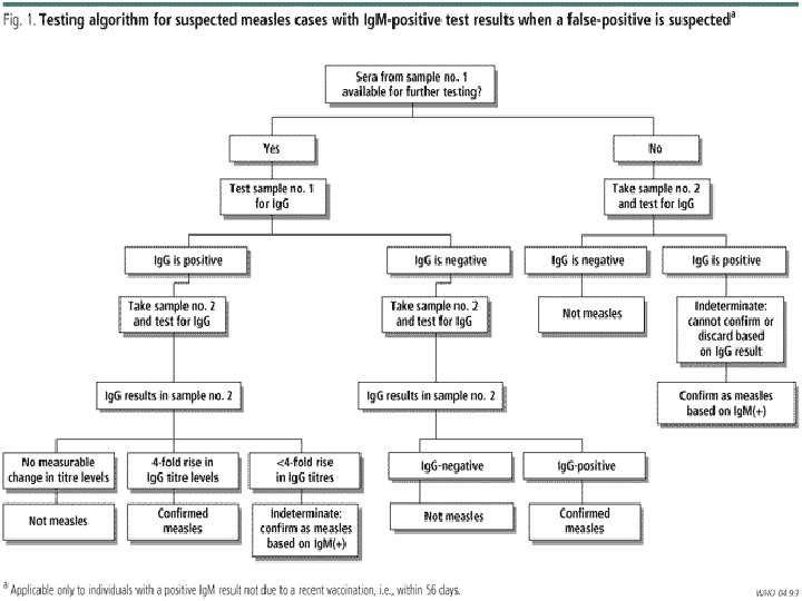
MEASLES ¨ DIAGNOSIS: Clinical picture. ¨ Laboratory confirmation: rarely needed. ¨ Prodrome: multinucleated giant cells can be demonstrated in smears of nasal mucosa. ¨ Abs: Ig. M detectable at least 1 month after rash onset. Acute-convelascent sera 4 fold increase ¨ Virus culture ¨ Leukopenia---» » Lekocytosis
MEASLES ¨ TREATMENT: No spesific antiviral thera- py. ¨ Supportive (antipyretics, bedrest, fluid intake) ¨ Treatment with oral Vit A reduces morbidity and mortality in children with severe measles in developing world. ¨ 6 mn-1 yr: 100. 000 U PO ¨ >1 yrs : 200. 000 U PO
MEASLES ¨ Hospitalized with measles and its complications ¨ Having risk factors: ¨ Evidence of vit A def ¨ Immundeficiency ¨ İmpaired intestinal absorbtion ¨ Severe malnutrition
MEASLES ¨ COMPLICATIONS: ¨ Acute otitis media ‘’most common’’ ¨ Pneumonitis ; interstisiel primer pneumonia/ secondary bacterial ¨ Encephalitis ¨ Exacarbation of undelying tbc-SSPE after 7/10 years ¨ Myocarditis-infrequate ¨ Appandicitis ¨ Laryngitis, tracheitis, bronchiolitis
MEASLES ¨ PREVENTION : Vaccine license in 1963 (12 -15 months/4 -6 years) ¨ Postexposure prophylaxia. Within 6 days of exposure, Ig (0. 25 ml /kg IM max 15 ml) within 72 hrs VACCINE
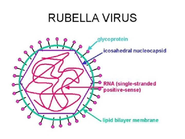
RUBELLA ¨ Rubella virus- RNA virus of genus Rubi- virus in the family Togaviridae. ¨ Distributed worldwide and affects both sexes equally ¨ Peak incidence; 5 -14 yr of age ¨ During clinical illness: the virus is present in nasopharyngeal secretions, blood, feces, urine
RUBELLA ¨ Virus has been recovered from the nasopha- rynx 7 days before the exanthem and 7 -8 days after its disappereance. ¨ The risk for congenital defects and disease is greatest with primary maternal infection during the first trimester.
RUBELLA ¨ CLINICAL MANIFESTATIONS : ¨ Incubation period: 14 -21 days, no prodro- mal phase or milder, 2/3 of infections are subclinical ¨ Congenital rubella syndrome ophtalmologic, cardiac, auditory, neurologic anomalies ¨ Most characteristical signs; retroauricular, postcervical and postoccipital LAP ; appears 24 hr before and 1 wk after rash.
RUBELLA ¨ Forsheimer spots : enantem appears in 20% of patients just before the onset of the rash on the soft palate ¨ Exanthem begins on the face spreads quickly. Discrete maculopapules. 2. day-pinpoint appereance. Mild itching. Pharengeal mucosa and the conjuctivae are slightly inflamed. Fever is low greade or absent. In older girls and women polyarthritis may occur
RUBELLA
Congenital RUBELLA
RUBELLA ¨ DIAGNOSIS : Clinical symptoms. Physical appearance. Serology : Ig. M (+) in the first day. Virus culture : nasopharynx, blood. ¨ TREATMENT: No spesific treatment, supportive. ¨ COMPLICATIONS: Encephalitis, trombocytopenia, rubella panencephalitis, arthritis
RUBELLA ¨ PREVENTION: Rubella vaccine ¨ Especially important for girls to have immunity to rubella before they reach childbearing age. ¨ The susceptable pregnant woman exposed to rubella for whom abortion is not an obtion Ig should be administered in a dose of 0. 55 ml/kg which reduces the attack rate but doesn’t eliminate the risk of fetal infection. ¨ Vaccine theoretically could prevent illness if administered within 3 days of exposure.
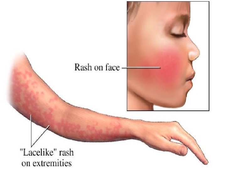
ERYTHEMA INFECTIOSUM (FIFTH DISEASE) ¨ Parvovirus B 19 ( discovered in 1975) member of Erythrovirus in the family Parvoviridae. ¨ DNA virus ¨ 5 -15 years ¨ Late winter and spring ¨ Transmission; respiratory route. ¨ Beningn selflimited exanthematous illness of childhood. ¨ Incubation period 4 -28 days (av : 16 -17 days)
Parvovirus B 19
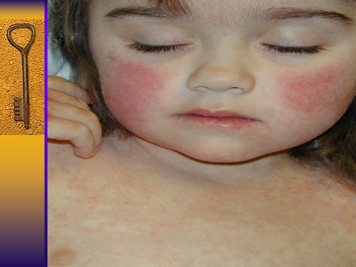
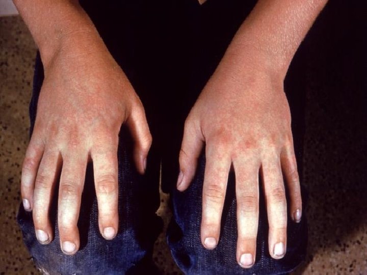
ERYTHEMA INFECTIOSUM ¨ Prodromal lesion phase ; mild, lowgrade fever , headache, URI signs. ¨ RASH: ¨ 1 st stage: erithematous facial flushing slapped ceak ¨ 2 nd stage: diffuse macular erthrema on the trunk and proximal extremities ¨ 3 rd stage: central clearing of macules (reticuler apperance). Resolves without desqumation. Prominent on extansor surface. Tends to wax over 1 -3 wks
ERYTHEMA INFECTIOSUM ¨ Recur with exposure to sunlight, heat, exercise, stress ¨ LAP, atypical papular, purpuric, vesicular rashes are described. ¨ DIAGNOSIS: clinical presentation ¨ Ig. M detection: 6 -8 wks ¨ Virus can’t be isolated by culture
ERYTHEMA INFECTIOSUM ¨ TREATMENT: No spesific antiviral thera- py. ¨ COMPLICATIONS: Arthralgies, arthritis, trompbocytopenic purpura, aseptic meningitis.
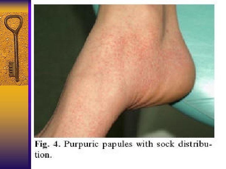
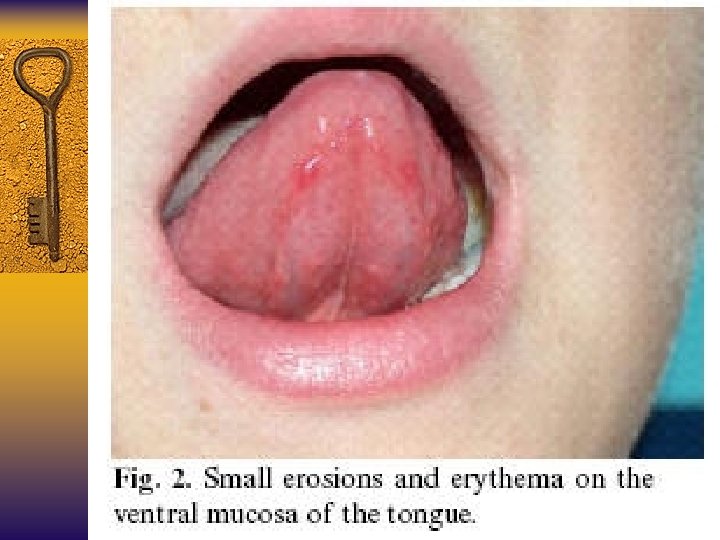
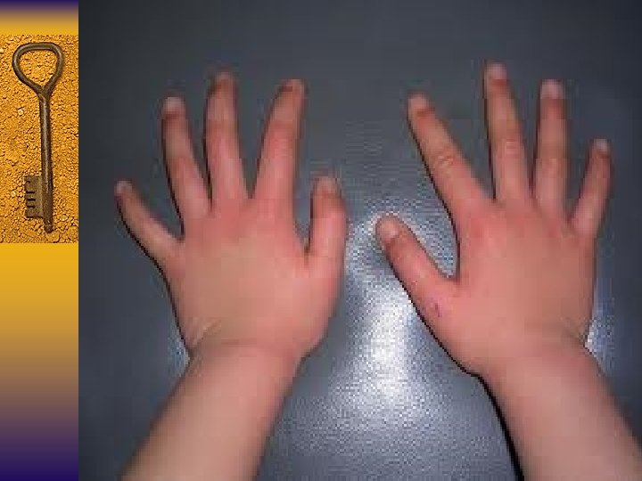
PAPULAR – PURPURIC GLOVES and SOCKS SYNDROME § Rare disease with a cutaneous involvement and with § § § § lesions in the oral cavity by parvovirus B 19 Affects children and young adults Appears in both sexes in spring and summer Edema and pruritus of hands and feet followed by a purpura at the same site Erythema and petechiae on the hard and soft palate Small erosions in the oral mucosa and tongue Commissural chelitis Nonspesific urethritis can appear
PAPULAR – PURPURIC GLOVES and SOCKS SYNDROME § Diagnosis is made by the clinical dermatological chracteristics and is confirmed by spesific serology for Parvovirus B 19 using ELISA or PCR § Supportive treatment
ROSEOLA INFANTUM (SIXTh DISEASE) ¨ Human Herpes Virus (HHV) type 6 was discovered in 1986 ( etiologic agent for most cases) ¨ HHV 7 was discovered 1990 in few cases ¨ HHV-6 and 7 belong to the ß- herpesvirus subfamily of herpesviruses ¨ Peak acquisition of primary HHV-6 infection 6 -15 months of age ( occur younger than 3 yr)
ROSEOLA INFANTUM
ROSEOLA INFANTUM ¨ Higher incidense during spring and fall months ¨ Incubation period, 5 -15 days (av: 10 days) ¨ Most adults excrete HHV-6 and 7 in saliva and may serve as primary sources for virus transmission to children ¨ Prodromal period is usually asymptomatic ; mild URI signs ¨ Mild cervical or less frequently occipital LAP may be noted ¨ Some chidren may have mild palpebral edema.
ROSEOLA INFANTUM ¨ Clinical illness is generaly heralded by high temparature. ¨ Some children may become irritable and anorexic. ¨ Seizures may occur in 15% of children. ¨ Rhinorrhea, sore throat, abdominal pain, vomiting and diarrhea ¨ Fever persists for 3 -7 days ; resolves abruptly
ROSEOLA INFANTUM
ROSEOLA INFANTUM ¨ A rash appears within 12 -24 hr of fever resolution. ¨ Rash. Rose colored, distinctive } on the trunk to neck, face, prox extremities ¨ 1 -3 days later the rash fades. ¨ The characteristic enanthem ( Nagayama spot) consist of the soft palate and the base of the uvula ¨ The enanthem may be present on the fourth day in 2/3 of patients with roseola
ROSEOLA INFANTUM ¨ DIAGNOSIS ¨ Spesific test for HHV 6 -7 ¨ Virus culture, PCR, Ag detection ¨ TREATMENT ¨ HHV-6 is inhibited by ganciclovir ( but not acyclovir. Foscarnet.
SCARLET FEVER ¨ Group A ß Hemolytic streptococcus is the cause Pyrogenic (erythrogenic) exotoxins (A, B, C) Responsible for the rash of scarlet fever. ¨ Infection may be spread by droplets, contact with skin lesions, transmitted by food, milk and water. ¨ Incubation period: 1 -7 days. (av: 3 days)
Group A ß Hemolytic streptococcus
SCARLET FEVER ¨ Onset in acute and is characterized by fever, vomiting, headache, toxicity, pharangitis and chills within 12 -48 hrs the typical rash appears. ¨ Temperature increases abruptly, may peak 39, 6 - 40 ºC on the 2 nd day and gradually returns to normal within 5 -7 days in untreated patients.
SCARLET FEVER ¨ The tonsils are hyperemic and edematous and may be covered with a gray white exudate. ¨ The tongue may be edematous and reddened. During the early days of illness the dorsum of the tongue has a white coat. WHITE STRAWBERRY TONGUE ¨ After several days the red tongue with prominant papillae. RED STRAWBERRY TONGUE
SCARLET FEVER ¨ THE PALATE AND UVULA MAY BE REDDENED AND COVERED WİTH PETECHIA. ¨ THE EXANTEM IS RED, PUNCTATIC OR FINELY PAPULAR. ¨ Appears initially in the axillers, groin and neck and generalized within 24 hrs. ¨ The area around the mouth is pale: CIRCUMORAL PALLOR ¨ Petechia may occur owing to capillary fragility
SCARLET FEVER
SCARLET FEVER ¨ Area of hyperpigmentation on the antecubi- tal fossae; PASTIA’S SIGN ¨ Small vesicular lesions in severe disease : MILIARY SUDAMINA ¨ Desquamation begins on the face over the trunk hands and feet ¨ Scarlet fever may follow infection of wounds, burns or skin infection
STRAWBERRY TONGUES
SCARLET FEVER ¨ DIAGNOSIS: ¨ Throat culture ¨ Current rapid Ag detection ¨ WBC ¨ ESR, CRP ¨ ASO, anti. DNA se B (+) (3 -6 wks)
SCARLET FEVER ¨ COMPLICATIONS : ¨ Sinusitis ¨ A. O. M. ¨ Mastoiditis ¨ Cervical adenitis ¨ Retropharingeal abscess ¨ Bronchopneumonia ¨ Menengitis ¨ Osteomyelitis ¨ Septic arthritis
SCARLET FEVER ¨ TREATMENT: Penicillin 10 days ¨ Single IM inj of a long active benzathine penicillin, erytromicin, clindamycin, first generation cephalosporins ¨ Successful treatment with shorter causes (5 days) of azithromycin or cefpodoxime has been reported.
VARICELLA (CHICKENPOX) ¨ Primary infection of VARICELLA ZOSTER VIRUS (VZV). HUMAN HERPES VIRIDAE ¨ Lifelong latent infection of sensory ganglion neurons reactivation of the latent infection HERPES ZOSTER (SHINGLES) ¨ Mild illness in childhood ¨ Occurs in winter-spring ¨ Within hauseholds of with a case of varicella transmission of VZV to susceptible individuals occurs at a rate of 80 -90%
VARICELLA VIRUS
VARICELLA ¨ Contagious from 24 -48 hr before the rash appears and until the vesicles are crusted (37 days after onset rash) ¨ Susceptible children may also acquire varicella after close direct contact with adults who have HZ. ¨ Transmitted respiratory secretions, fluid of skin lesions by airborne spread/through direct contact
VARICELLA ¨ Incubation period: 10 -21 days ( mean 15 days) ¨ Virus replicated in the resp tract primary (subclinical) viremia, widespread cutenous lesions occur during second viremia ¨ Prodromal symptom: fever, malasia, anorexia, headache, abdominal pain (24 -48 hr before the rash)
VARICELLA ¨ Varicella lesions appears on the scalp, face or trunk ¨ Prurutic erythamatous macules clear fluid vesicles (24 -48 hr) clouding and umblication. ¨ While initial lesions are crusting new crops form on the trunk and then the extremites.
VARICELLA ¨ Ulcerative lesions oropharenx and vagi- na, eyelids and conjuctivae ¨ Average number of varicella lesions 30 (10 -1500) ¨ VARYING STAGES OF DEVELOPMENT (MACULE, PAPULE, VESICLE). ¨ PRESENT AT THE SAME TIME.
VARICELLA
VARICELLA ¨ Progresivve varicella: visceral organ invol- vement, coagulopathy, severe hemorhage, . Highest in children with congenital cellular immune deficiency. ¨ High dose CST, HIV, malignancy etc. . .
VARICELLA ¨ DIAGNOSIS : Laboratory evaluation is not necessary ¨ Leukopenia (first 72 hr) lymphositosis ¨ Liver function tests are usually elevated ¨ Tissue culture method ( virus isolation) (710 days) ¨ VZV Ig. G Ab tests ; determine the immune status of individuals whose clinical history of varicella, is unknown.
VARICELLA ¨ TREATMENT : Antiviral treatment modifies the course: ACYCLOVIR : is not recommended routinely for uncomplicated cases ¨ Oral therapy: 20 mg/kg/dose (max: 800 mg) x 4 dose – 5 days Children older than 12 yrs, receiving aerosol steroids, having chronic cutaneous disease ¨ IV therapy: severe disease and immunocomprimised patients: 500 mg/m²/ every 8 hrs – 7 days
VARICELLA ¨ COMPLICATIONS : Mild varicella hepatitis ¨ Mild trombocytopenia ¨ Purpura, hemorrhagic vesicles, hematuria, GI bleeding ‘’rare’’ ¨ Nephritis, NS, HUS, arthritis, myocarditis, pericarditis, pancreatitis, orchitis ¨ Secondary bacterial infections (AGBHS, S. aureus) 5% ¨ Encephalitis and cerebellar ataxia; 5 yr , 20 yr 26 days after onset ¨ Pneumonia: very rare in children
VARICELLA ¨ PREVENTION : Vaccine : postexposure within 3 days. ¨ VZIG: postexposure prophylaxis for immunocomprimised children. Pregnant women. Newborn exposed to maternal varicella, whose mothers develop varicella , 5 days before to 2 days after delivery
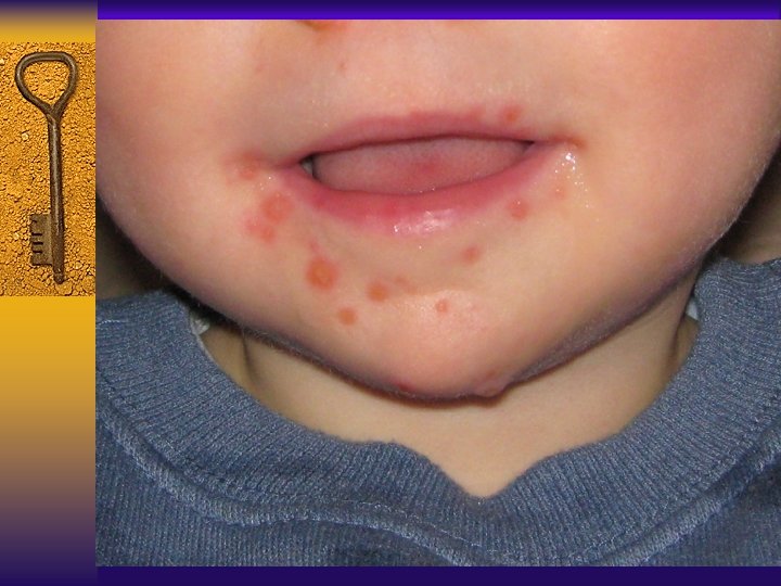
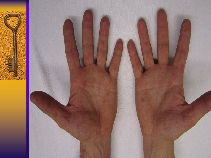
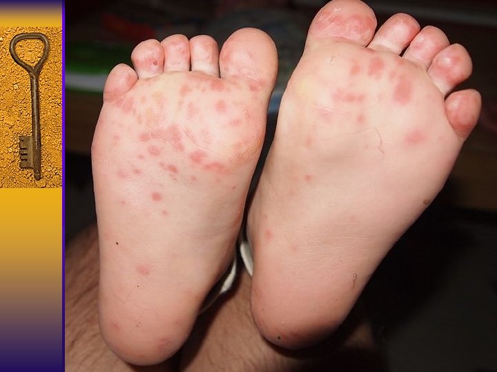
Hand Foot Mouth Disease ¨ Viral illness that usually affects infants and children younger than 5 years old ¨ Caused by viruses that belong to enterovirus genus – Coxsackievirus A 16 is the most common cause – Incubation period 3 -7 days – Usually starts with a fever, poor appetite, malaise and sore throat – 1 -2 days after fever starts painful sores usually develop in the mouth ( herpangina) → ulcers Skin rash usually on the palms of the hands and soles of the feet, also may appear on the knees, elbows, buttocs or genital area
Hand Foot Mouth Disease ¨ Spread from person to person by direct contact ¨ Viruses are found in the nose and throat secretions, fluid in blisters and stool of infected persons ¨ May be spread when infected pensons touch objects and surfaces that are then touched by others.
Hand Foot Mouth Disease ¨ Infected persons are most contagious during the first week of the illness ¨ Samples from the throat or stool may be collected for the diagnosis ¨ No spesific treatment
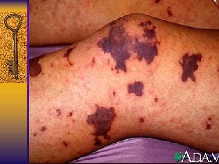
MENİNGOCOCCEMİA