CLASSIFICATION OF DENTAL CARIES DEFINITION Dental caries is


 SMOOTH SURFACE CARIES (PROXIMAL AND")


 A primary caries is one in which the lesion constitutes the initial")
 This type of caries is observed around the edges and under")


: Dentin caries. Extensive structural defect. Caries has penetrated up")


 This type of caries is a variant of rampant caries")


 system In this classification the shape and depth of")

 Class 1 restoration: include the structural defects of teeth")

 Class 2: cavity on single proximal surface of")


. Pit and fissure cavities b). Smooth surface cavities")
 G J MOUNT CLASSIFICATIN This new system defines")


- Slides: 58
CLASSIFICATION OF DENTAL CARIES
DEFINITION Dental caries is an irreversible microbial disease of the calcified tissues of the teeth, which results in demineralization of the inorganic portion and destruction of the organic substance of the tooth , which often leads to cavitation
1. BASED ON ANATOMICAL SITE 2. BASED ON PROGRESSION 3. BASED ON VIRGINITY OF LESION 4. BASED ON EXTEND OF CARIES 5. BASED ON TISSUE INVOLVEMENT 6. BASED ON PATHWAY OF CARIES SPREAD 7. BASED ON NUMBER OF TOOTH SURFACE INVOLVED
8. BASED ON CHRONOLOGY 9. BASED ON WHETHER CARIES IS COMPLETLY REMOVED OR NOT DURING TREATMENT 10. BASED ON TOOTH SURFACE TO BE RESTORED 11. BLACK’S CLASSIFICATION 12. WHO SYSTEM
1. BASED ON ANATOMICAL SITE OCCLUSAL (PIT AND FISSURE) SMOOTH SURFACE CARIES (PROXIMAL AND CERVICAL CARIES) Root surface caries ROOT CARIES
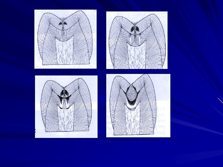
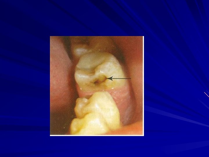
PIT AND FISSURE CARIES Highest prevalance of all caries bacteria rapidly colonize the pits and fissures of the newly erupted teeth Entry site may appear much smaller than actual lesion, making clinical diagnosis difficult. In cross section, the gross appearance of pit and fissure lesion is inverted V with a narrow entrance and a progressively wider area of involvement closer to the DEJ.
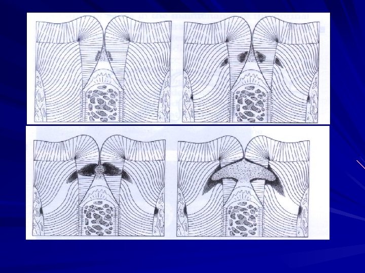
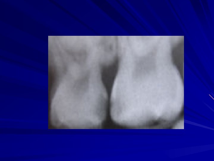
Smooth surface caries Lesion have a broad area of origin and a conical, or pointed extension towards DEJ. V shape with apex directed towards DEJ. After caries penetrate the DEJ softening of dentin spread rapidly and pulpally
ROOT SURFACE CARIES Root-surface caries is more common in older patients. Caries originating on the root is alarming because 1. it has a comparatively rapid progression 2. it is often asymptomatic 3. it is closer to the pulp 4, it is more difficult to restore
The cementum covering the root surface is extremely thin and provides little resistance to caries attack. Root caries lesions have less well-defined margins, tend to be U-shaped in cross sections, and progress more rapidly because of the lack of protection from enamel covering.
2. BASED ON PROGRESSION ACUTE CARIES ARRESTED CARIES CHRONIC CARIES
ACUTE CARIES Acute caries is a rapid process involving a large number of teeth. Pulp exposures and sensitive teeth are often observed in patients with acute caries.
CHRONIC CARIES These lesions are usually of long-standing involvement, affect a fewer number of teeth, and are smaller than acute caries. Pain is not a common feature because of protection afforded to the pulp by secondary dentin Pulp prognosis is hopeful in that the deepest of lesions usually requires only prophylactic capping and protective bases.
ARRESTED CARIES: Caries which becomes stationary or static and does not show any tendency for further progression Both deciduous and permanent affected With the shift in the oral conditions, even advanced lesions may become arrested. Arrested caries involving dentin shows a marked brown pigmentation and induration of the lesion Sclerosis of dentinal tubules and secondary dentin formation commonly occur
3. BASED ON VIRGINITY OF LESION INITIAL/PRIMARY RECURRENT/SECONDARY
PRIMARY CARIES(INITIAL) A primary caries is one in which the lesion constitutes the initial attack on the tooth surface. The designation of primary is based on the initial location of the lesion on the surface rather than the extent of damage.
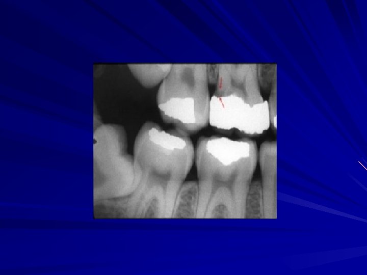
SECONDARY CARIES (RECURRENT) This type of caries is observed around the edges and under restorations. The common locations of secondary caries are the rough or overhanging margin and fracture of restoration It may be result of poor adaptation of a restoration, which allows for a marginal leakage, or it may be due to inadequate extension of the restoration.
4. BASED ON EXTENT OF CARIES INCIPIENT CARIES CAVITATION OCCULT CARIES
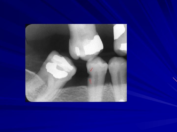
INCIPIENT CARIES The early caries lesion, best seen on the smooth surface of teeth, is visible as a ‘white spot’. Histologically the lesion has an apparently intact surface layer overlying subsurface demineralization.
These white spot lesion may be confused initially with white developmental defects of enamel formation on wetting the caries lesion disappear while the developmental defect persist
It is believed that increased fluid exposure encourages remineralization and slow down progress of the caries in the pit and fissure enamel while the cavitations continues in dentine, and the lesions become masked by a relatively intact enamel surface. These hidden lesions are called as fluoride bombs or fluoride syndrome.
5. Based on tissue involvement 1. Initial caries 2. Superficial caries 3. Moderate caries 4. Deep caries 5. Deep complicated caries
Dental caries can be divided into 4 or 5 stages Initial caries: Demineralization without structural defect. This stage can be reversed by fluoridation and enhanced mouth hygiene Superficial caries (Caries superficialis): Enamel caries, wedgeshaped structural defect. Caries has affected the enamel layer, but has not yet penetrated the dentin.
3. Moderate caries (Caries media): Dentin caries. Extensive structural defect. Caries has penetrated up to the dentin and spreads twodimensionally beneath the enamel defect where the dentin offers little resistance. 4. Deep caries (Caries profunda): Deep structural defect. Caries has penetrated up to the dentin layers of the tooth close to the pulp. 5. Deep complicated caries (Caries profunda complicata) : Caries has led to the opening of the pulp cavity (pulpa aperta or open pulp).
6. BASED ON PATHWAY OF CARIES SPREAD 1. FORWARD CARIES 2. BACKWARD CARIES
Decay starts in enamel then it involves the dentin. Wherever the caries cone in enamel is larger or at least the size as that of dentin, it is called forward decay However the carious process in dentin progresses much faster than in enamel, so the cone in dentin tends to spread laterally creating undermined enamel. In addition decay can attack enamel from its dentinal side. At this stage it becomes backward decay.
7. BASED ON NUMBER OF TOOTH SURFACE INVOLVED Simple Compound Complex A caries involving only one tooth surface A caries involving two surfaces of tooth A caries that involves more than two surfaces of a tooth
8. BASED ON CHRONOLOGY EARLY CHILDHOOD CARIES ADOLESCENT CARIES ADULT CARIES
EARLY CHILDHOOD CARIES Early childhood caries would include, two variants: Nursing caries and rampant caries. The difference primarily exist in involvement of the teeth[ mandibular incisors ] in the carious process in rampant caries as opposed to nursing caries.
NURSING CARIES RAMPANT CARIES n. Seen in infant and toddler n. Affects primary dentition in all ages, nincluding adoloscennce n. Affects primary and permanent dentition n. Mandibular incisors are not involved also affected ETIOLOGY n. Improper bottle n. MULTIFACTORIAL feeding n. Pacifier dipped in honey/other sweetner Frequent snacks Sticky refined CHO Decreased salivary flow
TEENAGE CARIES (ADOLESCENT CARIES) This type of caries is a variant of rampant caries where the teeth generally considered immune to decay are involved. The caries is also described to be of a rapidly burrowing type, with a small enamel opening. The presence of a large pulp chamber adds to the woes, causing early pulp involvement
ADULT CARIES With the recession of the gingiva and sometimes decreased salivary function due to atrophy, at the age of 55 -60 years, the third peak of caries is observed. Root caries and cervical caries are more commonly found in this group. Sometime they are also associated with a partial denture clasp.
9. BASED ON WHETHER CARIES IS COMPLETLY REMOVED OR NOT DURING TREATMENT RESIDUAL CARIES Residual caries is that which is not removed during a restorative procedure, either by accident, neglect or intention.
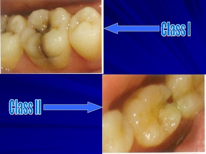
11. BLACK’S CLASSIFICATION Class 1 lesions: Lesions that begin in the structural defects of teeth such as pits, fissures and defective grooves. Locations include Occlusal surface of molars and premolars. occlusal two thirds of buccal and lingual surfaces of molars and premolars. Lingual surfaces of anterior tooth. Class 2 lesions: They are found on the proximal surfaces of the bicuspids and molars.
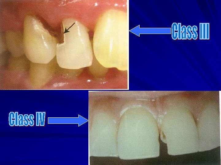
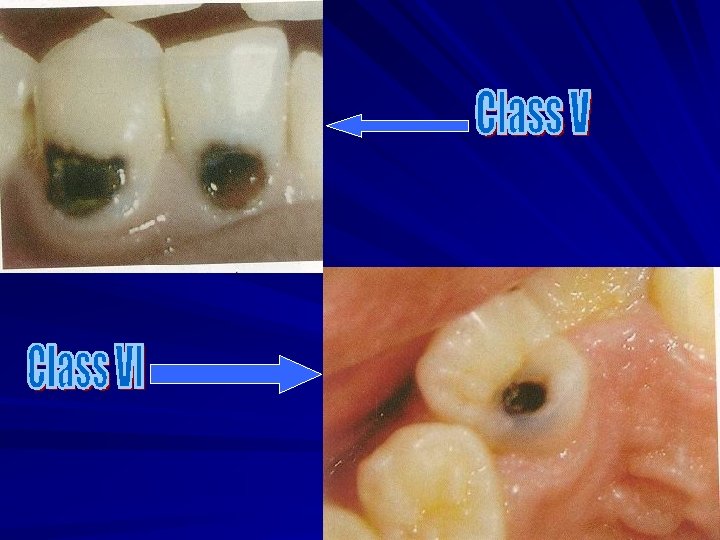
Class 3 lesions: Lesions found on the proximal surfaces of anterior teeth that do not involve or necessitate the removal of the incisal angle. Class 4 lesions: Lesions found on the proximal surfaces of anterior teeth that involve the incisal angle. Class 5 lesions: Lesions that are found at the gingival third of the facial and lingual surfaces of anterior and posterior teeth. Class 6 (Simon’s modification): Lesions involving cuspal tips and incisal edges of teeth.
12. World health organization (WHO) system In this classification the shape and depth of the caries lesion scored on a four point scale D 1. clinically detectable enamel lesions with intact (non cavitated) surfaces D 2. Clinically detectable cavities limited to enamel D 3. Clinically detectable cavities in dentin D 4. Lesions extending into the pulp
CLASSIFICATION S OF CAVITY PREPARATION
1. BASED ON TREATMENT&RESTORATION DESIGN(BLACK’S) Class 1 restoration: include the structural defects of teeth such as pits, fissures and defective grooves. Locations include Occlusal surface of molars and premolars. occlusal two thirds of buccal and lingual surfaces of molars and premolars. Lingual surfaces of anterior tooth. Class 2 restoration : They are found on the proximal surfaces of the bicuspids and molars.
Class 3 restoration : restoration on the proximal surfaces of anterior teeth that do not involve or necessitate the removal of the incisal angle. Class 4 restoration : restoration on the proximal surfaces of anterior teeth that involve the incisal angle. Class 5 restoration : restoration at the gingival third of the facial and lingual surfaces of anterior and posterior teeth. Class 6 (Simon’s modification): restoration involving cuspal tips and incisal edges of teeth.
2. Other modifications Charbeneu’s modification: a) Class 2: cavity on single proximal surface of bicuspids and molars b) Class 6: Cavities on both mesial and distal proximal surfaces of posterior teeth that will share a common occlusal isthmus c) Lingual surfaces of upper anterior teeth. d) Any other unusually located pit or fissure involved with decay.
3. Sturdevant’s classification Cavity Feature Simple cavity A cavity involving only one tooth surface A cavity involving two surfaces of tooth Compound cavity Complex cavity A cavity that involves more than two surfaces of a tooth
4. Finn’s modification of Black’s cavity preparation for primary teeth Class 1 : Cavities involving the pits and fissures of molar teeth and the buccal and lingual pits of all teeth. Class 2: cavities involving proximal surface of molar teeth will access established from the occlusal surface. Class 3: cavities involving proximal surfaces of anterior teeth which may or may not involve a labial or a lingual extension
Class 4: a restoration of the proximal surface of an anterior tooth which involves the restoration of an incisal angle. Class 5: cavities present on the cervical third of all teeth, including proximal surface where the marginal ridge is not included in the cavity preparation.
5. Baume’s classification a). Pit and fissure cavities b). Smooth surface cavities
6. Classification by Mount and Hume(1998) G J MOUNT CLASSIFICATIN This new system defines the extent and complexity of a cavity and at the same time encourages a conservative approach to the preservation of natural tooth structure. This system is designed to utilize the healing capacity of enamel and dentine.
The three sites of carious lesions: Site 1 Site 2 Site 3 Pits, fissures and enamel defects on occlusal surfaces of posterior teeth or other smooth surfaces Proximal enamel immediately below areas in contact with adjacent teeth The cervical one third of the crown or following gingival recession, the exposed root
The four sizes of carious lesions Size 1: Minimal involvement of dentin just beyond treatment by remineralization alone. Size 2: Moderate involvement of dentin. Following cavity preparation, remaining enamel is sound, well supported by dentin and not likely to fail under normal occlusal load. The remaining tooth structure is sufficiently strong to support the restoration.
Size 3: the cavity is enlarged beyond moderate. The remaining tooth structure is weakened to the extent that cups or incisal edges are split, or are likely to fail or left exposed to occlusal or incisal load. the cavity needs to be further enlarged so that the restoration can be designed to provide support and protection to the remaining tooth structure. Size 4: Extensive caries with bulk loss of tooth structure has already occurred.
Site Size Minimal 1 Moderate 2 Enlarged 3 Extensive 4 Pit/fissure 1 1. 2 1. 3 1. 4 Contact area 2 2. 1 2. 2 2. 3 2. 4 3. 1 3. 2 3. 3 3. 4 Cervical 3
THANK YOU