G V BLACKS CLASSIFICATION AND CLASS I CAVITY

G. V. BLACK’S CLASSIFICATION AND CLASS I CAVITY PREPARATION Presented By: Prakash Subedi


DEFINITION OF CAVITY PREPARATION � Cavity preparation is the mechanical alternation of defective, injured or diseased tooth in order to best receive a restorative material that will reestablish a healthy state for the tooth including esthetic correction when indicated, along with normal form and function. � The procedure of the preparing the tooth is the removal of the defective tooth structure. � Any remaining infected tooth structure may result of further carious progression, sensitivity or pain or fracture of the tooth and / restoration.

OBJECTIVES OF CAVITY PREPARATION � To remove all defects and provide necessary protection to the pulp, � To extend the restoration as conservatively as possible, � To form the tooth preparation so that under the force of mastication the tooth or the restoration or both will not fracture and the restoration will not be displaced, and � To allow for the esthetic and functional placement of a restorative material.

PRINCIPLES OF CAVITY PREPARATION ØGain access to caries. ØRemoval of all carious lesions. ØCut away all significantly unsupported enamel. ØExtended margins so that they are accessible for instrumentation and cleaning.

G. V. BLACK’S CLASSIFICATION OF CAVITY PREPARATION

Greene Vardiman Black • The man of the centuries • Father of Modern Dentistry • Raised dentistry from a trade to a profession and made it what it is today


Class I: All pit and fissure cavity preparation are class I and they are assigned to the three groups as follows � Cavity preparation on occlusal surface of premolars and molars. � Cavity preparation on occlusal two thirds of facial and lingual surfaces of molars. � Cavity preparation on palatal surface of maxillary incisors

� Class II : Cavity preparation on the proximal surface of posteriors involving 2 or more surfaces (i. e. MO, DO, MOD)

� Class III : Cavity preparation on the proximal surfaces of anterior teeth that do not involve the incisal angle (i. e. MB, ML, DB, DL) � Class IV : Cavity preparation on the proximal surfaces of anterior teeth that do involve the incisal angle (i. e. MIB, DIB, MIBL)

� Class V : Cavity preparation on the gingival third of the facial or lingual surfaces of all teeth (except pit-and-fissure lesions). � Class VI : Cavity preparation on the incisal edge of anterior teeth or the occlusal cusp heights of posterior teeth.

� Class VII : Cavity preparation on the labial surface of the anterior teeth

CLASS I CAVITY PREPARATION

STEPS IN THE CAVITY PREPARATION ØInitial tooth preparation: q. Obtaining Outline Form & initial depth q. Obtaining Primary Resistance Form q. Obtaining Primary Retention Form q. Obtaining Convenience Form ØFinal tooth preparation q. Removal of any remaining enamel pit or fissure, infected dentin, and/or old restorative material, if indicated q. Pulp protection, if indicated q. Secondary Resistance & Retention forms q. Procedures for finishing the external walls of the tooth preparation q. Final procedures: cleaning, inspecting & sealing

CLASS I CAVITY could be • Simple Occlusal cavity • Compound class I cavity (2 surfaces): Occluso-buccal cavity Occluso-lingual or palatal cavity • Complex class I cavity (more than 2 surfaces): Occluso-bucco-lingual cavity • Buccal or lingual pit • Palatal pit in maxillary incisor

Outline form: � The outline form of a class I cavity should describe a symmetrical design running along all pits, fissures, and angular grooves between the cusps and with a minimum width.

OUTLINE FORM – Smooth flowing, regular curves. No Sharp angles Angular irregularities in the outline are susceptible to fracture during condensation – a smooth flowing outline is easier to visualize and carve following condensation.

� � � In case of initial carious lesions, access is obtained by employing a small round bur (by giving a punch cut). In big carious lesions, access is obtained easily by breaking down the undermined enamel overlying the carious dentin, using a suitable size chisel. In either case, access is started at the most defective area of enamel, i. e. , a carious pit or fissure.

Enameloplasty done on terminal ends of shallow fissures to conserve tooth structure. ®procedure of reshaping the enamel surface with suitable rotary cutting instruments is termed enameloplasty. ®If one-third or less of the enamel depth is involved, the fissure may be removed by enameloplasty without preparing or extending the tooth preparation

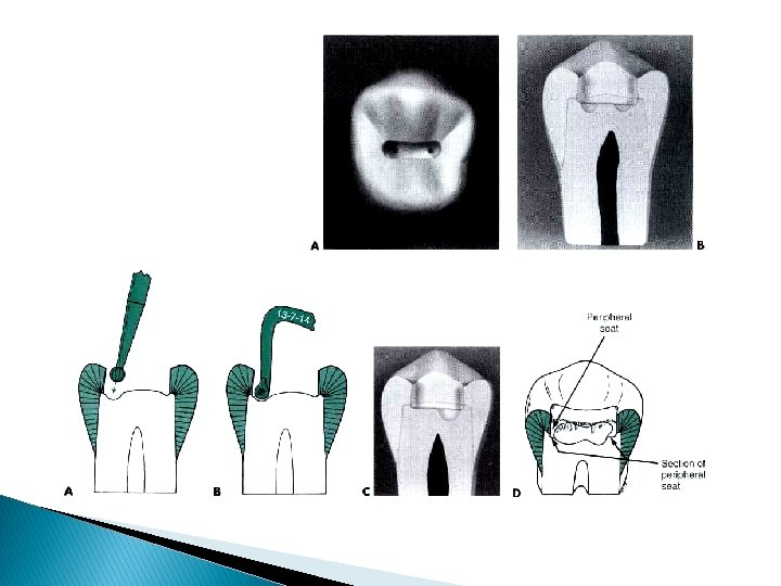
Resistance form: provided by: � Sufficient area or areas of relatively flat pulpal floor in sound tooth structure to resist forces directed in the long axis of the tooth and provide a strong, stable seat for the restoration � Minimal extension of external walls, which reduces weakening the tooth � Strong, ideal enamel margins � Sufficient depth (i. e. , 1. 5 mm) to result in adequate thickness of the restoration, providing resistance to fracture and wear
 Conservation of tooth structure is the basis for all cavity")
EXTENSIONS (Extension for Prevention) Conservation of tooth structure is the basis for all cavity preparations in order to preserve the strength of the tooth. However, sufficient extension of cavity preparations is necessary to ensure access (convenience form) for instrumentation, removal of defective tooth structure, insertion and finish of the restorative material, and maintenance of the restoration (prevention).

Ø The bur is held at a right angle to the involved surface of the tooth with light pressure and in-and-out direction is exerted. Cutting is continued until the Dentinoenamel Junction is reached.

Long axis of the tooth WRONG! Long axis of the crown CORRECT!

Extensions consist of: a. Caries and decalcifications b. Enamel unsupported by sound dentin c. Pits d. Major fissures and grooves e. Existing restorations f. Joining two outlines that come close together (i. e. , less than 0. 5 mm apart) eliminates defective tooth structure and eliminates areas (pits, fissures, etc. ) which are susceptible to recurrent caries and facilities oral hygiene procedures.

Bucco – lingual extension 1. Extend fully in areas of buccal and lingual grooves to terminate on smooth surfaces. 2. Extend minimally in areas of triangular ridges (optimal isthmus width is ¼ intercuspal distance or less) terminating on smooth surfaces. to allow a smooth tooth -restoration margin to be created (easier to finish and keep clean). to preserve the strength and function of the cusps while eliminating susceptible grooves or defective tooth structure (must be wide enough to allow condensation).
 1/4")
Isthmus just wide enough to accept instrumentation (Diameter of bur should be considered) 1/4 th intercuspal distance Not more than 1 – 1. 5 mm

ØIn a bucco-lingual direction, the cavity is extended just sufficient to eliminate the defective and susceptible tissues. ØThe lingual and the buccal wall should be parallel to the respective tooth surface.

Mesio-distal extension 1. Marginal ridge walls should be 1/2 distance from mesial and distal pit to the crest of each marginal. to preserve strength of marginal ridges.

3. Parallel to the contour of the marginal ridge to preserve a uniform bulk (strength) to the mariginal ridges. 2. Groove extensions are kept narrow (mesiodistally) where possible terminating on smooth tooth structure. to preserve strength of cusps while eliminating susceptible grooves and/or defective tooth structure (must be at least as wide as the narrowest condenser).
")
4. If marginal ridge is unsupported or very thin (less than 1. 6 mm) it should be included, resulting in a Class II preparation. If not included the marginal ridge may fracture. (amalgam will be stronger than the unsupported enamel)

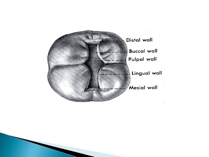
RESISTANCE AND RETENTION FORM A. Depth = 0. 1 -0. 2 mm into dentin. Depth of prepared external walls is 1. 5 – 2 mm. Minimum depth is required to provide sufficient bulk to prevent fracture and retain the amalgam. B. Pulpal floor 1. Smooth and flat Uniform thickness of restorative material. 2. Parallel to the occlusal plane resists occlusal stress and forces of condensation.

Pulpal floor mesio-distally is flat and perpendicular to the long axis

C. Buccal and lingual walls 1. Smooth and curved mesio-distally. 2. Smooth and straight pulpo-occlusally. Facilitates adaptation of amalgam and elimination of weak tooth structure.

3. Converge slightly pulpo -occlusally. 4. Diverge slightly pulpo -occlusally in buccal and lingual groove extensions (60). To provide mechanical lock or retention to the occlusal portion and create bulk at the margins. protects buccal and lingual surfaces from being undermined (RESISTANCE FORM).

Cavosurface angle of 90 – 100 are ideal. Marginal amalgam angle If less than 90 , unsupported ennalmel rods may remain which are liable to fracture If more, creates an acute amalgam margin that has tendency to fracture

D. Mesial and distal wall 1. Smooth and straight facilitates adaptation of amalgam and elimination of weak tooth structure. 2. Diverges slightly pulpo-occlusally (forms an obtuse angle with pulpal floor; not more than 10 ). protects marginal ridge from being undermined or weakened (enamel must be supported by dentin)

Removal of remaining Carious Dentin Ø In small size cavities, the carious dentin should Ø Ø have been removed during making the cavity extensions. In moderately deep and deep cavities, the carious dentin is peeled off carefully at the sides using large spoon excavators. Only light pressure in a direction parallel to that of the pulp is utilized. This is continued until a sound dentin floor is reached. Only defective areas are removed leaving round depressed areas in the wall. The level or position of entire wall shouldn’t be altered

Planning of Enamel Walls ØThe enamel walls of the cavity should be finished free from any loose, short, or undermined enamel, and trimmed to meet the tooth surface at a right cavo-surface angle. ØThis may be done by sharp and regular edged chisels and hatchets, plane fissure burs, stones, or sand-paper discs. ØAll sharp corners in enamel must be rounded, as they may contain short enamel rods.
")
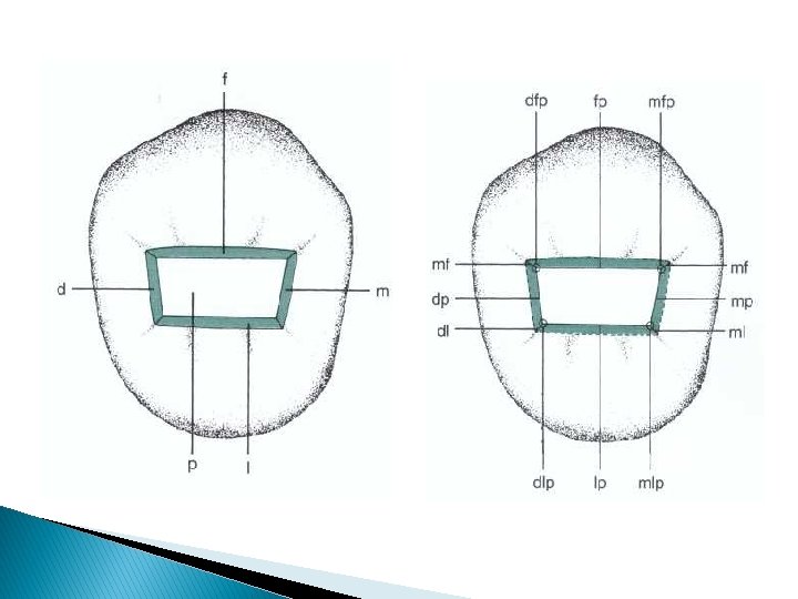
CAVITY FINISH A. Pulpo-occlusal line angle is well defined (no point angles are present) and follows general configuration of cavosurface outline. increases retention of the amalgam restoration and preparation is more easily visualized.
 2. Sound (well supported) easier to visualize")
B. Cavosurface margins 1. Sharp (well defined) 2. Sound (well supported) easier to visualize and carve. provides marginal integrity.

C. Cleanliness – cavity is free of debris and moisture. facilitates adaptation of amalgam to the cavity and improves the physical properties of the restoration by elimination of void or foreign material. A sharp explorer is then used to check the details of the prepared cavity and to loosen the tooth debris which are then blasted out with warm air.

Buccal and Lingual Extensions ØIn case of occluso-buccal and occluso-lingual cavities extensions are made through the fissures and towards the respective surfaces. ØThe cutting is done in dentin at the dentinoenamel junction using a bur until the occlusal ridge is undermined and removed.

After preparing occlusal cavity: Bur is held perpendicular to the pulpal floor & parallel to the long axis of the tooth crown. Moved towards the buccal/lingual direction along the fissure maintaining uniform depth until the bur reaches the buccal / lingual surface

Resistance form: �Keep the bur parallel to the buccal/ lingual surface of the corresponding groove so that the axial wall will follow the contour of the buccal / lingual surface at a uniform depth of 0. 5 mm inside the DEJ (0. 2 mm is permissible)

Resistance form: �Extend the lingual box gingivally to terminate at the buccal/lingual groove creating a flat gingival seat for resistance. �Gingival wall meets the tooth surface at 90° & the axial wall makes an obtuse angle with the pulpal floor �Axiopulpal line angle is rounded

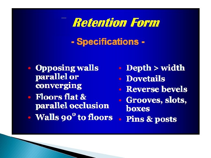
Retention form: � Mesial & distal walls of the B/L BOX are made parallel to each other with slight occlusal convergence. � Retentive grooves are then made in dentin along the axio -mesial and axio-distal line angles. The cavity walls and margins are finished as previously described.

Buccal Pit/ Palatal pit Cavities � The outline of these cavities usually describes a triangle with its base facing the gingival wall and its sides forming the mesial and distal walls.

ØAll walls are extended just enough to eliminate defective enamel and dentin. ØThe enamel walls are planed in the direction of enamel rods perpendicular to the axial wall. and ØAxial wall follows the contour of the buccal / lingual surface.

ØHoe excavators are used to smooth the axial wall and make it parallel with the external surface of the tooth. ØIt should be re-emphasize that the shape of the cavity will be governed by the extension of caries, accordingly the outline of these cavities may be a rounded or oval in shape.


- Slides: 57