DENTAL CARIES Department Of Conservative and Endodontics Dentistry


- Slides: 120

DENTAL CARIES Department Of Conservative and Endodontics Dentistry Explorer

NORMAL/SOUND TOOTH Dentistry Explorer

CONTENTS. • • • DEFINITION BRIEF HISTORY CLASSIFICATION ETIOLOGY HISTOPATHOLOGY MICROBIOLOGY EVIDENCE OF RELATION BETWEEN DIET AND DENTAL CARIES VACCINE CARIES ACTIVITY TEST CONCLUSION CARIOGRAM Dentistry Explorer REFERENCES

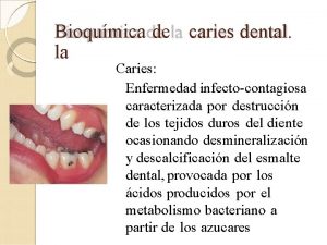
DEFINITION OF CARIES • CARIES/car·ies/ (kar´ēz) (kar´e-ēz); (noun) a Latin word meaning ‘rot/rotten’ or ‘decay’ also in Greek ‘ker’ means ‘death’ • DENTAL CARIES means ‘Rotten Teeth’ or ‘Tooth Decay’ • “Dental caries is an infectious microbiologic disease of the teeth that results in localized dissolution and destruction of the calcified tissues. ” –STURDEVANT’S • Layman understands dental caries as cavitation of/on the tooth. Dentistry Explorer

ALSO DEFINED AS…. . • “Dental caries is an irreversible microbial disease of calcified tissue of the teeth characterized by demineralization of the inorganic portion and destruction of the organic substance of the tooth, which often leads to cavitation. ” (SHAFER, HINE AND LEVY) – dietary carbohydrates are fermented by the bacteria forming an acid (low p. H) which causes demineralization, dissolution and destruction of calcified tissue of the teeth. Dentistry Explorer

DEFINITION……. . • “Dental caries, a bacterial infection, may be defined as a posteruptive pathological process of external origin, involving the softening of the hard dental tissue and proceeding to cavity formation. ” (dissolution of the hard dental tissues of an unerupted tooth which is not dental caries is tooth resorption) Dentistry Explorer

BRIEF HISTORY OF DENTAL CARIES • one of the most common of all disorders, second only to common cold • afflicted more humans longer than any other disease. • First appeared about 14000 years ago. From that time to the present, dental caries affected almost all human populations, at all socioeconomic levels, and at all ages • The first study about dental caries was published in 1870 by J. R. Mummery Dentistry Explorer

DENTAL CARIES HISTORY…… • Evidence from human skulls – 400’s – 1500’s • occlusal dental caries relatively uncommon – attrition outpaced occlusal caries • root caries predominate – 1600’s – 1800’s • more refined foods, sugar • new dental caries pattern – generally begin in pits & fissures of teeth – later on proximal surfaces (between teeth) – well-established by end of 1800’s in most developed countries Dentistry Explorer

DENTAL CARIES HISTORY • Throughout most of 1900’s – Dental caries experience • seen primarily in high-income countries • less prevalent in low-income world • likely related to diet • Late 1900’s – Dental caries experience • increase in some (not all) low-income countries • decrease in high-income countries among – children – young adults Dentistry Explorer

CLASSIFICATION • Dental caries can be broadly classified in 12 different headings: 1. Based on ANATOMICAL SITE 2. Based on PROGRESSION 3. Based on VIRGINITY OF LESION 4. Based on EXTENT OF CARIES 5. Based on TISSUE INVOLVEMENT 6. Based on PATHWAY OF CARIES SPREAD Dentistry Explorer

CLASSIFICATION Cont’d…. . 7. Based on NUMBER OF TOOTH SURFACE INVOLVED 8. Based on CHRONOLOGY 9. Based on WHETHER CARIES IS COMPLETELY REMOVED OR NOT DURING TREATMENT 10. Based on TOOTH SURFACE TO BE RESTORED 11. G. V. BLACK’S Classification. 12. WHO System 13. A new model for caries classification and management : (Julian Fisher and Michael Glick) Dentistry Explorer

1. BASED ON ANATOMICAL SITE A. OCCLUSAL SURFACE CARIES PIT AND FISSURE CARIES B. SMOOTH SURFACE CARIES a. PROXIMAL b. CERVICAL C. LINEAR ENAMEL CARIES D. ROOT SURFACE CARIES Dentistry Explorer

OCCLUSAL SURFACE CARIES PIT AND FISSURE CARIES • Highly prevalent among all other caries • Bacteria rapidly colonize the pits and fissures of the newly erupted teeth. • These early colonizers form a “bacterial plug” that remains in the site for long time (even the life of the tooth). • Type & Nature of the organisms prevalent in the oral cavity determine the type of organisms colonizing the pit & fissure. • Numerous gram positive cocci, especially dominated by S. sanguis are found in the newly Dentistry Explorer

OCCLUSAL SURFACE CARIES PIT AND FISSURE CARIES • The appearance of S. mutans in pits and fissures is usually followed by caries 6 to 24 months later. (Research by Anders Tylstrup &Ole Fejerskov) • Sealing of pits and fissures just after tooth eruption may lead to resistance to caries occurrence. • Caries expand as it penetrates into the enamel. • Shape (morphological variation) and depth of pit and fissures contributes to their high susceptibility to caries. Dentistry Explorer

DIFFERENT SHAPES OF PITS AND FISSURES • By NANGO (in 1960) – alphabetical description of fissures. U SHAPED V SHAPED Convergin g at the bottom Almost same width from top to bottom (in 14 % cases) IK SHAPED I SHAPED (in 19 % cases) Dentistry Explorer Extremely narrow slit with a large space at the bottom (in 26 % cases) INVERTED Y SHAPED (Pit caries predomin -ant)

SMOOTH SURFACE CARIES • Less favorable site for plaque attachment • Plaque usually attaches near the gingiva or under proximal contact. • In very young patients the gingival papilla completely fills the interproximal space under a proximal contact and is termed as col. • Crevicular spaces interproximally are less favorable habitat for S. mutans. • Consequently proximal caries is less likely to develop where this favorable soft tissue Dentistry Explorer

SMOOTH SURFACE CARIES • The proximal surfaces are susceptible to caries due to extra shelter provided for residing plaque in the proximal contact area. • Lesion have a broad area of origin and a conical, or pointed extension towards DEJ. • V shape with apex directed towards DEJ. • After caries penetrate the DEJ softening of dentin occurs and spreads rapidly and pulpally. Dentistry Explorer

Fig. Smooth Surface Caries Progression Dentistry Explorer

LINEAR ENAMEL CARIES • Linear enamel caries (Odontoclasia) is seen to occur in the region of the Neonatal Line of the maxillary anterior teeth. • The Neonatal Line (which represent a metabolic defect such as hypocalcaemia or trauma during birth) may predispose to caries, leading to gross destruction of the labial surface of the teeth. • Morphological aspects of this type of caries are atypical and results in gross destruction of Dentistry Explorer

ROOT SURFACE CARIES • More common in older patients. • The proximal root surface, near the cervical line, often is unaffected by the action of hygiene procedures (such as flossing) because it may have concave anatomic surface contours (fluting) and occasional roughness at the termination of the enamel. • These conditions, when coupled with exposure to the oral environment (as a result of gingival recession ), favor the formation of mature, caries-producing plaque and ultimately leads to proximal root-surface caries. • Caries originating on the root is alarming because 1. It has a comparatively rapid progression. 2. It is often asymptomatic. Dentistry Explorer

ROOT SURFACE CARIES 3. It is closer to the pulp. 4. It is more difficult to restore. • The root surface refers to cementum and allows plaque formation in the absence of good oral hygiene. • The cementum covering the root surface is extremely thin and provides little resistance to caries attack. • Root caries lesion do not have well-defined margins (tend to be Ushaped in cross sections) and progress more rapidly because of the lack of protection from as enamel covering and critical p. H for cemental dissolution is lower than for enamel. Dentistry Explorer

2. BASED ON PROGRESSION ACUTE CARIES light brown or gray pulp exposure and sensitivity often present their caseous consistency makes excavation difficult CHRONIC CARIES dark brown or leathery smaller than acute caries ARRESTED CARIES Dentistry Explorer

ACUTE CARIES • Rapid(fast spreading) process • Involve a large number of teeth at a time. • These lesions are lighter colored than the other types (light brown or grey) and their caseous consistency makes the excavation difficult. • Pulp exposures and sensitivity are often observed in patients with acute caries. • It has been suggested that saliva does not easily penetrate the small opening to the carious lesion, so there are little opportunity Dentistry Explorer

CHRONIC CARIES • Slow spreading • These lesions are usually of long-standing involvement, affect a fewer number of teeth, and are smaller than acute caries. • Pain is usually absent (because of protection afforded to the pulp by secondary dentin formation) • The decalcified dentin is dark brown and leathery. • Pulp prognosis is hopeful in that the deepest of lesions usually requires only prophylactic capping Dentistry Explorer

ARRESTED CARIES • Caries which becomes stationary or static and does not show any tendency for further progression • Both deciduous and permanent dentition are affected. • With the shift in the oral conditions, even advanced lesions may become arrested. • Arrested caries involving dentin shows a marked brown pigmentation and indurations of the lesion [the so called ‘eburnation of dentin’] • Sclerosis of dentinal tubules and secondary dentin formation commonly occur. • Commonly seen in caries of occlusal surface with large open cavity in which there is lack of food retention. • Also on the proximal surfaces of tooth in which the Dentistry Explorer adjacent approximating tooth has been extracted.

3. BASED ON VIRGINITY OF LESION INITIAL /VIRGIN/ PRIMARY CARIES RECURRENT / SECONDARY CARIES Dentistry Explorer

INITIAL /VIRGIN/ PRIMARY CARIES • A primary caries is one in which the lesion constitutes the initial attack on the tooth surface. • The designation of primary is based on the initial location of the lesion on the surface rather than the extent of damage. Dentistry Explorer

RECURRENT / SECONDARY CARIES • This type of caries is observed around the edges and under restorations. • The common locations of secondary caries are the rough or overhanging margin and fracture place in all locations of the mouth. • It may be result of poor adaptation of a restoration, which allows for a marginal leakage, or it may be due to inadequate extension of the restoration. • In addition caries may remain if there has not been complete excavation of the original lesion, which later may appear as a residual or recurrent Dentistry Explorer

4. BASED ON EXTENT OF CARIES INCIPIENT CARIES OCCULT CARIES CAVITATION Dentistry Explorer

INCIPIENT CARIES • The early incipient caries lesion, best seen on the smooth surface of teeth, is visible as a ‘ white spot ’. • Histologically the lesion has an apparently intact surface layer overlying subsurface demineralization. • Significantly such lesion can undergo remineralization and thus the lesion is not an indication for restorative treatment. • These white spot lesion may be confused initially with white developmental defects of enamel formation, which can be differentiated by their position [away from the gingival margin], their Dentistry Explorer

INCIPIENT CARIES • Also on wetting the caries lesion disappear while the developmental defect persist • It is believed that bite wing and OPG radiographs along with noninvasive adjuncts like fiber optic transillumination (FOTI), laser luminescence, electrical resistance method (ERM) are used for diagnosis of these occlusal lesions. • These lesion are not associated with microorganisms (different to those found in other carious lesion). Dentistry Explorer

OCCULT CARIES • Occult carious lesion are usually seen with low caries rate which is suggestive of increase fluid exposure. • It is believed that increased fluid exposure encourages remineralization and slow down progress of the caries in the pit and fissure enamel while the cavitations continues in dentin, and the lesions become masked by a relatively intact enamel surface. • These hidden lesions are called as fluoride bombs or fluoride syndrome. • Recently it is seen that occult caries may have its origin as pre-eruptive defects which are detectable only with Dentistry Explorer the use of radiographs.

CAVITATION • Once it reaches the dentinoenamel junction, the caries process has the potential to spread to the pulp along the dentinal tubules and also spread in lateral direction. • Thus some amount of sensitivity may be associated with this type of lesion. • This may be generally accompanied by cavitation Dentistry Explorer

5. BASED ON TISSUE INVOLVEMENT INITIAL CARIES SUPERFICIAL CARIES (Caries Superficialia) MODERATE CARIES (Caries Media) DEEP CARIES (Caries Profunda) DEEP COMPLICATED CARIES (Caries Profunda Complicata) Dentistry Explorer

INITIAL CARIES • Demineralization without structural defect. • This stage can be reversed by fluoridation and enhanced mouth hygiene Dentistry Explorer

SUPERFICIAL CARIES (Caries Superficialia) • Enamel caries, wedge-shaped structural defect. • Affected the enamel layer, but has not yet penetrated the dentin. Dentistry Explorer

MODERATE CARIES (Caries Media) • Dentin caries. • Extensive structural defect. • Caries has penetrated up to the dentin and spreads two-dimensionally beneath the enamel defect where the dentin offers little resistance. Dentistry Explorer

DEEP CARIES ( Caries Profunda) • Deep structural defect. • Caries has penetrated up to the dentin layers of the tooth close to the pulp. Dentistry Explorer

DEEP COMPLICATED CARIES (Caries Profunda Complicata) • Caries has led to the opening of the pulp cavity (pulpa aperta or open pulp). Dentistry Explorer

6. BASED ON PATHWAY OF SPREAD FORWARD CARIES v Caries cone in enamel is larger or at least the same size as that in dentin BACKWARD CARIES v. The carious process spreads rapidly in dentin laterally along DEJ resulting in undermined enamel(without dentin support) v. Thus decay can attack enamel from its dentinal side. Dentistry Explorer

7. BASED ON NUMBER OF TOOTH SURFACE INVOLVED q Given by CLIFFORD STURDEVANT SIMPLE CARIES • CARIES INVOLVING ONLY ONE SURFACE. COMPOUND CARIES • INVOLVING TWO SURFACES OF TOOTH. COMPLEX CARIES • INVOLVING MORE THAN TWO SURFACE OF TOOTH Dentistry Explorer

8. BASED ON CHRONOLOGY EARLY CHILDHOOD CARIES ADOLESCENT CARIES ADULT CARIES Dentistry Explorer

EARLY CHILDHOOD CARIES {ECC} Definition : • “The presence of one or more decayed(non cavitated or cavitated), missing( as a result of caries), or filled tooth surfaces in any primary tooth in a child 71 months of age or younger. ” American Academy of Pediatric Dentistry(AAPD) • AAPD also specifies that, in children younger than 3 years of age has any sign of smooth surface caries is indicative of Severe Early Childhood. Dentistry Caries (S-ECC). Explorer

ADOLESCENT CARIES • This type of caries is a variant form of rampant caries where the teeth generally considered immune to decay are involved. • The caries is also described to be of a rapidly burrowing type, with a small enamel opening. • The presence of a large pulp chamber leads to early pulpal involvement. • Mean age of occurrence = 11 -18 years (Shafer’s) Dentistry Explorer

ADULT CARIES • With the recession of the gingiva and sometimes decreased salivary function due to atrophy, at the age of 55 -60 years, the third peak of caries is observed. • Root caries and cervical caries are more commonly found in this group. • Sometime they are also associated with a partial denture clasp. Dentistry Explorer

9. BASED ON WHETHER CARIES IS COMPLETE REMOVED OR NOT DURING TREATMEN RESIDUAL CARIES Caries not removed during a restorative procedure, either by accident, neglect or intention. q RESIDUAL CARIES IN OLD AMALGAM RESTORATION q Sometimes a small amount of acutely carious dentin close to the pulp is covered with a specific capping material to stimulate dentin deposition, isolating caries from pulp. Dentistry Explorer

10. BASED ON SURFACE TO BE RESTORED O= OCCLUSAL SURFACE M= MESIAL SURFACE D= DISTAL SURFACE F= FACIAL SURFACE B= BUCCAL SURFACE L= LINGUAL SURFACE Dentistry Explorer

11. G. V. BLACK’S CLASSIFICATION • By GREENE VARDIMAN BLACK in early 1900’s. • Based on morphological consideration, treatment and restorative design. • Classified into 5 classes traditionally • And the 6 th class was added eventually by Simon. Dentistry Explorer Dr. G. V. Black (1836 A. D– 1915 A. D)

CLASS I LESIONS 1. Pits and fissures on the occlusal surface of molars and premolars. Dentistry Explorer

CLASS I LESIONS 2. Facial (buccal) and lingual pits of molars. Dentistry Explorer

CLASS I LESIONS 3. Lingual pits of maxillary incisors Dentistry Explorer

CLASS II LESIONS • Proximal (mesial or distal) surfaces of the premolars and molars involving two or more surfaces(i. e. MO, DO, MODB) Dentistry Explorer

CLASS III LESIONS • Proximal (mesial or distal) surfaces of incisors and canines (i. e. MB, ML, DB, DL ) Dentistry Explorer

CLASS IV LESIONS • Proximal (mesial or distal) surfaces of incisors and canines and also involving the incisal angle (i. e. MIB, MIBL, DIB, ) Dentistry Explorer

CLASS V LESIONS • Gingival 1/3 rd (Cervical third) of the facial and lingual surfaces of anterior and posterior tooth. Dentistry Explorer

CLASS VI LESIONS ( by SIMON) • Incisal edges of anterior teeth • Cusp tips of posterior teeth Dentistry Explorer

12. WHO SYSTEM Ø In this classification the shape and depth of the caries lesion scored on a four point scale D 1 surfaces Clinically detectable enamel lesions with intact (non- cavitated) D 2 Clinically detectable cavities limited to enamel D 3 Clinically detectable cavities in dentin Lesions extending into the pulp D 4 Dentistry Explorer

A new model for caries classification and management : The FDI World Dental Federation Caries Matrix (Julian Fisher and Michael Glick) JADA 2012; 143(6): 546 -551 q. Horizontal Axis depicts the extent of the caries lesion and pathology. q Vertical Axis depicts the Level of caries Information : Level 1 = WHO DMFT system. Level 2 = ADA Caries Classification System. Level 3 = International Caries Detection and Assessment System (ICDAS) Dentistry Explorer

RADIATION CARIES • Radiography is frequently associated with xerostomia due to a. decreased salivary secretion, b. Increase in viscosity of saliva and c. Low p. H • Decreased salivary secretion may lead to a rampant form of caries, • Three types of defects due to irradiation 1. Lesion usually encircling the neck of teeth - amputation of crowns may occur and - occlusal surface and incisal edges wear away. 2. Begins as brown to black discoloration of tooth. Dentistry Explorer 3. Spot depression which spreads from any surface.

etiology of dental caries Dentistry Explorer

THEORIES OF DENTAL CARIES EARLY THEORIES : 1. THE LEGEND OF THE WORM q Sumerian text (cuneiform text) q Discovered on a clay tablet from Euphrates Valley (lower Mesopotamian area) in 5000 B. C. q This text also focuses on creation of the heavens, the earths, the marshes, and later created the worm. q The remedy for toothache at that time was – mix beer, the plant SA-KIL-BIR and oil together was put on the tooth thrice. Dentistry Explorer

THEORIES OF DENTAL CARIES 2. • • • ENDOGENOUS THEORIES a. HUMORAL THEORY By Greek Physicians Contains 4 elemental humors of the body – blood , phlegm, black bile and yellow bile Galen – “ Dental caries is produced by internal action of acrid and corroding humors” Hippocrates – “ The stagnation of juices in the tooth was the cause of toothache” Aristottle – “ Greek diet (such as fig ) which adhere to tooth contribute to tooth decay” b. VITAL THEORY • At the end of 18 th century. • This theory states that teeth are an integral part of the body and they were vitally affected by and in turn affected the body. Dentistry Explorer • The tooth decay originated like bone gangrene from within the tooth

THEORIES OF DENTAL CARIES 3. EXOGENOUS THEORIES : A. Chemical (Acid) theory : • In 17 th and 18 th Century. • Tooth are destroyed by acids formed in the oral cavity • Robertson (in 1835)- “Dental decay was caused by acid formed by fermentation of food particles around teeth” • Microorganism was not recognized yet at that Dentistry Explorer

B. PARASITIC (SEPTIC) THEORY • Antoni van Leeuwenhoek (1632 -1723) – “ microorganism were associated with the carious process” • Dubos (in 1954) – “microorganisms ‘animal culae’ can have toxic and destructive effect on tissue” • Erdl , Ficinus observed filamentous parasites in the membrane (enamel cuticle) removed from the teeth. • Dental caries developed due to infiltration and decomposition of the enamel cuticle, the Dentistry Explorer

C. MILLER’S CHEMICO-PARASITIC THEORY • By Willoughby D. Miller(in 1889) • This postulates that oral bacteria act on sugar to produce acid which demineralizes the inorganic component of enamel, resulting in the development of a carious lesion. Dentistry Explorer Willoughby D. Miller (1853 A. D. – 1907 A. D. )

D. PROTEOLYTIC THEORY • By Gottlieb (in 1947), Frisbie and Nuckolls (in 1947) and Pincus (in 1950). • They described caries like lesion that were initiated by proteolytic activity at slightly alkaline p. H, and considered that the process involved depolymerization and liquification of the organic matrix of enamel. • Gottlieb – “microorganism invade the organic pathways(lamellae) of the enamel” Dentistry Explorer

E. PROTEOLYSIS CHELATION THEORY • Schatz et al (in 1955) • Implies simultaneous microbial degradation of the organic component (hence, proteolysis) and the dissolution of the minerals of the tooth by the process of chelation. • Chelate (in Greek means claw) means that compounds that are able to bind metallic ions. And the process is called Chelation. • This postulates that enamel is demineralized by chelating agents at neutral p. H. • Protein breakdown products as well as lactic acid are some chelating agents known to exist in nature. Dentistry Explorer

F. THE SUCROSE CHELATION THEORY • By Egglers-Lura (in 1967) • Proposed that the sucrose itself, and not the acid derived from it, can cause dissolution of enamel by forming an ionized calcium saccharate. • Calcium saccharates and calcium complexing intermediates require inorganic phosphate, which is subsequently removed from the enamel by phosphorylating enzymes. Dentistry Explorer

G. SULFATASE THEORY • By Pincus (in 1950) • Bacterial sulfatase hydolyzes the ‘mucoitin sulfate’ of enamel and ‘chondroitin sulfate’ of dentin producing sulfuric acid that, in turn, causes decalcification of the dental tissue. Dentistry Explorer

H. COMPLEXING AND PHOSPHORYLATION THEORY • It demonstrate that an uptake of phosphate by plaque bacteria occurs during aerobic and anaerobic glycolysis and the synthesis of polyphosphates. • The high bacterial utilization of phosphate in plaque causes a local disturbance in the phosphate equilibrium in the plaque and the tooth enamel resulting in loss of inorganic phosphate from enamel. • Soluble calcium complexing compound produced by bacteria causes further tooth Dentistry Explorer

KEYES TRIAD • By Keyes and Jordan (in 1960) • Based on interaction of 3 primary factors. They are : a. Susceptible host(tooth) b. Cariogenic plaque Bacteria (dental plaque) c. Local substrate(diet) Dentistry Explorer

NEWBRUN TETRALOGY • By Newbrun (in 1982) • New factor “TIME” was added MICROORGANISMS SA LIV SA LI VA A TOOTH TIME SA LIV A FOOD SA LIV A IES R A C Dentistry Explorer

CURRENT CONCEPT OF DENTAL CARIES • Multi-factorial Origin. • Includes two factors in etiology of caries 1. Primary (Essential) Factors. 2. Secondary Factors. 1. The primary essential factors includes: a. Host b. Microbial Flora c. Substrate d. Time Dentistry Explorer

2. Secondary Factors Includes: Plaque Substrate Tooth • • • Oral hygiene q Bacteria in plaque produces an Oral microflora organic acids to dissolve Saliva- p. H, composition, flow, buffer Fluoride in plaque organic phase. Diet q Major role in caries production Transmissibility Type of carbohydrates q Chelators and proteolytic enzymes Chemical composition of food are produced as by products. Physical characteristics of food q Chelators binds to calcium ions. Oral clerance q Proteolytic enzyme breakdown the Frequency of eating organic matrix. Sugar intake and frequency • • Fluofide concentration Carbonate and citratelevel Age of tooth q has major role in caries production Morphology of tooth Trace elements Nutrition Saliva Dentistry Explorer Composition of enamel

FEJERSKOV & MANJI in 1990 Personal Factor Oral & Nutritional Factors Dentistry Explorer

Thank you Dentistry Explorer

Classification by Mount and Hume (1998)- G. J. Mount Classification • • This new system defines the extent and complexity of a cavity and at the same time encourages a conservative approach to the preservation of natural tooth structure. This system is designed to utilize the healing capacity of enamel and dentine. The three sites of carious lesions: Site 1 - Pits, fissures and enamel defects on occlusal surfaces of posterior teeth or other smooth surfaces Site 2 - Proximal enamel immediately below areas in contact with adjacent teeth Site 3 - The cervical one third of the crown or following gingival recession, the exposed root The four sizes of carious lesions Size 1: Minimal involvement of dentin just beyond treatment by remineralization alone. Size 2 : Moderate involvement of dentin. Following cavity preparation, remaining enamel is sound, well supported by dentin and not likely to fail under normal occlusal load. The remaining tooth structure is sufficiently strong to support the restoration. Size 3 : the cavity is enlarged beyond moderate. The remaining tooth structure is weakened to the extent that cups or incisal edges are split, or are likely to fail or left exposed to Dentistry occlusal. Explorer or incisal load. the cavity needs to

Histopathology of dental caries Dentistry Explorer

HISTOPATHOLOGY OF DENTAL CARIES • Can been studied through the use of ground section of teeth that are usually between 60 µm to 100 µm thickness. Dentistry Explorer

HISTOPATHOLOGY OF ENAMEL CARIES • Usually small carious lesion. • Caries of the enamel is preceded by the formation of microbial(dental) plaque. • Smooth surface caries appears as an area of decalcification beneath the dental plaque as smooth chalky white, opaque area often termed as “white spot”. Dentistry Explorer

HISTOPATHOLOGY OF ENAMEL CARIES • The small carious lesion (incipient caries lesion) in human enamel causes minimal damage to the outer smooth surface but considerable demineralization present below the surface. • Longitudinal section under light microscope • Divided into 4 zones starting the inner advancing front of the lesion Zone 1 : The Translucent Zone 2 : The Dark Zone. Dentistry Explorer Zone 3 : The Body of The

ENAMEL CARIES (GROUND SECTION) Dentistry Explorer

ZONE 1 : THE TRANSLUCENT ZONE • Lies at advancing front of the enamel lesion • First recognizable zone of alteration from normal enamel • ½ of the total lesion is translucent zone. • Not always present. • Pore volume (under Dentistry Explorer

ZONE 2 : THE DARK ZONE • Lies adjacent and superficial to the translucent zone. • Positive zone (always present and on polarized light this zone shows positive birefringence) • Pore volume (under polarized Dentistry Explorer

ZONE 3 : THE BODY OF THE LESION • Lies between surface layer and dark zone. • Area of greatest demineralization. • Pore volume (under polarized light) = 5% (periphery) 25 % (center) Dentistry Explorer

ZONE 4 : THE SURFACE ZONE • Relatively unaffected by caries (only partial demineralization) • Surface of enamel is relatively immune to caries (due to hypermineralization because of saliva contact, and higher surface F-content) • also pore volume is lower than the body Dentistry Explorer

HISTOPATHOLOGY OF DENTINAL CARIES • Caries advancement in dentin proceeds through 3 stages 1) demineralization of dentin (by weak organic acids). 2) degeneration and dissolution of organic material of dentin , mainly collagen fibers (type I). 3) bacterial invasion after the loss of structural integrity caused due to 1) and 2). Dentistry Explorer

EARLY DENTINAL CHANGES • In early carious dentinal lesion the zones are clearly distinguished. Dentistry Explorer

Dintinal changes q From inner to outer - 5 different zones are (accn to Sturdevant) Zone 1: Normal Dentin Zone 2: Sub transparent Dentin Zone 3: Transparent Dentin Zone 4: Turbid Dentin Zone 5: Infected Dentin Dentistry Explorer

ADVANCED DENTINAL CHANGES Ovoid necrotic debris on liquefaction foci Dentistry Explorer

Zone of DENTINAL CARIES • Beginning pulpally at the advancing edge of the lesion adjacent to the normal dentin, 5 zones are described Zone 1: Zone of Fatty Degeneration of Tomes’ Fibers Zone 2: Zone of Dentinal Sclerosis Zone 3: Zone of Decalcification of Dentin Zone 4: Zone of Bacterial Invasion Dentistry Explorer

Zone 1: Zone of Fatty Degeneration of Tomes’ Fibers • The most advancing front of dentinal caries • Characterized by the presence of a layer of fat globules ; hence stains red with sudan red. • Significance: 1) fat layer leads to impermeability of the dentinal tubules (DT) – trying to prevent further invasion of carious lesion 2) favors sclerosis of dentin in zone 2. Dentistry Explorer

Zone 2: Zone of Dentinal Sclerosis • Layer of sclerotic dentin which appears white in transmitted light • Calcification of Dentinal Tubule as a reaction of vital pulp and vital dentin to carious invasion , so as to prevent further penetration of microorganisms. • Formation of this zone is minimal in rapidly progressing caries, and • Prominent in slow caries. Dentistry Explorer

Zone 3: Zone of Decalcification of Dentin • This zone lies above the zone of sclerotic dentin • Initial decalcification of only the walls of the Dentinal Tubule(DT). • Presence of PIONEER BACTERIA- first of the microorganisms penetrating DT before there is any clinical evidence of caries. • Bacteria present in individual DT are in pure form (i. e. either completely cocci or completely bacilli; not in mixed form) Dentistry Explorer

Zone 4: Zone of Bacterial Invasion • In a layer above zone 3. • Characterized by the presence of microorganisms • In early stage of caries- acidogenic microorganisms • In deeper layer- proteolytic microorganisms replace acidogenic bacteria • Supports the hypothesis of caries development: - Initiation (by acidogenic bacteria) and Dentistry Explorer

Zone 4: Zone of Bacterial Invasion • During initiation phase- in the early stage when caries is not deep , acidogenic bacteria predominant which utilizes carbohydrate for their metabolism • Later in progression phase – as the caries goes deeper , less and less of carbohydrate substrate available , hence acidogenic bacteria are replaced by proteolytic microorganisms which uses dentinal protein for their metabolism. Dentistry Explorer

Zone 5: Zone of Decomposed Dentin • Most superficial zone of early dentinal caries. • No recognizable structure in decomposed dentin • Collagen and minerals seem to be absent • Great number bacteria dispersed in this decomposed granular matter. Dentistry Explorer

MICROBIOLOGY OF DENTAL CARIES Dentistry Explorer

MICROBIOLOGY OF DENTAL CARIES • 300 - 400 microflora are present in human mouth. • Clarke (1924) – first to associate bacteria with dental caries. 1. first to isolate MS from human dental caries 2. first to produce caries in extracted teeth. • MS : - A gram + facultative anaerobe characterized by 8 serotypes, a-h. • Seven species of Streptococcus mutans are isolated of which S. mutans (serotype c, e, f) and S. sobrinus (serotype d, g) are found in human. Dentistry Explorer

Criteria for Cariogenicity Listed by NEWBURN (in 1983) • An organism must be acidogenic. • An organism must be aciduric. • An organism must exhibit tropism for teeth. • An organism must utilize refined sugar (sucrose). Dentistry Explorer

Carbohydrate metabolism by MS PTS • PES + CHO PYRUVATE + P-CHO • Carbohydrate must me tranported across the membrane and phosphorylated before metabolism by 1 Multiple Sugar Metabolism (MSM) system 2 Transport via the Phosphoenolpyruvate(PEP) 3 Sugar Phosphotransferase system (PTS) Dentistry Explorer

WINDOW OF INFECTIVITY of MS • Caufield, 1993 • 19 -31 months • 6 -12 years Dentistry Explorer

WHY S. mutans SUCCEDED ? • Three factors 1 ability to adhere to other bacteria and tooth surface. 2 ability to rapidly metabolize nutrients(CHO) 3 ability to survive in acidic environment. Dentistry Explorer

DIET AND DENTAL CARIES Sugars : • Arch criminal SUCROSE Physical properties of foods & cariogenecity: • Texture effect salivary flow rate • Improve cleansing action, reduce retention & increase saliva flow may reduce dental caries • Acidic food prolonged contact with tooth DEMINERALIZATION. Dentistry Explorer

EVIDENCE OF RELATION BETWEEN DIET AND DENTAL CARIES Dentistry Explorer

WARTIME STUDIES • Decrease in caries rate was seen during and after the World War II due to sugar restriction during this period. • In the postwar years restriction to sugar supply was eased & sugar became more readily available rise in caries rate. Dentistry Explorer

HOPE WOOD HOUSE STUDY • Hopewood House (NSW, Australia) for 10 years. • SULLIVAN –in 1958; HARRIS –in 1963 • Diet sugar & refined carbohydrates were excluded. • Dental surveys ages 3 & 14 years. Carbohydrates given : • • Whole wheat bread Soya beans Wheat germ Oats Rice Potatoes Molasses etc Dentistry Explorer

HOPE WOOD HOUSE STUDY ü ü ü Dairy products Raw vegetables Fruits Nuts Vegetarian diet with adequate protein, fats, minerals & vitamins • Fluoride content of water was insignificant • No tea was consumed Dentistry Explorer

RESULTS AND CONCLUSION Results : At the end of 10 years • 13 yr old children had mean DMF = 1. 6 with 53 % being caries free • General child population 10. 7 only 0. 35% being caries free. • Children’s oral hygiene was poor with prevalent gingivitis in 75% cases Conclusion : • Dental caries can be reduced to by Spartan diet & without beneficial influence of fluoride & in presence of unfavorable oral hygiene. Dentistry Explorer

VIPEHOLM STUDY • GUSTAFSSON et al – in 1954 • In 1939 Swedish Government requested the Royal Medical Board to investigate the measures that should be followed to reduce dental disease in Sweden. • Study Vipeholm study at Vipeholm hospital, Lund an institution for mentally disabled individuals diet & dental caries • 436 patients 6 experimental groups & one control group. • Control group low carbohydrate, high fat diet free from refined sugar. Dentistry Explorer

Experiment Groups In Study • SUCROSE GROUP 300 gm sucrose at mealtime • BREAD GROUP 345 gm of sweet bread containing 50 gm sugar. • CHOCOLATE GROUP 300 gm sugar with meals 100 gm supplemented by 65 gm of milk chocolate between meals during 2 nd year • CARAMEL GROUP 22 caramels daily in 2 portions • 8 TOFFEE GROUP 8 toffees in 2 portions • 24 TOFFEE GROUP 24 toffees between meals Dentistry Explorer

Conclusion : VIPEHOLM STUDY 1. Risk of sugar increasing caries activity is great if sugar is consumed in a form with a strong tendency to be retained. 2. Risk is greatest sugar is consumed between meals 3. Increase in caries activity due to intake of sugar rich foodstuff consumed in a manner favoring caries lesion disappears on withdrawal of such foodstuff from diet 4. Carious lesion may continue to appear despite the avoidance of refined sugar, maximum restriction of natural sugars & total dietary carbohydrates. 5. Risk of increase in caries activity is intensified with an increase in duration of sugar clearance from saliva. Dentistry Explorer

TURKU SUGAR STUDY • Turku, Finland • By Scheinin & Makinen et al in 1975 A. D. • 2 year study. Aim : To Compare cariogenicity of Sucrose, Fructose & Xylitol. 125 Young Adults 3 Groups 1 -- Sucrose Group (35 people) 2 – Xylitol Group (52 people) 3 – Fructose Group (38 people) Dentistry Explorer

RESULT : TURUKU STUDY 1 st year : • Sucrose Group = Fructose Group (Caries Prevalence) • Xylitol Group almost no caries 2 nd year : • Caries continued to increase in sucrose group • Unchanged in fructose group • Xylitol some white spots had been remineralized to Dentistry Explorer a point that they could not be scored.

HEREDITARY FRUCTOSE INTOLERENCE • In 1959– Froesch • Inborn error of fructose metabolism transmitted by autosomal recessive gene. • Metabolic error deficiency of hepatic fructose-1 phosphate which inhibits fructose phosphorylation. • C/F pallor, nausea, vomiting, coma & convulsion following ingestion of fructose containing products • Reduced caries experience seen in such patients as compared to control group of same age Dentistry Explorer

CARIES VACCINE • A Vaccine currently under development to treat dental caries by inoculating against bacteria commonly known to contribute to their formation, particularly Streptococcus mutans. (Mosby Dental Dictionary, 2 nd Edition) Dentistry Explorer

CARIES ACTIVITY TEST CARIES ACTIVITY Ø Refers to the increment of active lesion (new and recurrent lesions) over a stated period of time. Ø Caries activity is a measure of speed of progression of a carious lesion. CARIES SUSCEPTIBILITY Ø Refers to the inherent tendency of the host and target tissue, the tooth, to be affected by the caries process. Ø It is the resistance of a tooth to a caries producing environment. CARIES ACTIVITY TEST Ø Measures the degree to which the local environment challenges(e. g. dietary effect on microbial growth and metabolism) favors the probability of carious lesions. Dentistry Explorer

CARIES ACTIVITY TESTS AND METHODS. 1) MICROBIAL TESTS FOR MUTANS STREPTOCOCCI DETECTION 2) MICROBIAL TESTS FOR LACTOBACILLI DETECTION 3) SNYDER TEST 4) ALBAN TEST 5) REDUCTASE TEST 6) SWAB TEST 7) FOSDICK CALCIUM DISSOLUTION TEST (ENAMEL SOLUBILITY TEST) 8) SALIVARY BUFFER CAPACITY 9) DIP-SLIDE MEASUREMENT OF SALIVARY YEAST 10) PLAQUE PH MEASUREMENT 11) DEWAR TEST Dentistry Explorer 12) CARIOGRAM

Sequale of Dental Caries DENTAL CARIES Extensive Dental Caries ACUTE/CHRONIC PULPITIS Pulpal Involvement ACUTE/CHRONIC PERIODONTITIS Periapical Penetration PERIAPICAL ABSCESS PERIAPICAL GRANULOMA Periapical Tissue Destruction PERIODONTAL CYST OSTEOMYELITIS/ PERIOSTITIS CELLULITIS Bone Involvement and Destruction ABSCESS Spread of Infection and Disability Dentistry Explorer

ETIOLOGY OF DENTAL CARIES Salivary flow and component Proteins , antibacterial components and agents Fluoride, calcium and phosphate Protective dietary components Remineralization and Inhibition of demineralization. Reduced salivary function Bacteria : Mutans streptococci, lactobacilli Dietary component frequency and carbohydrate Demineralization and Inhibition of remineralization. Dentistry Explorer
 What is conservative force
 Katz classification of premolars
 Mount hume classification
 Mounts classification
 Mounts classification
 Gj mount classification
 Force conservative et non conservative
 Force conservative et non conservative
 The force of gravitation is conservative or nonconservative
 Dentobuff strip test
 S shaped matrix
 Crown preparation burs names
 Skirt in tooth preparation
 Skirts in cast gold restoration
 Pediatric class 2 prep
 Classification of instruments in conservative dentistry
 Turbid dentin
 Arch criminal in caries
 Dental caries
 Turbid dentin in carious tooth
 Sonic and ultrasonic in endodontics
 Smear layer adalah
 Difference between inlay and amalgam
 Access cavity preparation grossman
 Mouse hole effect in endodontics
 Grossman endodontics
 Creekside endodontics
 Access cavity krasner and rankow
 Diaket
 Obturating materials
 Maf endo
 Chapter 54 endodontics
 Perennialism characteristics
 Work energy theorem
 Conservative comparative and superlative
 Conservative policies under reagan and bush
 Definition of a conservative vector field
 Force conservative
 Activity sheet 1 conservative moderate or speculative
 3-5 exonuclease vs 5-3 exonuclease
 Activity sheet 3 stock market calculations answer key
 Forze conservative
 Forze conservative e dissipative
 Principal of curriculum construction
 Conservation of energy
 Conservative america in the ascent
 Conservative cavity preparation
 Forte conservative si neconservative
 Why electric force is conservative
 Conservative criminology
 Is thrust a conservative force
 Deletion biology
 Conservative mixing
 Conservative spectrum
 Conservative tide
 Plate boundary project
 Lavoro momento torcente
 The conservative order
 A conservative movement emerges
 Caries risk assessment form
 Dental formula of human
 Cervical burnout
 Caries ad pulpam penetrans
 Gv blacks classification
 Zone of caries
 Millers liquefaction foci
 Frank caries
 Que es caries
 Caries stabilization
 Detal caries
 Difoti caries detection
 Modified bass
 Teoria del glucogeno de la caries
 Teoria del glucogeno de la caries
 Texas health steps quick reference guide
 Caries mesial incipiente
 Hidden caries
 Rairena
 Caries control restoration
 Medical model of caries management
 Non cutting dental instruments
 Rotary instrument in dentistry
 Pulp protection liners and bases
 Bonding system materials
 What is a polar molecule
 Pubh4401
 Detroit mercy dental school tuition
 Teeth disking
 Theories of mineralization of calculus
 Define pediatric dentistry
 Classification of denture stomatitis
 Budapest dentistry university
 Squint test in dentistry
 Clinical risk management in dentistry
 Sleep dentistry baxter
 Post dam area in complete denture
 Polymer in dentistry
 Semi critical instruments in dentistry
 Flexibility in dental materials
 Cold working in dentistry
 Los dientes
 Tools of public health dentistry
 Sprue former
 Indirect retainer fulcrum line
 Restorative instruments in dentistry
 Realeff effect ppt
 A point angle is the junction of
 Askov in public health dentistry
 Askov dental health education
 Dr br vacher
 Black's classification
 Furcation classification
 Final tooth preparation stage
 Multiple circlet clasp
 Boxing wax uses
 Dental composite classification
 Silver alloy in dentistry
 Non operating dental auxiliaries
 Unicompositional amalgam
 Lathe cut alloy
 Deflasking
