PEDIATRIC OPERATIVE DENTISTRY cont Principles of cavity preparation
")


 � � EXTENSION FOR Cavity design dictated")


- Slides: 38
PEDIATRIC OPERATIVE DENTISTRY (cont. )
Principles of cavity preparation
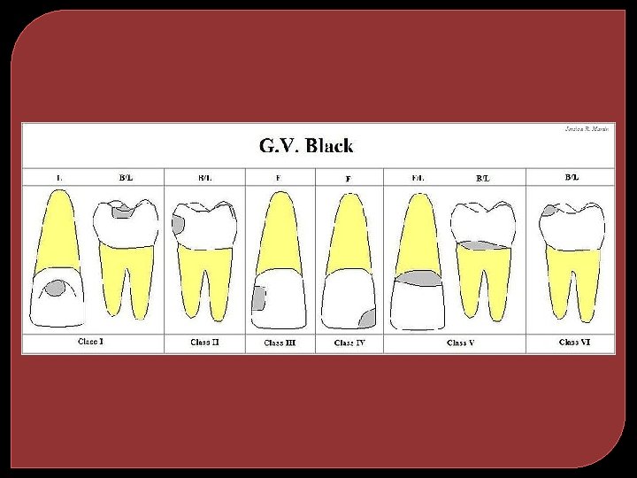
Black’s classification CLASS I CAVITY � All pit and fissure cavities � Occlusal surfaces of posterior teeth � Occlusal 2/3 of buccal and lingual surfaces of premolars & molars � Lingual surfaces of anterior teeth.
Class ii cavity �All proximal surface cavities on the premolars and molars.
Class iii cavity �All proximal surface cavities on the incisors and canines which do not involve the removal and restoration of the incisal angle.
Class iv cavity �All proximal surface cavities on the incisors and canine which involve the removal and restoration of the incisal angle.
Class v cavity �All gingival cavities located within the gingival one third of the tooth. These may be either on the facial or lingual Gingival one third of the tooth
Class vi cavity �CAVITIES ON THE INCISAL EDGES AND CUSP TIPS OF ALL TEETH.
Blacks concept extension for prevention 1. 2. 3. 4. 5. 6. 7. Obtaining Outline form Obtaining Resistance form Obtaining Retention form Obtaining Convenience form Removal of infected dentin Finishing enamel walls Debridement /toilet of the cavity
Outline form �Area of tooth surface or enamel margin to be included in the finished cavity �It can be. External outline form Internal outline form
Resistance form � Shape & configuration of the cavity that best enables both the restoration & tooth to withstand occlusal forces without fracture
Retention form � Factors of cavity design that prevent the restoration from being displaced.
Convenience form � Shaping the cavity to facilitate access for instrumentation , for condensation, adaptation and finishing
Removal of infected dentin � Elimination of any infected carious tooth structure or faulty restoration left in the cavity preparation
Finishing enamel walls � To place the margins on smooth tooth structure � To have smooth walls and rounded angles � To facilitate placement and finishing of restorative materials
Cleaning of the cavity � Removing all chips or loose debris that have accumulated � Drying the cavity � Making a final complete inspection of the preparation for any remaining infected dentin, unsound enamel margin
PREPARATION OF CLASS I CAVITY The extension & depth of the cavity will be determined by the amount & location of caries & preoperative occlusal anatomy. Every effort should be made to retain as much wellsupported enamel as possible.
PREPARATION OF CLASS I CAVITY Cont. � The maximum intercuspal cavity width should be one-quarter to one-third of the intercuspal width. � 0. 5 mm pulpally to the DEJ to provide sufficient bulk of amalgam to withstand occlusal forces. � Pulpal floor should be flat & smooth � Internal line angles should be rounded to reduce any stresses in the set amalgam
PREPARATION OF CLASS I CAVITY Cont. � The extension is made, buccal or lingual cavity walls should be straight & either parallel or converging occlusally � The extension should be cut 0. 5 mm into dentine & should extend gingivally to include the developmental pits. � Retention grooves can be place in dentine if considered necessary.
PREPARATION OF CLASS I CAVITY Cont. � The ‘isthmus’ area where the extension meets the occlusal section can be rounded or beveled to increase the bulk of amalgam. as it is subjected to heavy stresses during lateral movements
Class II Cavity Outline: The outline follows the fissure pattern so as to prevent secondary caries occurring adjacent to the restoration. A smooth flowing outline reduces stress & permits better of the amalgam. Isthmus: This should be between 1/4 of the intercuspal distance (approximately 1. 5 mm)
CLASS II CAVITY Cont. Depth: This should be 0. 5 mm below dentinoenamel junction or 1. 5 mm from the cavosurface Internal angles: All the internal angles should be rounded so as to limit stress & to ensure that amalgam can be easily packed into these regions. Pulpal floor: Pulpal floor should be slightly concave.
CLASS II CAVITY Cont. Buccal & lingual walls: should be converging so making the cavity retentive. Also, the. cavosurface angle needs to be a right angle to ensure maximum strength at the enamelamalgam junction. Gingival floor: should be located just below the contact area with the adjacent tooth but supragingivally.
CLASS II CAVITY Cont. Axial wall: The width of the floor of the box should be approx. 1 mm. follows external contour of tooth Buccal & lingual walls: These should be convergent, parallel to the appropriate external surface & make a cavosurface angle of 90 degree. Axio-pulpal line angle: Rounded to give maximum thickness of amalgam with minimum stress in this area
CLASS II CAVITY Cont. � Retention � Occlusal grooves- made to enhance retention. dovetail -It should be made including all carious areas & shapes should be such that it locks the occlusal portion of filling
CLASS II CAVITY Cont.
DIAGRAM ILLUSTRATING THE INCREASED DANGER OF PULP EXPOSURE WHEN THE GINGIVAL WALL IS CARRIED TOO DEEPLY
Recent Concept CONSTRUCTION WITH CONSERVATION (OLD CONCEPTPREVENTION) � � EXTENSION FOR Cavity design dictated by site & extent of lesion. No need to extend cavity into the “caries free” area. � Biologically active restorative material which assists remineralization & healing of remaining tooth structure. � ONLY Irretrievable/ degenerated/ broken down tooth surface to be removed. � Completely control plaque accumulation by eliminating surface cavitation as a result of caries.
Modifications required for cavity preparation in primary & young permanent teeth
REASONS FOR PRESERVATION OF PRIMARY TEETH �Necessary till the succedaneous teeth replace them �Mastication of food �Preservation and increase in arch length �Development of speech and phonetics �Prevention of any infection or caries to the permanent teeth �Prevention of malocclusion of permanent teeth �Esthetics
Difference between primary & permanent teeth � Primary teeth have thinner enamel & dentin than permanent teeth. � The pulps of primary teeth are larger in relation to crown size than permanent pulps
� The pulp horns of primary teeth are closer to outer surface of the tooth than permanent pulps � In primary teeth, the enamel rods of the gingival third of the crown extend in an occlusal direction from dentin-enamel junction � Primary teeth demonstrates greater constriction of the crown and have a more prominent cervical contour than permanent teeth
� Primary teeth have broad, flat proximal contact areas. � Primary teeth are whiter than their permanent successors. � Primary teeth have relatively narrow occlusal surface in comparison with their permanent successors.
Factors to be considered while restoring the primary teeth � Smaller � Thin tooth dimension of deciduous teeth enamel � Broad contact areas � Proximity � Narrow of pulp chamber to outer tooth surface occlusal table
Class I cavities � For small carious lesions, the isthmus should not be more than 1/3 the intercuspal distance. � Pulpal � Use floor should be flat of preventive resin restoration
CLASS II CAVITY Cont. CLASS II CAVITY Proximal box of deciduous teeth • Box converges occlusally • Minimal flare to prevent weakening of enamel walls • Isthmus 1/4 th to 1/5 th of intercuspal width • Rounded Axio-pulpal angle grooved to increase retention • No bevel in gingival seat • Depth minimal to prevent pulp exposure at cervical constriction • Wide gingival floor
THANK YOU