FURCATION INVOLVEMENT AND ITS MANAGEMENT FURCATION It can


")


 ROOT TRUNK LENGTH SHORT TRUNK LONG TRUNK ► Less attachment")


- Slides: 36
FURCATION INVOLVEMENT AND ITS MANAGEMENT
FURCATION It can be defined as: an area of complex anatomic morphology that may be difficult or impossible to be debrided by routine periodontal instrumentation. FURCATION INVOLVEMENT • It means the progress of inflammatory disease that result in attachment loss which affect the bifurcation or trifurcation of multirooted teeth.
DIAGNOSIS CLINICAL PROBING RADIOGRAPHIC VIEW
ETIOLOGY Prolonged presence of microbial dental plaque. Extent of attachment loss on furcation depends on presence of these factors: 1. local anatomic factors ►Root trunk length ►Root morphology ► Root length ► Interradicular dimension ► anatomy of furcation 2. Local developmental anomalies: ►cervical enamel projections 3. Dental caries 4. Pulpal death
CLASSIFICATION Glickman`s Classification(1953)
CLASS I INCIPIENT FURCATION This is an early lesion. The pocket is suprabony, involving the soft tissue. There is slight bone loss in the furcation area. Radiographic change is not usual since bone loss is minimal. A periodontal probe will detect root outline or may sink into a shallow V-shaped notch into the crestal area
CLASS I INCIPIENT FURCATION The level of bone loss allows for the insertion of the periodontal probe into the concavity of the root trunk
CLASS II PATENT FURCATION In this, bone is destroyed in one or more aspects of the furcation, but a portion of the alveolar bone and periodontal ligament remain intact, permitting only partial penetration of the probe into the furca. Radiographs may or may not reveal this type of furcation.
CLASS II PATENT FURCATION The level of bone loss allows for the insertion of a periodontal probe into the furcation area between the roots.
CLASS III COMMUNICATING OR THROUGH AND THROUGH FURCATION This type of probe penetrates completely from one side to the other side characterized by severe bone destruction in the furcation area. It is clearly shown in the radiographs as a radiolucent area in between the roots, especially in the lower molars.
CLASS IV AS IN CLASS III, BUT THE GINGIVAL TISSUES RECEDE APICALLY SO THAT FURCATION IS CLEARLY VISIBLE.
HAMP/NYMAN/LINDHE: 1975 I: HORIZONTAL BONE LOSS < 3 MM II: HORIZONTAL BONE LOSS >3 MM III: THROUGH & THROUGH
TARNOW/ FLETCHER 1984 A- vertical destruction of bone upto 1/3 rd of the interradicular height (0 -3 mm) B-vertical destruction of bone upto 2/3 rd of inter-radicular height (4 -7 mm) C- vertical destruction beyond the apical third (>7 mm)
LOCAL ANATOMIC FACTORS 1) ROOT TRUNK LENGTH SHORT TRUNK LONG TRUNK ► Less attachment has to be lost before furcation involved. ► More attachment has to be lost before furcation involved. ► Accessible to treatment more than tooth with long trunk. ► Less accessible to treatment and plaque control.
ROOT LENGTH AND ROOT FORM ROOT LENGTH ROOT FORM ► is directly related to ► Dilacerations quantity of attachment supporting the tooth. ► Teeth with long roots if furcation exposed there will be sufficient attachment & tooth could stay in furcation longer period. ► Curvature ► Grooves
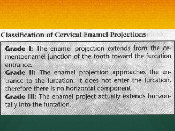
RIDGES, CONCAVITY & PROJECTIONS ANATOMY OF FURCATION: ► Bifurcation ridges. ► Concavity in the roof of the furcation (dome) ► Presence of accessory canals CERVICAL ENAMEL PROJECTIONS: ► High prevalence in 2 nd Max. & Mand. Molars. ► It`s presence may affect plaque control and may accelerate furcation attachment loss.
OBJECTIVE OF TREATMENT The elimination of the microbial plaque from the exposed surfaces of the root complex. The establishment of an anatomy of the affected surfaces that facilitates proper self-performed plaque control.
THERAPEUTIC CLASSES OF FURCATION CLASS I DEFECTS Conservative periodontal therapy Oral hygiene , scaling and root planning CLASS II Flap surgery with odontoplasty and osteoplasty CLASS II TO IV ADVANCED DEFECTS Periodontal surgery Endodontic therapy Restoration of tooth
NON-SURGICAL ROOT PREPARATION Scaling & root planing Most effective in grade I and shallow grade II. Deeper sites respond less favorably
In most situations, it results in the resolution of the inflammatory lesion in the gingiva.
ANTIMICROBIALS Adjunct to scaling and root planning Chlorhexidine Tetracycline fibers No clinically significant difference in clinical parameters after irrigation
OPEN DEBRIDEMENT Greater calculus removal than closed Ultrasonic Narrow furcations Dome of furcation Surgical access and increased operator experience significantly enhance calculus removal in molar furcation.
OSSEOUS SURGERY Most effective in grade II furcation Osteoplasty and ostectomy techniques Remove the lip of defect to reduce horizontal depth Bone ramps into the furcation to enhance plaque control Reduce probing depths
ROOT RESECTION Grade II or grade III CONTRAINDICATIONS Inadequate bone support Fused roots Inoperable endodontically Patient considerations
HEMISECTION IS THE SPLITTING OF A TWO ROOTED TOOTH INTO TWO SEPARATE PORTIONS MANDIBULAR MOLARS Grade III furcation Need widely separated roots Soft tissue positioned below level of pulp chamber
HEMISECTION
ROOT SEPARATION Root separation involves the sectioning of the root complex and the maintenance of all roots
REGENERATION OF FURCATION DEFECTS Guided tissue regeneration Predictable outcome of GTR therapy was demonstrated only in degree II furcation involved mandibular molars less favorable results have been reported in other types of furcation defects GTR could be considered in areas with isolated degree II furcation defects
OSSEOUS GRAFTING Autogenous bone Allografts Freeze dried bone Demineralized Freeze dried bone Alloplasts Hydroxyapatite Non-porous Porous Bioglass
EXTRACTION Attachment loss is so extensive that no root can be maintained If tooth/gingival anatomy will not allow proper plaque control For endodontic or restorative reason Osseointegrated implant substitute
PROGNOSIS Hirshfeld and Wasserman. “A long term survey of tooth loss in 600 treated periodontal patients. ” J Perio 1978 600 patients followed an average of 22 years with recall every 4 -6 months 1464 molars initially diagnosed with furcation invasion 70% survival of furcated molars
PATIENTS FACTORS Determine patient`s goals and expectations Screen for local, behavioral and systemic factors; Oral hygiene Compliance Stress Intraoral Accessibility Uncontrolled Diabetes Smoking Healing response to Previous Therapy
THANKYOUYOU