FURCATION INVOLVEMENT TREATMENT OF FURCATIONAND TREATMENT INVOLVED TEETH


- Slides: 22

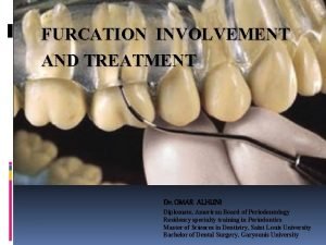
FURCATION INVOLVEMENT TREATMENT OF FURCATIONAND TREATMENT INVOLVED TEETH Dr. OMAR ALHUNI Diplomate, American Board of Periodontology Residency specialty training in Periodontics Master of Sciences in Dentistry, Saint Louis University Bachelor of Dental Surgery, Garyounis University

Gingivitis: inflammation of gingiva soft tissues Periodontitis: inflammation of deeper structures plus destruction of periodontium The destruction of periodontal tissues progresses in the apical direction affecting all periodontal tissues The progress of periodontal disease results in attachment loss sufficient enough to affect the bifurcation or trifurcation of multirooted teeth.

�Terminology �Anatomy �Etiology �Classiffication �Diagnosis �Differential Diagnosis �Prognosis �Treatment

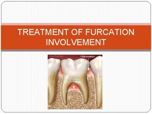
Terminology � Furcation: area between individual root cones � Root cone: divided region � Root trunk: undivided region � Root complex: portion of tooth apical to the CEJ

Anatomy Teeth with furcations: Maxillary Premolar Maxillary Molar Mandibular Molar Maxillary Premolars: 40% of cases have 2 roots Furcation in middle or apical third of root Mean distance to furcation from CEJ ~7 mm

Anatomy Maxillary molars 1 st and 2 nd molars have 3 roots 1 st molar has shorter root trunk than 2 nd CEJ to Furcations for 1 st molar �Mesial ~3 mm �Buccal ~4 mm �Distal ~5 mm Buccal furcation more narrow than mesial and distal Mesial-the furcation entrance is located more palatally. Distal – located at midpoint of tooth in buccal –palatal dimension

Anatomy Mandibular molars: Two roots w/ mesial root larger than distal �Mesial root more vertical �Distal root projects to the D Root trunk on 1 st shorter than 2 nd �Buccal =3 mm �Lingual =4 mm

Etiology Primary Factor: bacterial plaque Contributing Factors: Iatrogenic Factors TFO Furcation Location Thickness of Overlying Gingiva and Bone Cementicles Cervical Enamel Projections: 50% of mandibular 2 molar Enamel Pearls 8% of maxillary 2 molar Intermediate bifurcation ridge 73% of mandibular molar Accessory pulp canals: 28% of molar

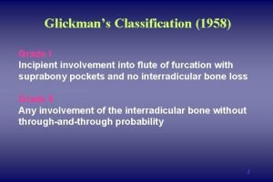
CLASSIFICATION Glickman Classification – horizontal probing �Grade 1 – incipient, pocket formation into furcation fluting, interradicular bone is intact. �Grade 2 – moderate, loss of interradicular bone but not through and through �Grade 3 – through and through, gingival tissue occludes orifices �Grade 4 – exposed, high and dry Tarnow & Fletcher – vertical probing � Subclass A – vertical loss 0 -3 mm � Subclass B – vertical loss 4 -6 mm � Subclaass C – vertical loss > 6 mm

HAMP CLASSIFICATION 1975 Degree I- horizontal penetration into furcation <3 mm Degree II- horizontal penetration into furcation >3 mm Degree III- Through-and through furcation

Diagnosis Clinical Assessment: The Naber's probe is used to detect and measure the involvement of furcaton Radiographic Assessment: intraoral periapical radiographs and vertical “bitewing” radiographs for detection of furcation invasion.

Differential Diagnosis Pulpal pathosis: Vitality must always be tested Endodontic tx fails to resolve after 2 months then defect associated with marginal periodontitis Trauma from occlusion: Occlusal interferences may cause inflammation and tissue destrauction Occlusal adjustment always precedes perio therapy

PROGNOSIS: Prognosis of involved tooth depends on several factors like: General condition of the patient. �Poor results in smokers Tooth type and degree of furcation involvement. �maxillary premolars with furcation involvement = poor or hopeless prognosis Tooth or root morphology �Teeth with long root trunks and short roots = poor or hopeless prognosis Operator’s skill and experience

Treatment Objectives for Tx: Eliminate of the microbial plaque from the exposed surfaces of the root complex Establish anatomy of the affected surfaces that facilitate proper selfperformed plaque control Tx : Options Sc. Rp ( Nonsurgical) furcation plasty (surgical) GTR (Mand molars) Tunnel preparation Root resection Extraction

Sc. Rp Nonsurgical Treatment Results in resolution of inflammation Re-establish normal gingival anatomy

Furcation plasty Resective tx to eliminate the defect Odontoplasty and osteoplasty Used mainly at buccal and lingual furcations Steps: �Release flap for access �Remove inflammatory soft tissue and Sc. Rp �Odontoplasty eliminating horizontal defect and opening furcation �Recontour alveolar bone �Apically position flap

GTR Regeneration: Reproduction or reconstitution of a lost or injured part (Bone Fill) Principles of GTR space creation clot stabilization wound protection Position Paper Most studies reported favorable results in II mandibular Class furcations.

Tunnel Preparation Treatment for deep Class II and Class III mand molars �Best Tx for short trunks, wide seperation angle, long divergence Includes surgical exposure of the entire furcation �Allows for easy cleaning for pt Increases risk for root sensitivity and root caries

Root Separation and Resection(RSR) Root separation �Involves sectioning of the root complex and maintaining all roots Root resection �Involves sectioning w the removal of 1 -2 roots GENERAL GUIDELINES: �Remove the root that will eliminate the furcation �Remove the root with the greatest amount of bone and attachment loss. �Remove the root with the greatest number of anatomic problems.

Extraction: Considered when loss of support is extensive Restore w/ implant if possible Fugazzotto , 2001:

Class I : Scaling and root planing Furcation plasty Class II: Scaling and root planing Furcation plasty GTR (mandibular molars) Tunnel preparation Root resection Extraction/implant placement Class III: Tunnel preparation Root resection Extraction/implant placement

Dr. OMAR ALHUNI Diplomate, American Board of Periodontology Tel: 092 -382 -9123 Email: omar 4 huni@yahoo. com
 Cul de sac furcation involvement
 Furcation of teeth
 Through and through furcation
 Glickman classification of furcation
 Furcation entrance
 Furcation plasty definition
 Father involvement in early childhood
 Panther involvement network
 Controlled emotional involvement
 Open and closed skills examples
 Pacing continuum
 Gaisce personal skill ideas
 Youth involvement
 Classify the
 Theory of social judgement (sherif) menyatakan bahwa
 Tarnside curve of involvement
 Panther involvement network
 Panther involvement network
 Define active community participation
 Brazil ww2 involvement
 Alliances in ww1
 Total member involvement pdf
 Epstein's six types of parent involvement

