Dental Caries Definition CARIES Latin word meaning rot


 Cavitation")


 System In this classification the shape and depth of the")


 • Attributed to")
 • Most commonly")
")


 MILLER’s CHEMOPARASITIC THEORY / ACIDOGENIC THEORY • Willoughby D Miller 1890")


 plaque acidogenicity • Depends on")
 Buffering & neutralizing factors • Intrinsic– weak org acids---AA, proteins & phosphates in")


 ü suggested that bacteria produce acid if CHO substrate is")


 Tooth • Composition • Morphologic characteristics • position B) Saliva •")
 TOOTH FACTOR 1) COMPOSITION • Influence the initiation & rate of progression •")

 MORPHOLOGIC CHARACTERISTICS –Presence of deep, narrow occlusal fissures or B & L pits")

 POSITION ü Malaligned, out of position, rotated teeth ü difficult to cleanse and")
 SALIVA FACTOR 1) COMPOSITION • varies • The inorganic phase of enamel consists")
 p. H ü The p. H at which any particular saliva ceases to")

 Quality of saliva • Mild increase or decrease in flow – has little")

 VISCOSITY Salivary viscosity is not important – Miller § Numerous cases show free")
 ANTIBACTERIAL PROPERTY Lysozyme…. . hydrolytic enzyme in saliva • can lyse many cariogenic")
 IMMUNOGLOBULINS • Secretory Ig. A – Resistant to proteolytic enzymes – Agglutinating activity")
 DIET FACTOR 1) Physical form Diet of primitive man : • Consisted of")


 definitely increases the caries activity • The")

- Slides: 115
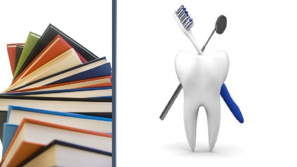
Dental Caries
Definition • “CARIES” Latin word meaning “rot” or “decay”. • WHO defines as a localised post eruptive, pathological process of external origin, involving softening of hard tooth tissues and proceeding in to formation of cavity.
• Dental caries is an infectious irreversible microbial disease of the calcified tissues of the teeth, characterized by demineralization of the inorganic portion & destruction of the organic substance of the tooth, which often leads to cavitations. (shafer’s)
Classification Pit and Fissure Caries or Occlusal Caries According to the morphology or anatomical site of the lesion: Smooth Surface Caries Linear Enamel Caries Root Caries
Acute Dental Caries According to the dynamics with regard to rate of carious progression: Chronic Dental Caries
Early Childhood Caries Nursing Bottle Caries Based on Chronology : Rampant Caries Adolescent Caries Adult Caries
Incipient Caries Based on Severity: Occult Caries(Hidden Caries) Cavitation
Arrested Caries Based on Progression: Recurrent or Secondary Caries Radiation Caries
G. V. BLACK’S CLASSIFICATION Class 1 lesions: Class 1 • Involving pits, fissures and defective grooves. Class 2 lesions: • Involving proximal surfaces of the bicuspids and molars. Class 2 Class 3 lesions: • Involving proximal surfaces of anterior teeth that do not involve incisal angle. Class 3
Class 4 lesions: • Lesions found on the proximal surfaces of anterior teeth that involves the incisal angle. Class 4 Class 5 lesions: • Lesions that are found at the gingival third of the facial and lingual surfaces of anterior and posterior teeth. Class 5 Class 6 (Simon’s modification): • Lesions involving cuspal tips and incisal edges of teeth. Class 6
World Health Organization (WHO) System In this classification the shape and depth of the caries lesion is scored on a four point scale D 1. clinically detectable enamel lesions with intact (non cavitated) surfaces D 2. Clinically detectable cavities limited to enamel D 3. Clinically detectable cavities in dentin D 4. Lesions extending into the pulp
Pit and Fissure Caries Seen on : -Occlusal surface of molars and premolars -Buccal and lingual surface of molars -Palatal surface of max incisors • Pits and Fissures with high steep walls and narrow bases are more prone to caries-poor self cleansing features • Appear brown or black, soft and catch a fine explorer tip
• Enamel bordering it appear bluish white as it is undermined • Presence of large carious lesion with tiny point of opening (mistaken idea of “INTERNAL CARIES”-phenomena of tooth getting decayed from inside outward)
Smooth Surface Caries Seen on: -proximal surfaces -gingival third of buccal and lingual surfaces • Preceded by formation of microbial plaque • Proximal caries begins just below the contact point • Initially appears as a faint white opacity without loss of enamel continuity-well demarcated • Later becomes roughened due to superficial decalcification of enamel
Root caries Hazen defined as “a soft, progressive lesion that is found anywhere on the root surface that has lost connective tissue attachment and is exposed to the oral environment”. • Seen in older age groups with significant gingival recession and exposed roots. • Previously called as “caries of cementum”. • Katz and his associates, studied the distribution pattern, and found that teeth frequently affected are first mand molars>mand premolars>max cuspids • Proximal surfaces are more affected in max arch and buccal surfaces in mand arch The Prevalence and Intra-oral Distribution of Root Caries in an Adult Population, Caries Res 1982, 16: 265 -272.
• Initiates on mineralized cementum and dentin • Evidence suggest that microorganisms involved in root caries are different from coronal caries, being filamentous rather than coccal • Microorganisms invade cementum along sharpey’s fibers • Cementum forms in concentric layers has lamellated apperance, microorganisms spread laterally in various layers Stamm and Banting, studied effect of fluoride on root caries and found, lifelong consumption of fluoridated water is capable of significantly reducing root caries which is becoming a growing dental public health problem in adult population. J Am Dent Assoc. 1990 Feb; 120(2): 143 -9
Acute dental caries • Rapid clinical course • Results in early pulp involvement • Two chronological periods commonly affected are 4 -8 yrs and 11 -18 yrs • Initial entrance of lesion remains small-saliva cannot enter the small opening, to neutralize the acid formed
Acute dental caries • Occurs frequently in children and young adults-dentinal tubules are large and open and show no sclerosis • Process is so rapid, little time for reparative dentin deposition seen • Dentin is light yellow • Pain is an apt feature
Rampant caries • Sudden, rapid and almost uncontrollable destruction of teeth, affecting the surfaces of teeth that are relatively caries free. • Includes-proximal and cervical surfaces of mandibular incisors also. • Caries increment of 10 or more new carious lesions over one year.
• Affects both Primary dentition and Permanent dentition of teenagers. • Carious process is so severe that only root stumps are left • Dietary factors affecting oral flora, oral substrate and physiological factors affecting saliva- significant role
Nursing bottle caries (nursing caries, baby bottle syndrome, bottle mouth syndrome) • Attributed to prolonged use of: -nursing bottle containing milk or milk formula, fruit juice or sweetened water -Sugar or honey-sweetened pacifiers • Clinically seen as widespread carious destruction of deciduous teeth
Nursing bottle caries (nursing caries, baby bottle syndrome, bottle mouth syndrome) • Most commonly 4 max incisors followed by first molars then cuspids • Absence of caries in mand incisors distinguishes this from rampant caries
Adolescent caries • Acute caries attack above 11 -18 yrs of age, (later period) • Seen on surfaces relatively considered immune to caries • Seen as small opening in enamel with extensive undermining • Rapid progression-no time for reparative dentin formation
Chronic dental caries • Progresses slowly and involves pulp in much later phase • Entrance to lesion is much larger • Deep brown in color • Much destruction of tooth surface • Minimum softening of dentin
• Moderate lateral spread of caries at DEJ • Slow progress of lesion-allows sufficient time for : -sclerosis of dentinal tubules -secondary dentin deposition • Pain not common-protection to pulp by secondary dentin
Recurrent caries • Occurs in the immediate vicinity of a restoration. • Usually seen near the gingival margins of class 2 and class 5 restorations and rarely on class 1 restorations. • Soft at the margin of filling and detected with an explorer
Recurrent caries • Due to poor adaptation of filling material to the cavity produces “leaky margin”. • Besic in 1943 studied the fate of bacteria sealed in dentinal tubules and noted that lactobacilli died out, while streptococci persisted.
Arrested caries • Caries becoming static or stationary and does not show any tendency for further progression. • Both dentitions affected • Appears as brown stained, polished and is hard • “Eburnation of dentin”- caries on occlusal surfaces characterized by a large open cavity and superficially softened and decalcified dentin is gradually burnished taking up brown color. • Sclerosis of dentinal tubules and secondary dentin formation seen
Radiation caries • Development of rampant caries in patients undergoing radiation therapy in head and neck region. • Increased viscosity and low p. H of saliva seen after irradiation • Del Regato in 1939 observed xerostomia as a complication of radiation therapy and postulated that caries development may be due to modification of saliva secretion • Frank et al (1965)and Baden(1970) explained 3 forms of dental defects as a consequence of irradiation: Ø Caries like lesion: -completely encircling the neck of tooth leading to crown amputation Ø Brown-black discoloration Ø Spot depression-spreads from incisal or occlusal edges to labial and lingual surfaces.
Incipient caries • Seen as a white spot • Contrasts well with more transparent enamel • Caries attack changes the optical behavior of affected enamel-becomes opaque as porous enamel scatters more light than sound enamel • Requires air-drying to become visible • Usually confused with white developmental defects of enamel, so differentiated by: Ø Position-usually away from gingival margin Ø Shape-unrelated to plaque accumulation Ø Symmetry-usually affect the contralateral tooth Ø On wetting-carious lesion disappears while developmental defects persists.
Occult caries • Also known as hidden caries • A lesion not clinically diagnosed but detected only on radiographs • Acc to Seow in 2000, prevalence range from 0. 8% in premolars in 1415 yrs and 50% in 20 yrs. • “Fluoride syndrome or fluoride bombs”-it is believed that increased fluoride exposure encourages remineralization, slowing down caries progression in enamel while cavitation continues in dentin and the lesion becomes masked by a relatively intact enamel surface.
EPIDEMIOLOGY Caries in Prehistoric man • Caries is considered a disease mainly of modern civilization, as prehistoric humans rarely suffered from it. • Caries in such persons was seen to occur below the contact areas & also on the CEJ.
Caries in Modern man • Occurs universally. • No human being is immune. • Commonest infection to occur in oral cavity. • Directly linked to dietary habits of modern man – processed, soft food. • Primitive man’s diet was coarse, raw and mostly uncooked.
FACTORS AFFECTING CARIES PREVALENCE RACE: People living in same geographical area but belonging to different race have differing caries incidence. Chinese, Blacks, Indians have lesser caries incidence than the Caucasian whites.
AGE: Dental caries more prevalent in children up to 12 years. Incidence decreases somewhat in younger and middle age group. Incidence increases again by the older age.
GENDER: Incidence of caries is significantly higher in females than males. This may be due to the fact that teeth in females erupt earlier compared to males.
FAMILIAL: Hereditary pattern is evident. Children of parents with low caries experience also show lesser caries incidence and vice versa.
ETIOLOGY OF DENTAL CARIES • early theories Worm theory Endogenous theories Chemical theory Parasitic theory • modern theories Acidogenic theory Proteolytic theory Proteolysis chelation theory Sucrose chelation theory Auto-immune theory Sulfatase Theory
Early Theories 1. The Legend of Worms – • Earliest mention is from ancient Sumerian text (5000 BC) known as the “Legend of worms”. • Caries is caused by worms which drank blood of the teeth & fed on roots in the jaws.
2. ENDOGENOUS THEORIES Humoral Theory • Greek physicians • Dental caries is produced by internal actions of acids & corroding humors & an imbalance in these humors resulting in disease. Blood (sanguine) Phlegm (phlegmatic) Black bile (melancholic) Yellow bile (choleric)
Vital Theory of tooth decay • Hippocrates, Galen • Proposed that tooth decay originated like a bone gangrene, from within the tooth itself. • Penetration of caries into dentin and pulp without detectable catch on the surface • Hunter, 1778, proposed that caries arose secondary to pulpal inflammation.
3. CHEMICAL THEORY Parmly, 1820 s. • He observed that caries affected externally not internally. • He proposed that an unidentified chymal agent was responsible. Robertson, 1835 • Supported Parmly • DC was caused by acid formation by fermentation of food particles around the teeth.
4. PARASITIC THEORY / SEPTIC THEORY Erdl, 1843. • He observed filamentous parasites (parasitische vegetabil) assoc. with carious lesions. Fincus, 1847 • Described filamentous organisms in enamel cuticle & carious lesions. … denticolae
Parasitic theory was disseminated by 2 German physicians Leber & Rottenstein 1867 • Caries initiation is by an undefined acid which rendered enamel porous & facilitated the formation of a bed of filamentous microrg. on the enamel surface. Underwood & Miles 1881 • Combined actions of germs & acids • Acc. To them organisms 1 st attacked the organic material & feeding upon it created an acid that removed lime salt.
Modern Theories 1) MILLER’s CHEMOPARASITIC THEORY / ACIDOGENIC THEORY • Willoughby D Miller 1890 • blend of 2 theories • States that caries is produced by acids produced by microorganisms of the mouth.
hypothesis Ø DC is a chemoparasitic process consisting of 2 stages: * Decalcification of enamel & dentin which leads to total destruction ( preliminary stage). ** Dissolution of the softened residue (subsequent stage). Ø The acid which affects the primary decalcification is derived from the fermentation of starches & sugars lodged in the retaining centers of the teeth.
Ø experiments to prove his theory Ø Bread & sugar when incubated in vitro with saliva at body temp. , there was acid production within 48 hours enough to decalcify sound dentin. Ø He isolated many microorg from the oral cavity, many of which were acidogenic & some proteolytic. Since many forms produce lactic acid, he believed that caries was not caused by a single organism , but by a variety of them.
Evidences ü Acid was present within deep carious lesions. ü Bread or sugar incubated at 37°C with saliva could decalcify the entire crown of a tooth. ü There’s correlation btwn cariogenic bacteria in saliva & caries activity. ü Inoculation of acidogenic bacteria in gnotobiotic rats on CHO diet induced caries. ü p. H of cavities was lower than those of normal teeth
Drawbacks ü Unable to explain the predilection of specific sites on a tooth to caries. ü Initiation of smooth surface caries. ü why some populations are caries free ü phenomenon of arrested caries.
Role of 3 factors in caries process. ORAL MICROORGANISM in acid production & proteolysis. CHO SUBSTRATE ACID which causes dissolution of tooth minerals
ORAL MICROFLORA Salivary & Crevicular fluid components Dietary CHOs Localized acid production Demineralization of inorganic & Dissolution of organic DENTAL CARIES Miller’s theory - Schematic representation
ROLE OF CHO o Primitive men who had relatively low caries index manifested an increase in caries incidence after exposure to civilized / refined diet. o Presence of readily fermentable CHO is resp for their loss of caries resistance ü Miller – when teeth was incubated with saliva & bread or sugar resulted in decalcifn. ü There was no effect when meat & fat was used in place of CHO. ü Cane sugar or cooked starches produced acids but little was formed when raw starch was substituted
Volker & Pinkerton • Reported the production of acid from mixtures of either sucrose or starch incubated with saliva with no difference in acid production btwn raw & refined sugarcane.
Cariogenicity of dietary CHO varies i. Frequency of ingestion ii. Physical form iii. Chemical composition iv. v. Route of administration Presence of other food constituents
Sticky, solid CHO ---- more cariogenic than liquid form Soft retentive foods ---- more cariogenic Refined, pure CHO --- more cariogenic than crude form CHO that clear rapidly from mouth by saliva & swallowing ---- less cariogenic than those which are slowly cleared Meals rich in fat, protein or salt, decreases oral retentiveness of CHO Polysacch are less easily fermented by plaque bacteria than mono & disacch.
ROLE OF MICROORGANISMS Miller – ü demonstrated the presence of microorg within the tubules of decayed teeth mainly cocci & leptothrix. ü He laid the foundation for the role of acids elaborated by bacteria in caries production. Goadby, 1990 – o isolated a gram + ve bacillus from carious dentin & termed it B. necrodentalis. o These played a role in decalcification of tooth enamel & dentin. o Later he changed --- & stated that streptococci --- active cause.
Gies & Klinger, 1915 – studied extensively on various bacteria assoc. with DC. Mc. Intosh, James & Lazarus-Barlow, 1922 – § isolated bacteria – Bacillus odontolyticus frm carious dentin. § These organisms were capable of lowering the p. H to the degree that enamel was softened. Clarke, 1924 Ø isolated S mutans from teeth found to be in the early stages of disease.
R Bunting – reported bacillary forms. § Acc. to him DC is a truly infective disease with B. acidophillus as a specific etiologic factor. § He noted that there was a spontaneous cessation of caries coincident with the disappearance of B. acidophillus from the mouth either thru prophylactic, therapeutic or dietetic control. Harrison, 1940 – ü Concluded an apparent relationship of lactobacilli with initial caries & streptococci with more advanced lesions.
Florestano, 1942 – ü Cultured organisms from the saliva of carious & non carious persons & studied their acidogenic potential. ü acidogenic streptococci & staphylococci were isolated from both the groups. ü Their acid production & presence in large no. suggested a role in DC equal to that of lactobacilli.
MICROBIAL FLORA & DENTAL CARIES • • • S mutans S salivarius S mitior S milleri S oralis S sanguis Lactobacillus acidophilus L casei Actinomyces viscosus A naslundii L. fermentum & S. lactis ---were not able to induce caries Not all organisms are cariogenic & caries will not occur in the absence of microorganisms
Localization of microflora related to caries Pit & fissure S mutans S sanguis Lactobacillus species Actinomyces species Smooth surface S mutans S salivarius Root surface A. viscosus A. naeslundi S mutans S sanguis Deep dentinal caries Lactobacillus species A. naeslundi
LACTOBACILLI Ø Gram +ve, non-spore forming rods Ø Grows under micro aerophilic condn. Ø L. fermentum & L. brevis Ø L. casei & L. acidophilus Ø Acidogenic and aciduric Ø Multiply in low p. H of plaque & carious lesions Ø Detected in Deep fissures and dentinal lesions, favoring their retention Ø Synthesize intra & extra cellular polysaccharides Role in progression of caries
ACTINOMYCES – Gram +ve, filamentous, anerobes – acidogenic – A. naeslundi & A. viscosus (facultative anaerobes) – A. israelii & A. odontolyticus (strict anaerobes) – Role in initiation of root lesions – Most common in subgingival microflora
VEILLONELLA – Gram -ve cocci – Anticariogenic – Lack key enzymes for energy production –. ’. Do not utilize sugars as energy source – Utilizes lactic acid → converting to Propionic acid
Streptococcus mutans – Clarke, 1924 – Catalase -ve, gram +ve cocci – Synthesizes insoluble polysaccharides frm sucrose – More aciduric than other streptococci – No colonization in absence of teeth – Can live on p. H as less as 4. 2 – capable of inducing caries – able to synthesize extracellular dextrans & levans
Metabolism of S mutans • Substrate – Sucrose • Can be used: – Intracellularly – Extracellularly • By conversion of sucrose to adhesive extracellular CHO polymers by cell bound & extracellular enzymes.
Internalization is accompanied by direct phosphorylation of sucrose by glycolytic pathway ↓ Lactic acid production & Degradation of sucrose to glucose & fructose (Invertase)
• Sucrose which doesnot enter the cell, maybe used for the extracellular synthesis of CHO polymers. • S mutans polymerize the glucose & fructose moieties of sucrose to synthesize glucans & fructans. Glucans • Dextrans – less resistant to enzymatic attack • Mutans – imp constituent of fibrillar plaque & is less soluble & more resistant to enzymatic attack Fructans • Highly soluble • Degraded by plaque bacteria • Reservoir of fermentable sugars Enzymes responsible are glycosyl & fructosyltransferases
• Lipoteichoic acid: – Highly negatively charged compounds – Adhesiveness of bacteria
• S. sanguis – Occlusal fissures – Low cariogenicity • S. salivarius – Not found in dental plaque – Adheres well to epithelial surface & not to hard tissues. – Produce levans (water soluble polymer of fructose) • S. mitior – Soft, round & brown-black colonies
ROLE OF ACIDS • Exact mech of CHO degradation to form acids in oral cavity by bacterial action is unclear • Occurs through enzymatic breakdown of sugar • Acids formed are chiefly lactic acid • aspartic acid, butyric acid, acetic acid, propionic acid, glutamic acid etc.
Role of Dental Plaque • Contributory factor for initiation of DC • Dental plaque is a soft, unmineralized, bacterial deposit which forms on teeth & dental prosthesis that are not adequately cleaned • An important component of dental plaque is acquired pellicle, which forms just prior to or concomitantly with bacterial colonization and may facilitate plaque formation • The microbiology of dental plaque include 3 groups of micro-organisms: • Streptococci • Actinomyces • Veillonellae
• Miller, stated the role of foods & acids produced by their degradation, he thought that plaque protected enamel against attack by carious process. • G V Black, 1899 regarded plaque to be important in caries process • mere presence of plaque does not necessarily mean caries will occur
Stephan curve • Within 1 -3 min of rinsing with glucose or sucrose soln, p. H falls from 6. 5 to below 4. 5 to 5. In about 10 -30 min, p. H returned to normal • If there’s a intake of glucose or sucrose during this period there’s a further decrease in p. H requiring more time to return to normal. • Stephan’s curve is a graphical representation of the plaque p. H plotted against time after 10% glucose rinse.
• At neutral & slightly acidic p. H levels ------there’s No Demineralization • At a certain p. H value, the environment of the tooth is no longer capable of preventing dissolution & is c/a Critical p. H, which is about 5. 3 to 5. 5. • Below critical p. H------ Demineralization takes place. • Caries inactive & caries free individuals have similar stephan curves • Their resting plaque p. H is close to neutrality (p. H 7)
• Individuals with slight caries activity, started with a resting p. H slightly below 7. after glucose rinse, plaque p. H drops to 5 & then returns to normal. • Individuals with extremely high caries activity, have resting p. H of 5. 5. after glucose rinse, plaque p. H drops below 4 & returns to normal after 60 min. • Low resting plaque p. H in caries active ppl could be due to acid production from intracellular polysaccharides.
Factors that determine the shape of stephan’s curve 1) plaque acidogenicity • Depends on cariogenic bacterial composition, diet, thickness & diffusion properties of plaque 2) Conc & form of CHO • Low conc--- acid prod reaches max. • More conc--- delayed fall in p. H & longer time is taken to return to normal. • The p. H responses to equal conc of glucose, maltose & sucrose are similar while fructose is markedly less. • Cooked starch if retained for sufficient time with enough amylase activity, is as acidogenic as sucrose
3) Buffering & neutralizing factors • Intrinsic– weak org acids---AA, proteins & phosphates in plaque • Extrinsic--- salivary buffers like bicarbonates 4) CHO clearance • Rapid clearance rate--- CHO challenge to plaque is less---. ’. Less p. H response
Two hypotheses concerning the pathogenicity of plaque • Nonspecific plaque hypothesis – Promotes the universal presence of potential pathogens in plaque – Assumes that all accumulations of plaque are pathogenic – Requires a therapeutic goal that completely eliminates plaque in all patients – unrealistic and not achievable
• Specific plaque hypothesis - Walter Loesche – Accumulation of plaque is not always assoc with disease – Plaque is assumed pathogenic only when sign of diseases are present – Provides a new scientific basis for the treatment of caries – Because only a limited number of microorganisms are capable of caries production, it is aimed at elimination of the specific pathogenic organisms – The goal of therapy is to suppress the cariogenic plaque and to replace them with pathogen-free plaques
Current hypothesis for plaque bacteria Neutral p. H S. sanguis S. oralis Stress Environmental change Ecological shift Acid production Low p. H S mutans lactobacilli Excess sugar Ecologic plaque hypothesis Remineralization Disease Demineralization
2. THE PROTEOLYTIC THEORY • Organic or protein elements are the initial pathway of invasion by microorganisms • It means that the organic component is lost 1 st by proteolysis , followed by loss of inorganic content that has been made more susceptible to acid attack.
Gottlieb 1944 • Postulated that caries is a proteolytic process : the microorganisms invade the organic pathways & destroy them in advance. Acid formation is accompanied by proteolysis • Proteolytic enzymes attack enamel lamellae, tufts, rod sheaths, and walls of dentinal tubules • Proteolytic organisms produce yellow pigment… characteristic of caries
Manley and Hardwick (1951) ü suggested that bacteria produce acid if CHO substrate is available or cause proteolysis if its not available ü. ’. Either acid productn or proteolysis can occur initially ü They proposed 2 types of carious lesions v. Microorg invade through lamellae → attack enamel & involve dentine before clinical evidence of caries v. No lamellae present → theres alteration of enamel produced thru Decalcification of enamel by acids → Chalky appearance of teeth
Drawbacks • No evidence to support the claim that initial attack on enamel is proteolytic. • Gnotobiotic rats– caries can occur in the absence of proteolytic organisms.
3. Proteolysis – Chelation theory Schatz et al 1995 • Theory states that a simultaneous microbial degradation of the organic components & the dissolution of the minerals of the tooth by the process known as chelation. • Chelation is a process involving the complexing of a metallic ion to a complex subst thru a covalent bond which results in a highly stable, poorly dissociated or weakly ionized compound.
Acc. to this…. Ø DC is a bacterial destruction of teeth where initial attack is on the organic components of enamel. Ø The breakdown products of this organic matter has chelating properties & thereby dissolve the minerals in enamel. Ø This results in the formation of substs which form chelates with the mineralized components of enamel Ø &. ’. decalcify enamel at a neutral or even alkaline p. H.
DRAWBACKS: • Whether the initial attack of DC is on the org. & inorg portion of enamel by stating that both maybe attacked simultaneously.
4. THE SUCROSE - CHELATION • Egglers-Lura 1967 • Proposed that sucrose itself & not the acid derived from it, can cause dissolution of enamel by forming an ionized Ca saccharate • ie, it forms chelating agent & forms Ca sacch, thus removing Ca from enamel Drawback § Sucrose is rapidly metabolized to acid & polysacch. § Hence its not present for sufficient time to chelate & § Ca sacch is formed only at a high p. H
5. Auto-immune theory Burch and Jackson, 1966, analyzed caries epidemiologic data & suggested that genes, partly inherited and partly mutational determine whether a site on a tooth is at risk Few odontoblasts damaged by autoimmune mechanism ↓ Defense capacity of enamel and dentin altered ↓ Dental caries
6. Sulfatase theory Pincus, 1950 Bacterial sulfatase hydrolyses the ‘Mucotin Sulfate’ of enamel and ‘Chondroitin Sulfate’ of dentin to produce sulfuric acid that in turn causes decalcification of dental tissue • hypothesis is unlikely : – Concentration of sulfated polysaccharides in enamel is very small – not readily accessible as a substrate for enzymatic degradation
MULTIFACTORIAL CONCEPTS OF CARIES ETIOLOGY Interplay of 4 factors Microorganisms Host and teeth CARIES Substrate Time Caries tetralogy – Newbrun 1982
• Caries requires a susceptible host, a cariogenic flora and a suitable substrate that must be present for a sufficient length of time • The mere presence of microorganisms & substrate at a given point on a tooth surface is insufficient to establish a caries in all individuals • Variations in caries are due to the presence of a no. indirect or contributing factors
Host factors A) Tooth • Composition • Morphologic characteristics • position B) Saliva • Composition • p. H • Quantity • Viscosity • Antibacterial factors C) Diet • Physical factors • Local factors D) Systemic conditions
(A) TOOTH FACTOR 1) COMPOSITION • Influence the initiation & rate of progression • Armstrong & Malherbe reported significant diff in F content of sound & carious teeth.
• Surface enamel more resistant than subsurface enamel. • Surface – tends to accumulate more F, Zn, Fe & Pb. Its highly mineralized • Subsurface – More carbonate, Mg & Na • Decreased enamel density & permeability with age
2) MORPHOLOGIC CHARACTERISTICS –Presence of deep, narrow occlusal fissures or B & L pits predispose to development of dental caries –Tend to trap food, bacteria & debris • Attrition leads to flattening of the inclined planes providing less opportunity for food entrapment in the fissures – less predisposition to caries – • • • Certain surfaces are more prone to decay Mand 1 st M; O > B > M > D > L Max 1 st M; O > M > L > B > D Max LI; Lingual surface
– Most susceptible permanent teeth; mand 1 st M – max 1 st M – mand & max 2 nd M – Least susceptible; mand I & C – Evidences indicates that alteration of tooth struc by disturbances in formn or in calcification is of secondary importance ü Influences the rate of progression
3) POSITION ü Malaligned, out of position, rotated teeth ü difficult to cleanse and tend to favor the accumulation of food debris ü This in susceptible persons is sufficient to cause caries
(B) SALIVA FACTOR 1) COMPOSITION • varies • The inorganic phase of enamel consists of crystalline hydroxyapatite essentially in the form of Ca & PO 4 complexes – These complexes usually dissociate as p. H drops – Under normal circumstances saliva is supersaturated wrt enamel apatite • Prevents enamel from dissolving • Tend to precipitate apatite on the surface of enamel
2) p. H ü The p. H at which any particular saliva ceases to be saturated with Ca & PO 4 is referred to as critical PH ü Below this value the inorg. material of the tooth may dissolve ü Critical p. H = 5. 5 Sellman 1949, studied buffer capacity of saliva & its relation to DC… he found that the total amt of acid needed to reduce the salivary p. H to a given level was always greater for saliva of caries resistant person.
• Buffering capacity of saliva is a very significant property that affects the DC process • Bicarbonate – important salivary buffer – It can buffer rapidly by losing carbon dioxide – Diffuses into dental plaque easily & neutralize the acid formed frm CHO by the organism – Brings p. H to 9 – Higher the salivary flow rate, greater buffering capacity
3) Quality of saliva • Mild increase or decrease in flow – has little significance • Total or near-total reduction adversely affects DC – exacerbation of dental caries – Rapid accumulation of plaque – Decreased removal of bacteria and food debris
• Normal salivary secretion: 700 -800 ml/day • Decrease salivary flow may be due to: – Sjogren’s syndrome – Therapeutic radiation to head and neck region – Surgical removal of salivary glands – Drugs like atropine – Conditions like DM, Parkinsonism – Congenital absence/malformation of salivary glands – Acute viral infections involving the salivary glands – Anxiety, mental stress, depression
4) VISCOSITY Salivary viscosity is not important – Miller § Numerous cases show free of caries even when saliva was extremely viscous § Patients with an abundant, thin, watery saliva often exhibited rampant caries • Some showed high caries incidence with thick mucinous saliva • Viscosity is due to mucin content derived from submaxillary, sublingual & accessory glands but its significance in DC is not clear
5) ANTIBACTERIAL PROPERTY Lysozyme…. . hydrolytic enzyme in saliva • can lyse many cariogenic as well as noncariogenic streptococci • Found that lysozyme activity is significantly greater in a group of caries free children than in caries susceptible group
6) IMMUNOGLOBULINS • Secretory Ig. A – Resistant to proteolytic enzymes – Agglutinating activity against oral isolates of α – hemolytic streptococci • Inhibits adherence and prevents colonization • Ingestion of inactivated S mutans cells by germ free rats elicits salivary antibody formation as does the consumption of large no. of S mutans cells by human subjects
(C) DIET FACTOR 1) Physical form Diet of primitive man : • Consisted of food containing great amt of roughage, which cleanses the teeth from adherent debris during masticatory excursions. • Presence of sand & soil in incompletely cleaned vegetables induce attrition of occlusal & proximal surfaces…. causing reduction in decay
Diet of modern man : • Soft, refined food that tend to cling to the teeth & are not removed as they lack roughage. • Reduction in mastication due to softness of the diet.
• Mastication of food reduces the no. of cultivable oral microorganisms. • CHO content of the diet is universally accepted factor in DC. • Can be voluntarily altered as caries preventive measure
Physical properties of food, ü that improve the cleansing action & ü reduce the retention of food within the oral cavity & ü increase salivary flow are to be encouraged in every day diet
• The prevalence of caries in native population was low & their diet did not contain any sucrose other than small amounts found in fruits & veg. • As their diets changed to include products containing sugar, their prevalence increased.
conclusion • An increase in CHO(mainly sugar) definitely increases the caries activity • The risk of caries is greater if the sugar is consumed in a form that will be retained on the surface of teeth • The risk of sugar increasing caries activity is greatest if the sugar is consumed between meals • Upon withdrawal of the sugar rich foods, the increased caries activity rapidly disappears This study showed that the physical form of CHO is much more imp. in cariogenicity than the total amnt of sugar ingested
Histopathology of Dental Caries