Cell Structure and Function Learning Target I can


 – Observed sliver of cork")

Theodor Schwann, German biologist • (1839) Matthias Schleiden, German botanist “")


 membrane -- A surrounding protective membrane •")


 • Include fungi,")


 help molecule or ions")


 • Example is sodium-potassium")


- Slides: 73
Cell Structure and Function
Learning Target I can discuss the history and components of the cell theory.
Cells • http: //www. youtube. com/watch? v=RU 5 Ym c 00 S 4 w
Cells • A cell is the smallest living unit of life • Most are microscopic
Discovery of Cells • Robert Hooke from England (1665) – Observed sliver of cork – Saw “row of empty boxes” – Coined the term cell because it reminded him of monk’s cells
Discovery of Cells • Anton van Leeuwenhoek – From Holland – Observed tiny moving organisms in pond water – Animalcules
Cell theory • (1838)Theodor Schwann, German biologist • (1839) Matthias Schleiden, German botanist “ all living things are made of cells” • (1855) Rudolf Virchow “all cells come from pre-existing cells”
Principles of Cell Theory • All living things are made of cells • Cells are the basic units of structure and function in living things. • New cells are produced from existing cells.
Cell Size
Cells Have Large Surface Area-to-Volume Ratio
Characteristics of All Cells • Cell (Plasma) membrane -- A surrounding protective membrane • Cytoplasm – cell contents in thick fluid • Control center with DNA
Learning Target I can discuss the history and components of the cell theory.
Learning Target I can analyze the similarities and differences between prokaryotic and eukaryotic cells.
Cell Types • Prokaryotic • Eukaryotic
Review • What are the characteristics of life?
Prokaryotic Cells • • First cell type on earth Small with simple structure Possess all characteristics of life Cell type of Bacteria and Archaea
Prokaryotic Cells • No membrane bound nucleus • Nucleoid = region of genetic material (DNA) • Organelles not bound by membranes
Eukaryotic Cells • Nucleus bound by membrane; contains genetic material (DNA) • Include fungi, protists, plant, and animal cells • Possess many organelles Protozoan
Learning Target • I can analyze the similarities and differences between prokaryotic and eukaryotic cells.
Learning Target • I can describe the functions of all major cell organelles.
Composite Animal Cell
Composite Plant Cell
Cell Structure • Eukaryotic cell is divided into two major parts: – Nucleus – Cytoplasm • the portion of the cell outside the nucleus; organelles are suspended in this
Nucleus • Control center of the cell • Found in plant and animal cells • Surrounded by a double membrane • Contains nearly all cell’s DNA • Chromatin
Nuclear Envelope • Separates nucleus from rest of cell • Double membrane • Has nuclear pores to allow movement of materials into and out of the nucleus
DNA • Hereditary material stored in the nucleus • Chromosomes contain the DNA which contain the instructions for controlling the cell’s functions • Most of the time the DNA is coiled into chromatin • Chromosomes are seen as coiled strands inside the nucleus is the genetic material seen in the nucleus
Nucleolus • • Found inside the nucleus Most cells have 2 or more Directs synthesis of RNA Produces ribosomes
Ribosomes • • • Small particles of RNA and protein Located all over the cell Site of protein synthesis Assembles amino acids into proteins Can be free or attached
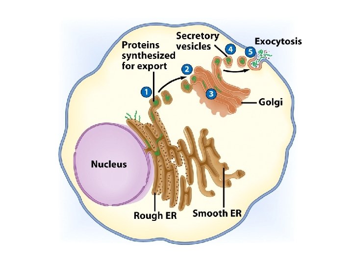
Endoplasmic Reticulum • Helps move substances within cells • Network of interconnected membranes • Two types – Rough endoplasmic reticulum – Smooth endoplasmic reticulum
Rough Endoplasmic Reticulum • Has ribosomes attached to surface – Manufacture proteins – Not all ribosomes are attached to rough ER. Some are free in the cytoplasm • May chemically modify proteins from ribosomes
Smooth Endoplasmic Reticulum • No attached ribosomes • Has enzymes that help build molecules – Carbohydrates – Lipids
Golgi Apparatus • Involved in synthesis of plant cell wall • Modifies, sorts and packages proteins and other materials from the ER for storage in the cell or for secretion outside of the cell.
Golgi Apparatus Function 1. Molecules come in vesicles 2. Vesicles fuse with Golgi membrane 3. Molecules may be modified by Golgi 4. Molecules pinched-off in separate vesicle 5. Vesicle leaves Golgi apparatus 6. Vesicles may combine with plasma membrane to secrete contents
Lysosomes • Contain digestive enzymes • Functions – Aid in cell renewal – Use enzymes to break down food and worn out cell parts – Digests invaders
Vacuoles • Membrane bound storage sacs • More common in plants than animals • Store – Water – Food – Wastes
Mitochondria • Have their own DNA • Bound by double membrane
Mitochondria • Produces the energy a cell needs to carry out its functions • Break down fuel molecules (cellular respiration) – Glucose – Fatty acids • Release energy – ATP
Chloroplasts • Capture energy from the sun and convert it into chemical energy • Photosynthesis
Microfilaments/Microtubules • Located all over the cell • 3 functions: – mechanical support – anchor organelles – help move substances
A = actin, IF = intermediate filament, MT = microtubule
Cilia & Flagella • Provide motility • Cilia – Short – Used to move substances outside human cells • Flagella – Whip-like extensions – Found on sperm cells
Cilia & Flagella Structure • Bundles of microtubules • Continuous with plasma membrane
Centrioles • Pairs of microtubular structures • Play a role in cell division
Learning Target • I can describe the functions of all major cell organelles.
Learning Target • I can explain how the cell membrane controls movement of substances both into and out of the cell and within the cell.
Cell Membrane • • Also known as the plasma membrane Contains cell contents Double layer of phospholipids & proteins Controls what enters or exits the cell
Phospholipids • Polar – Hydrophilic head – Hydrophobic tail • Interacts with water
Cell Walls • Found in plants, fungi, & many protists • Surrounds plasma membrane; provides support and protection for the cell
Cell Wall Differences • Plants – mostly cellulose • Fungi – contain chitin
Cytoplasm • Viscous fluid containing organelles • components of cytoplasm – – Interconnected filaments & fibers Fluid = cytosol Organelles (not nucleus) Storage substances
Learning Target • I can explain how the cell membrane controls movement of substances both into and out of the cell and within the cell.
Learning Targets • I can describe and contrast these types of cell transport: osmosis, diffusion, facilitated diffusion and active transport. • I can predict the effect of osmosis within cells based on the type of solution surrounding the cell.
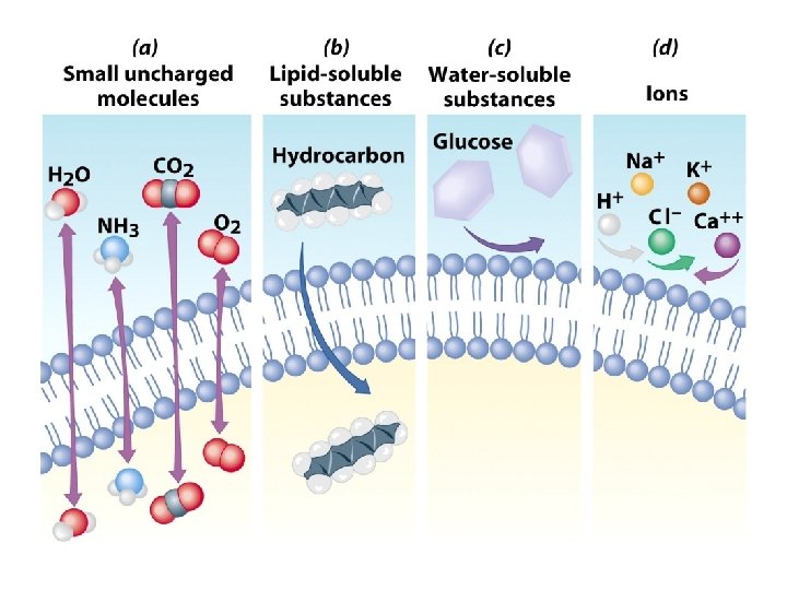
Movement Across the Plasma Membrane • Materials move through the plasma membrane in two ways: – Passive transport – Actively transport
Passive Transport • No energy required • Move due to gradient – differences in concentration, pressure, charge • Move to equalize gradient – Materials move from areas of high concentration to areas low concentration (downhill)
Types of Passive Transport 1. Diffusion 2. Osmosis 3. Facilitated diffusion
Diffusion • Molecules move to equalize concentration
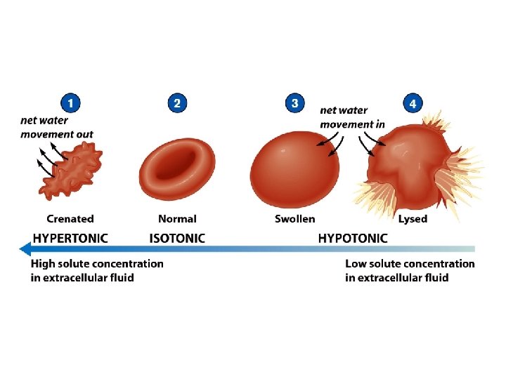
Osmosis • Special form of diffusion • Fluid flows from lower solute concentration • Often involves movement of water – Into cell – Out of cell
Solution Differences & Cells • solvent + solute = solution • Hypotonic – Solutes in cell more than outside – Outside solvent will flow into cell – Cell will swell • Isotonic – Solutes equal inside & out of cell • Hypertonic – Solutes greater outside cell – Fluid will flow out of cell – Cell will shrink
Facilitated Diffusion • Selectively permeable membrane • Channels (are specific) help molecule or ions enter or leave the cell • Channels usually are transport proteins (aquaporins facilitate the movement of water) • No energy is required
Process of Facilitated Transport • Protein binds with molecule • Shape of protein changes • Molecule moves across membrane
Membrane Proteins 1. Channels or transporters – Move molecules in one direction 2. Receptors – Recognize certain chemicals
Membrane Proteins 3. Glycoproteins – Identify cell type 4. Enzymes – Catalyze production of substances
Active Transport • Molecular movement • Requires energy (against gradient) • Example is sodium-potassium pump
Endocytosis • Movement of large molecules – Particles – Organisms • Movement is into cells • Types of endocytosis – bulk-phase (nonspecific) – receptor-mediated (specific)
Process of Endocytosis • Plasma membrane surrounds material • Edges of membrane meet • Membranes fuse to form vesicle
Forms of Endocytosis • Phagocytosis – cell eating • Pinocytosis – cell drinking
Exocytosis • Reverse of endocytosis • Cell discharges material
Process of Exocytosis • Vesicle moves to cell surface • Membrane of vesicle fuses • Materials expelled
End Chapter 5
Learning Targets • I can describe and contrast these types of cell transport: osmosis, diffusion, facilitated diffusion and active transport. • I can predict the effect of osmosis within cells based on the type of solution surrounding the cell.