Cell Structure and Function Cell Structure All Cells


 Ø Function – isolates the cell’s genetic material, DNA l DNA")


 Continuous with the outer membrane")
 l l The ER is continuous with the outer membrane of")
 • Network of flattened membrane sacs create")

 l l l Tubular membrane structure Continuous with")


 l l Move whole cells or materials")

 Ø Function – site of photosynthesis Ø Structure l l 2")


- Slides: 51
Cell Structure and Function
Cell Structure Ø All Cells have: an outermost plasma membrane l genetic material in the form of DNA l cytoplasm with ribosomes l
1. Plasma Membrane • All membranes are phospholipid bilayers with embedded proteins • The outer plasma membrane isolates cell contents l controls what gets in and out of the cell l receives signals l
2. Genetic material in the form of DNA l l Prokaryotes – no membrane around the DNA Eukaryotes – DNA is within a membrane
3. Cytoplasm with ribosomes l l Cytoplasm – fluid area inside outer plasma membrane and outside DNA region Ribosomes – make proteins
Why Are Cells So Small? Ø Cells need sufficient surface area to allow adequate transport of nutrients in and wastes out. Ø As cell volume increases, so does the need for the transporting of nutrients and wastes.
Why Are Cells So Small? Ø However, as cell volume increases the surface area of the cell does not expand as quickly. l If the cell’s volume gets too large it cannot transport enough wastes out or nutrients in. Ø Thus, surface area limits cell volume/size.
Prokaryotic Cell Structure Ø Prokaryotic Cells are smaller and simpler in structure than eukaryotic cells. Typical prokaryotic cell is _____ l Prokaryotic cells do NOT have: l • Nucleus • Membrane bound organelles
Prokaryotic Cell
TEM Prokaryotic Cell
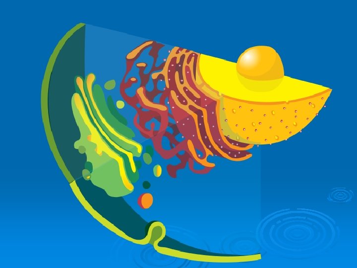
Eukaryotic Cells Ø Structures in all eukaryotic cells l l l Nucleus Ribosomes Endomembrane System • Endoplasmic reticulum – smooth and rough • Golgi apparatus • Vesicles l l Mitochondria Cytoskeleton
NUCLEUS CYTOSKELETON RIBOSOMES ROUGH ER MITOCHONDRION CYTOPLASM SMOOTH ER CENTRIOLES GOLGI BODY PLASMA MEMBRANE LYSOSOME VESICLE Fig. 4 -15 b, p. 59
Nucleus (4. 5) Ø Function – isolates the cell’s genetic material, DNA l DNA directs/controls the activities of the cell • DNA determines which types of RNA are made • The RNA leaves the nucleus and directs the synthesis of proteins in the cytoplasm at a _______
Nucleus Ø Structure l Nuclear envelope • Two Phospholipid bilayers with protein lined pores l l Each pore is a ring of 8 proteins with an opening in the center of the ring Nucleoplasm – fluid of the nucleus
Nuclear pore bilayer facing cytoplasm Nuclear envelope bilayer facing nucleoplasm Fig. 4 -17, p. 61
Nucleus Ø DNA is arranged in chromosomes l l Chromosome – fiber of DNA with proteins attached Chromatin – all of the cell’s DNA and the associated proteins
Nucleus Ø Structure, continued l Nucleolus • Area of condensed DNA • Where ribosomal subunits are made l Subunits exit the nucleus via nuclear pores
ADD THE LABELS
Endomembrane System Ø Series of organelles responsible for: Modifying protein chains into their final form l Synthesizing of lipids l Packaging of fully modified proteins and lipids into vesicles for export or use in the cell l And more that we will not cover! l
Structures of the Endomembrane System Ø Endoplasmic Reticulum (ER) Continuous with the outer membrane of the nuclear envelope l Two forms - smooth and rough l Ø Transport vesicles Ø Golgi apparatus
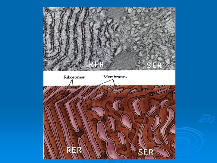
Endoplasmic Reticulum (ER) l l The ER is continuous with the outer membrane of the nuclear envelope There are 2 types of ER: • Rough ER – has ribosomes attached • Smooth ER – no ribosomes attached
Endoplasmic Reticulum Ø Rough Endoplasmic Reticulum (RER) • Network of flattened membrane sacs create a “maze” l RER contains enzymes that recognize and modify proteins • Ribosomes are attached to the outside of the RER and make it appear rough
Endoplasmic Reticulum Ø Function RER • Proteins are modified as they move through the RER • Once modified, the proteins are packaged in transport vesicles for transport to the Golgi body
Endomembrane System Ø Smooth ER (SER) l l l Tubular membrane structure Continuous with RER No ribosomes attached Ø Function SER l Lipids are made inside the SER • fatty acids, phospholipids, sterols. . l Lipids are packaged in transport vesicles and sent to the Golgi
Golgi Apparatus Ø Golgi Apparatus l Stack of flattened membrane sacs Ø Function Golgi apparatus l l Completes the processing substances received from the ER Sorts, tags and packages fully processed proteins and lipids in vesicles
Golgi Apparatus
Transport Vesicles Ø Transport Vesicles l l Vesicle = small membrane bound sac Transport modified proteins and lipids from the ER to the Golgi apparatus (and from Golgi to final destination)
Endomembrane System Ø Putting it all together l DNA directs RNA synthesis RNA exits nucleus through a nuclear pore ribosome protein is made proteins with proper code enter RER proteins are modified in RER and lipids are made in SER vesicles containing the proteins and lipids bud off from the ER
Endomembrane System Ø Putting it all together ER vesicles merge with Golgi body proteins and lipids enter Golgi each is fully modified as it passes through layers of Golgi modified products are tagged, sorted and bud off in Golgi vesicles …
Endomembrane System Ø Putting it all together Golgi vesicles either merge with the plasma membrane and release their contents OR remain in the cell and serve a purpose Another animation
Lysosomes Ø The lysosome is an example of an organelle made at the Golgi apparatus. l Golgi packages digestive enzymes in a vesicle. The vesicle remains in the cell and: • Digests unwanted or damaged cell parts • Merges with food vacuoles and digest the contents
Lysosomes Ø Tay-Sachs disease occurs when the lysosome is missing the enzyme needed to digest a lipid found in nerve cells. l As a result the lipid accumulates and nerve cells are damaged as the lysosome swells with undigested lipid.
Mitochondria Function – synthesis of ATP Ø l 3 major pathways involved in ATP production 1. Glycolysis 2. Krebs Cycle 3. Electron transport system (ETS)
Mitochondria
Mitochondria TEM
Cytoskeleton
Cytoskeleton Ø Intermediate filaments l l Present only in animal cells of certain tissues Fibrous proteins join to form a rope-like structure • Provide internal structure • Anchor organelles in place.
Cilia and flagella (structures for cell motility) l l Move whole cells or materials across the cell surface Microtubules wrapped in an extension of the plasma membrane (9 + 2 arrangement of MT)
Plant Cell Structures Ø Structures found in plant, but not animal cells l l Chloroplasts Central vacuole Other plastids/vacuoles – chromoplast, amyloplast Cell wall
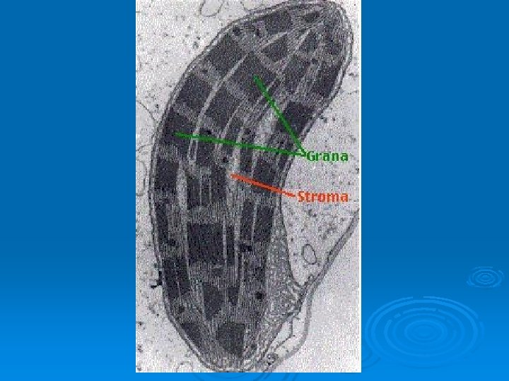
Chloroplasts (4. 14) Ø Function – site of photosynthesis Ø Structure l l 2 outer membranes Thylakoid membrane system • Stacked membrane sacs called granum l l Chlorophyll in granum Stroma • Fluid part of chloroplast
Plastids/Vacuoles in Plants Ø Chromoplasts – contain colored pigments • Pigments called carotenoids Ø Amyloplasts – store starch
Central Vacuole Ø Function – storage area for water, sugars, ions, amino acids, and wastes l Some central vacuoles serve specialized functions in plant cells. • May contain poisons to protect against predators
Central Vacuole Ø Structure l l l Large membrane bound sac Occupies the majority of the volume of the plant cell Increases cell’s surface area for transport of substances cells can be larger
Cell Wall Ø Function – provides structure and protection l l Ø Never found in animal cells Present in plant, bacterial, fungus, and some protists Structure l l l Wraps around the plasma membrane Made of cellulose and other polysaccharides Connect by plasmodesmata (channels through the walls)
Typical Plant Cell
Typical Plant Cell –add the labels
Origin of Mitochondria and Chloroplasts Ø Both organelles are believed to have once been free-living bacteria that were engulfed by a larger cell.
Proposed Origin of Mitochondria and Chloroplasts Ø Evidence: l l l Each have their own DNA Their ribosomes resemble bacterial ribosomes Each can divide on its own Mitochondria are same size as bacteria Each have more than one membrane