Universit degli Studi di Messina UOC di Cardiochirurgia
- Slides: 79
Università degli Studi di Messina UOC di Cardiochirurgia Azienda Ospedaliera Universitaria Policlinico “G. Martino” dir. Prof. Roberto Gaeta PROTESI VALVOLARI CARDIACHE - corso integrato malattie apparato cardiovascolare e respiratorio - Prof R Gaeta Chirurgia Cardiaca (SSD Med 23)
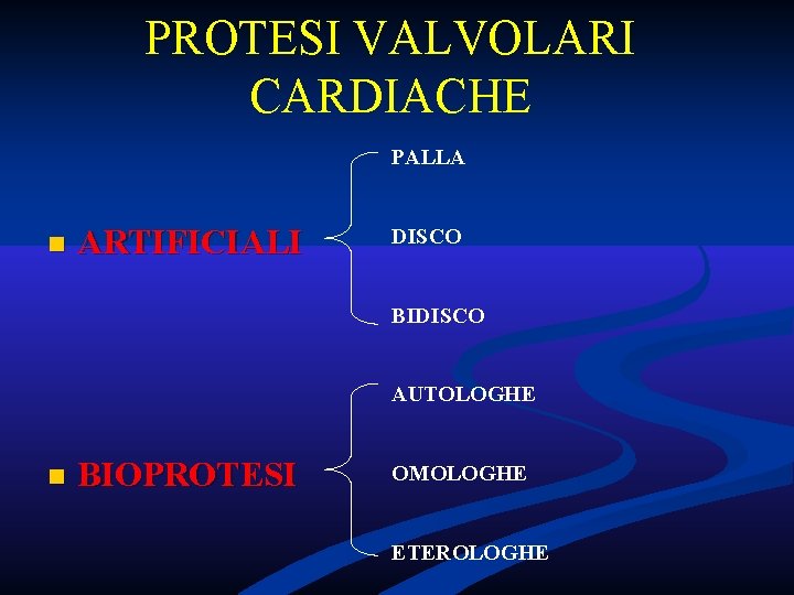
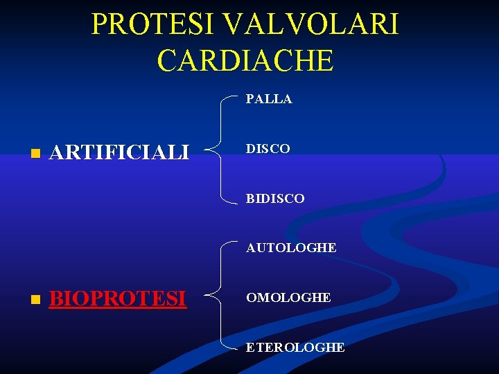
PROTESI VALVOLARI CARDIACHE PALLA n ARTIFICIALI DISCO BIDISCO AUTOLOGHE n BIOPROTESI OMOLOGHE ETEROLOGHE
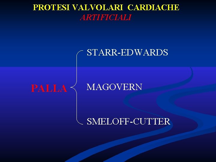
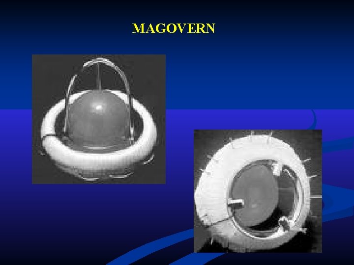
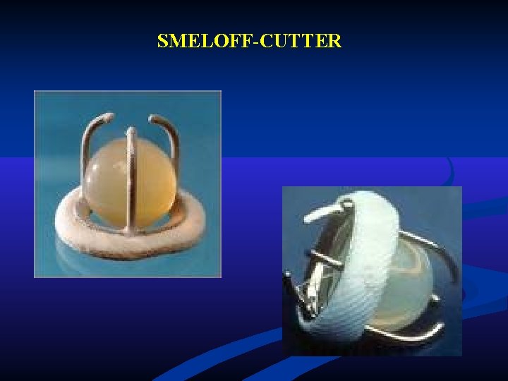
PROTESI VALVOLARI CARDIACHE ARTIFICIALI STARR-EDWARDS PALLA MAGOVERN SMELOFF-CUTTER
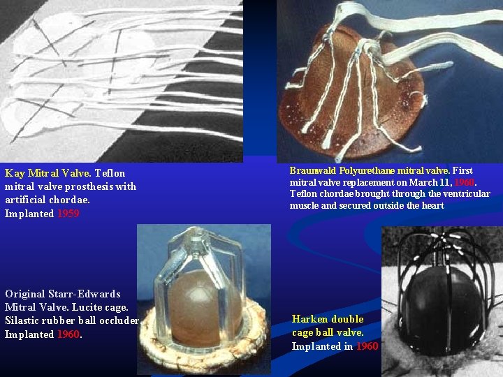
Kay Mitral Valve. Teflon mitral valve prosthesis with artificial chordae. Implanted 1959 Original Starr-Edwards Mitral Valve. Lucite cage. Silastic rubber ball occluder. Implanted 1960. Braunwald Polyurethane mitral valve. First mitral valve replacement on March 11, 1960. Teflon chordae brought through the ventricular muscle and secured outside the heart Harken double cage ball valve. Implanted in 1960
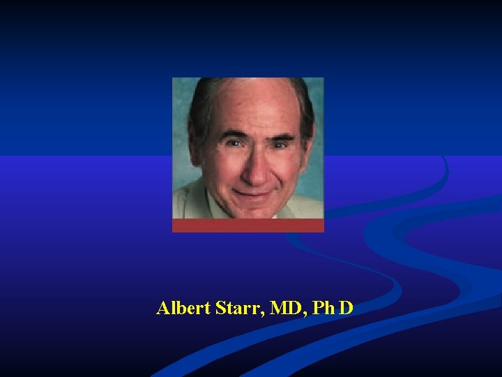
Albert Starr, MD, Ph D
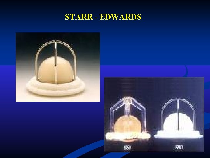
STARR - EDWARDS
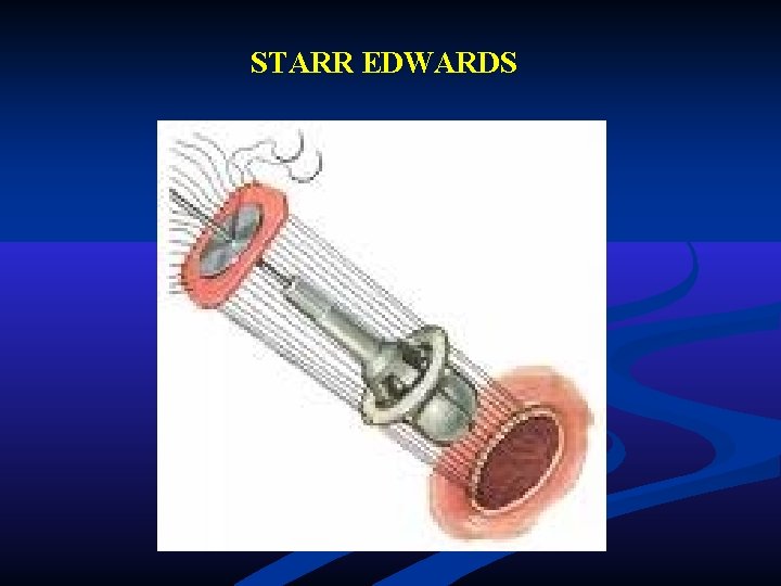
STARR EDWARDS
MAGOVERN
SMELOFF-CUTTER
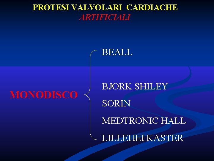
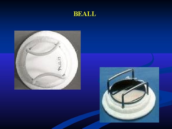
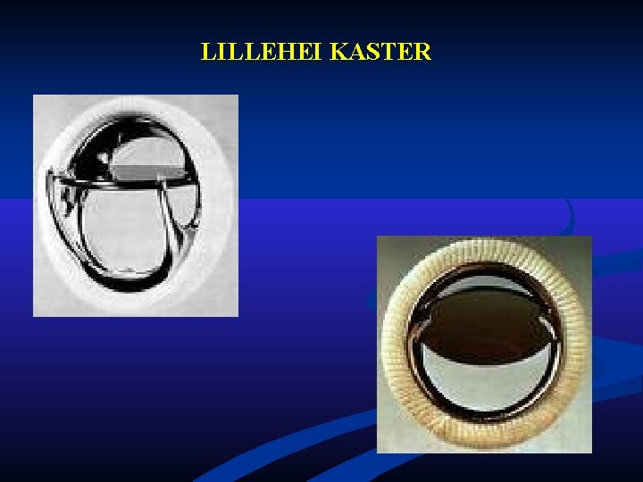
PROTESI VALVOLARI CARDIACHE ARTIFICIALI BEALL MONODISCO BJORK SHILEY SORIN MEDTRONIC HALL LILLEHEI KASTER
BEALL
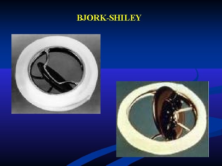
BJORK-SHILEY
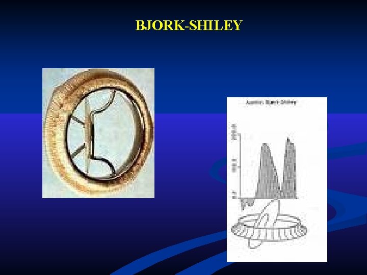
BJORK-SHILEY
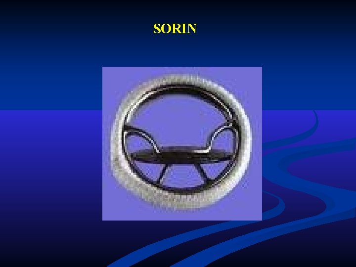
SORIN
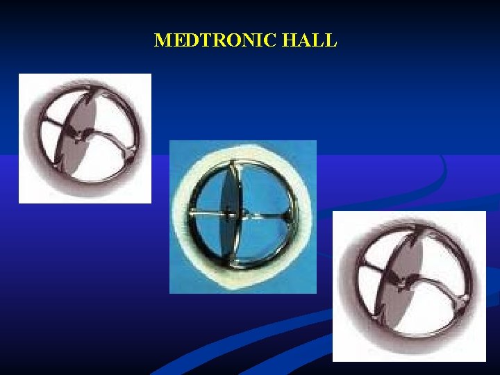
MEDTRONIC HALL
LILLEHEI KASTER
PROTESI VALVOLARI CARDIACHE ARTIFICIALI ST. JUDE BIDISCO CARBOMEDICS DUROMEDICS SORIN
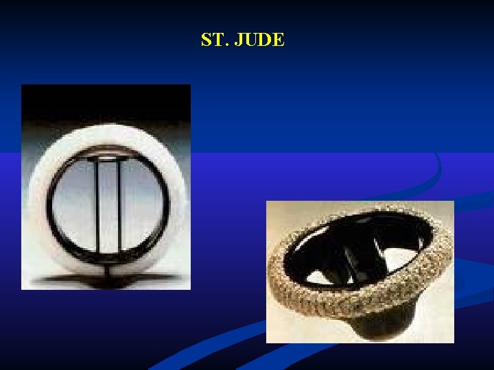
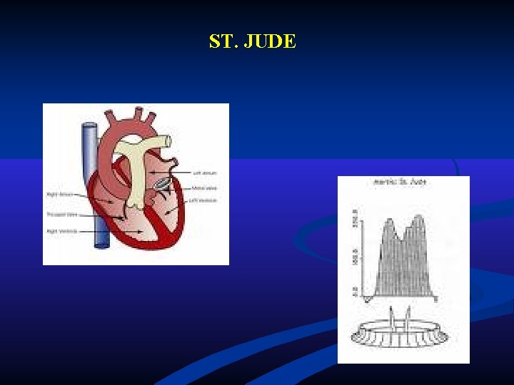
ST. JUDE
ST. JUDE
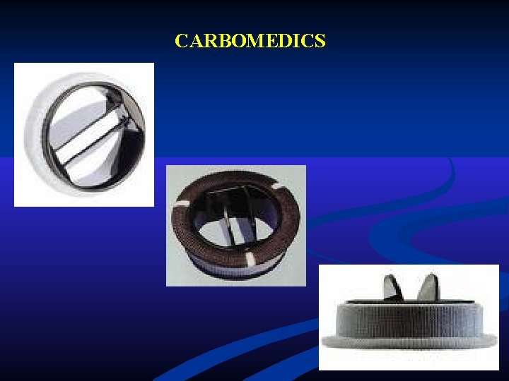
CARBOMEDICS
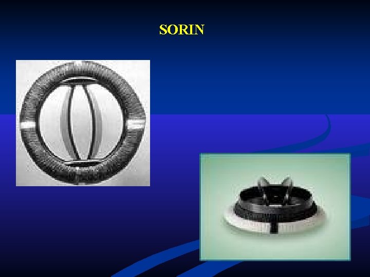
SORIN
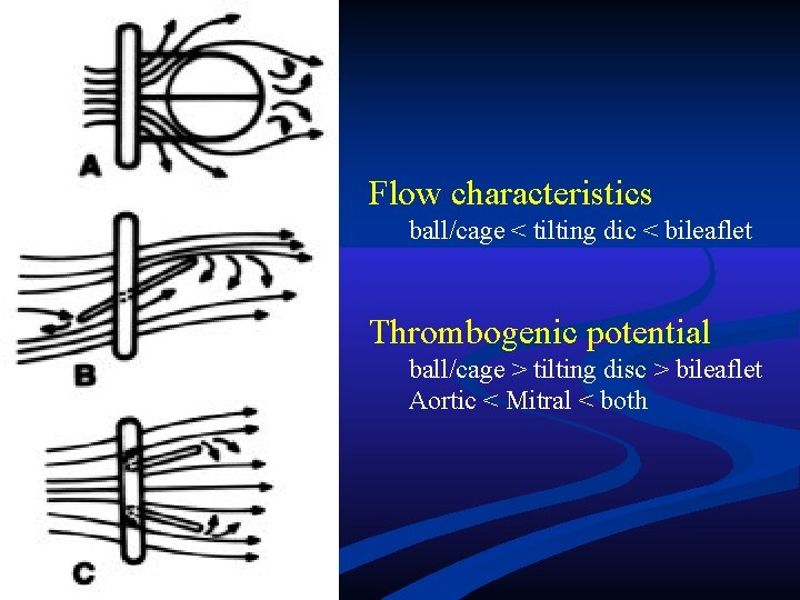
Flow characteristics ball/cage < tilting dic < bileaflet Thrombogenic potential ball/cage > tilting disc > bileaflet Aortic < Mitral < both
PROTESI VALVOLARI CARDIACHE PALLA n ARTIFICIALI DISCO BIDISCO AUTOLOGHE n BIOPROTESI OMOLOGHE ETEROLOGHE
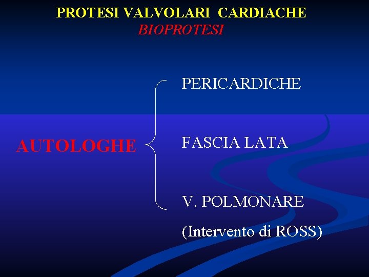
PROTESI VALVOLARI CARDIACHE BIOPROTESI PERICARDICHE AUTOLOGHE FASCIA LATA V. POLMONARE (Intervento di ROSS)
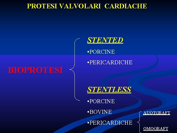
PROTESI VALVOLARI CARDIACHE STENTED • PORCINE BIOPROTESI • PERICARDICHE STENTLESS • PORCINE • BOVINE AUOTGRAFT • PERICARDICHE OMOGRAFT
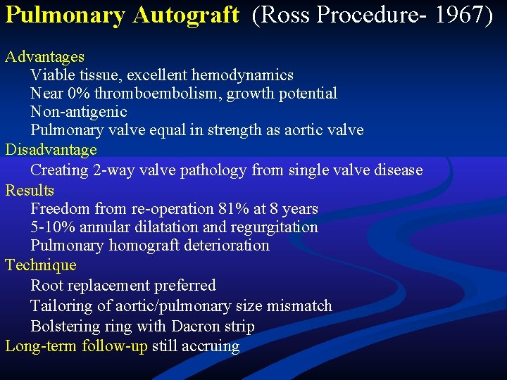
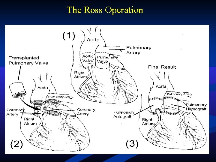
Pulmonary Autograft (Ross Procedure- 1967) Advantages Viable tissue, excellent hemodynamics Near 0% thromboembolism, growth potential Non-antigenic Pulmonary valve equal in strength as aortic valve Disadvantage Creating 2 -way valve pathology from single valve disease Results Freedom from re-operation 81% at 8 years 5 -10% annular dilatation and regurgitation Pulmonary homograft deterioration Technique Root replacement preferred Tailoring of aortic/pulmonary size mismatch Bolstering with Dacron strip Long-term follow-up still accruing
The Ross Operation
PROTESI VALVOLARI CARDIACHE BIOPROTESI OMOLOGHE HOMOGRAFT (cadavere)
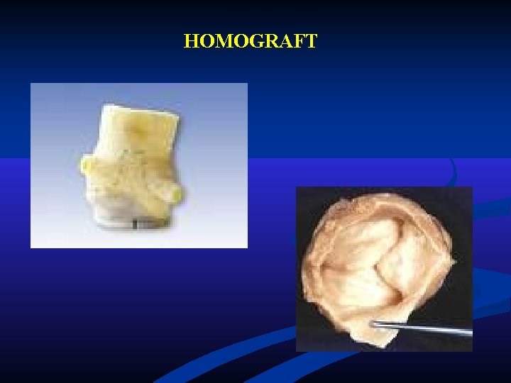
HOMOGRAFT
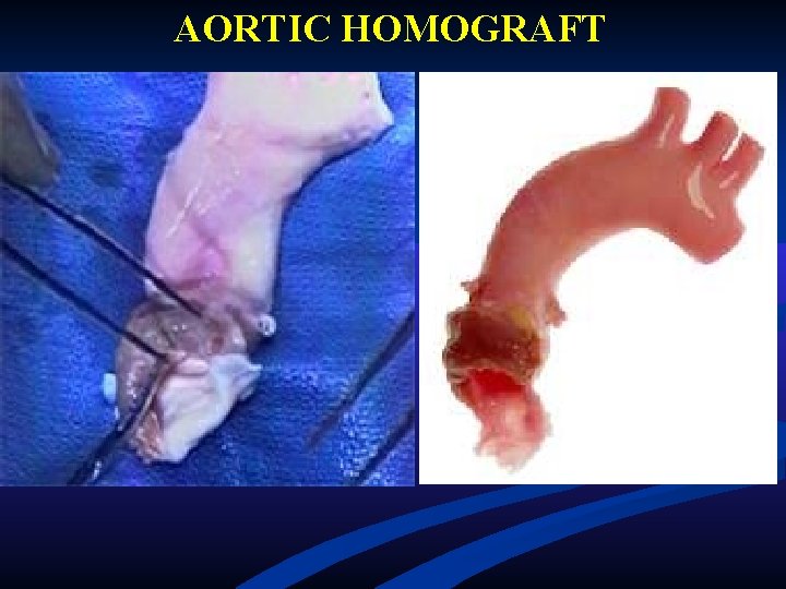
AORTIC HOMOGRAFT
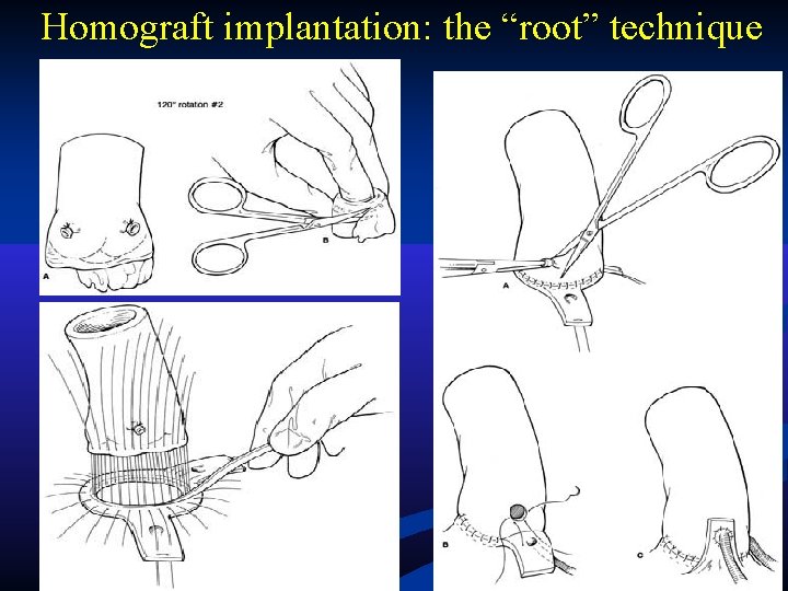
Homograft implantation: the “root” technique
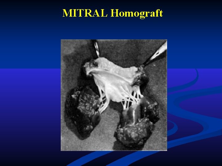
MITRAL Homograft
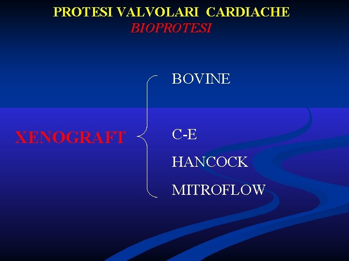
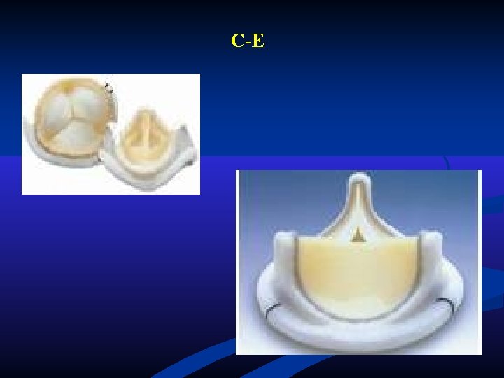
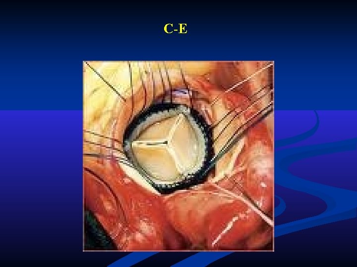
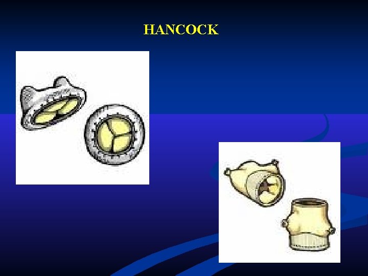
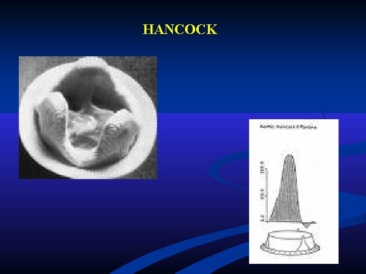
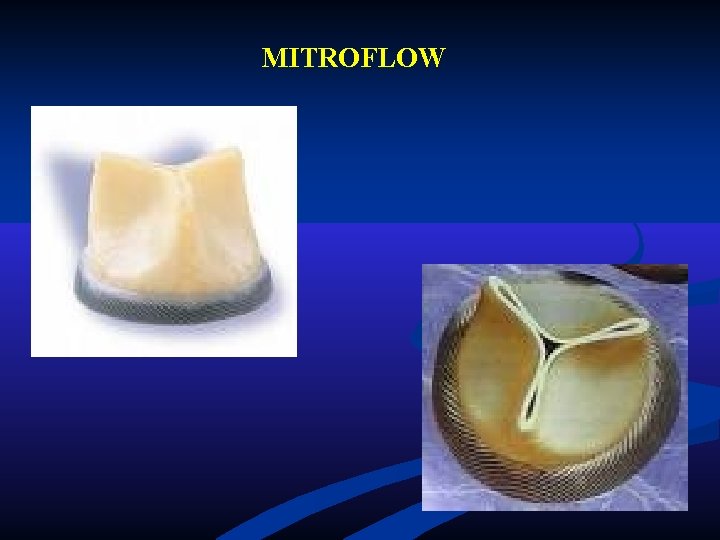
PROTESI VALVOLARI CARDIACHE BIOPROTESI BOVINE XENOGRAFT C-E HANCOCK MITROFLOW
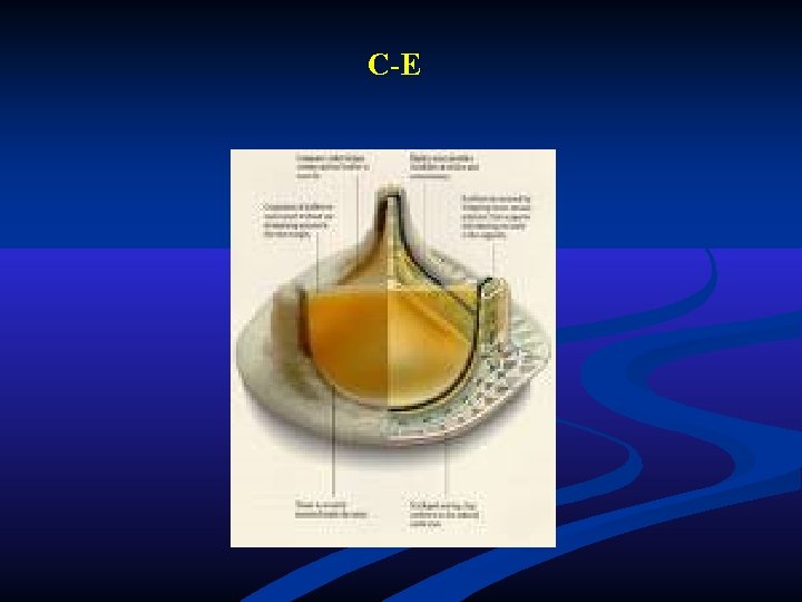
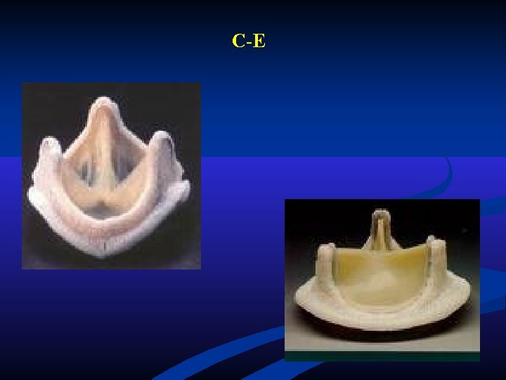
C-E
C-E
C-E
C-E
HANCOCK
HANCOCK
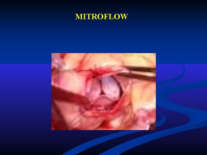
MITROFLOW
MITROFLOW
PROTESI VALVOLARI CARDIACHE ARTIFICIALI • ANTICOAGULAZIONE • LUNGA DURATA • RUMORE
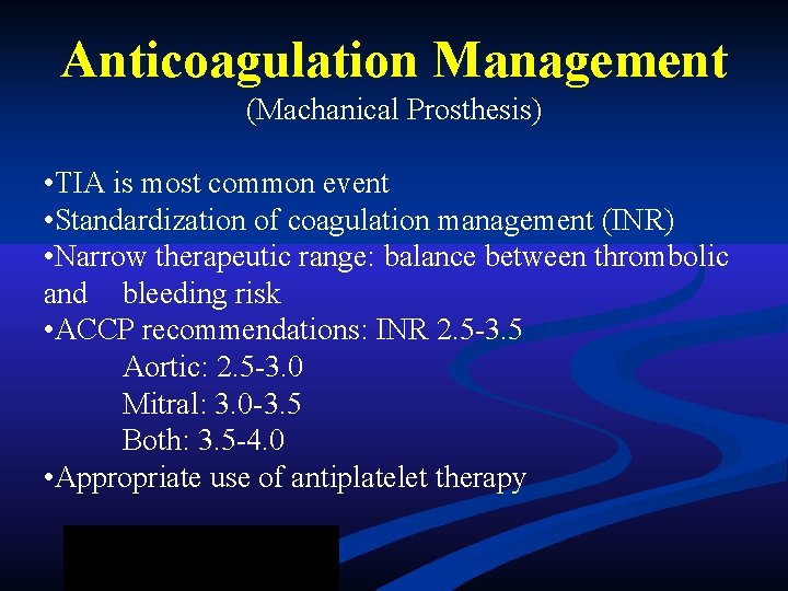
Anticoagulation Management (Machanical Prosthesis) • TIA is most common event • Standardization of coagulation management (INR) • Narrow therapeutic range: balance between thrombolic and bleeding risk • ACCP recommendations: INR 2. 5 -3. 5 Aortic: 2. 5 -3. 0 Mitral: 3. 0 -3. 5 Both: 3. 5 -4. 0 • Appropriate use of antiplatelet therapy
PROTESI VALVOLARI CARDIACHE BIOLOGICHE • NO ANTICOAGULAZIONE • DURATA MEDIA (10 -12 anni) • SILENZIOSE
PROTESI VALVOLARI CARDIACHE • TROMBOEMBOLIA • INFEZIONI • LEAK
The Perfect Valve • Excellent hemodynamics • Non-thrombogenic • Durable • Unrestricted availability • Easily implantable • Silent function • Low cost
Valvulopatia Mitralica
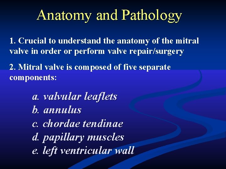
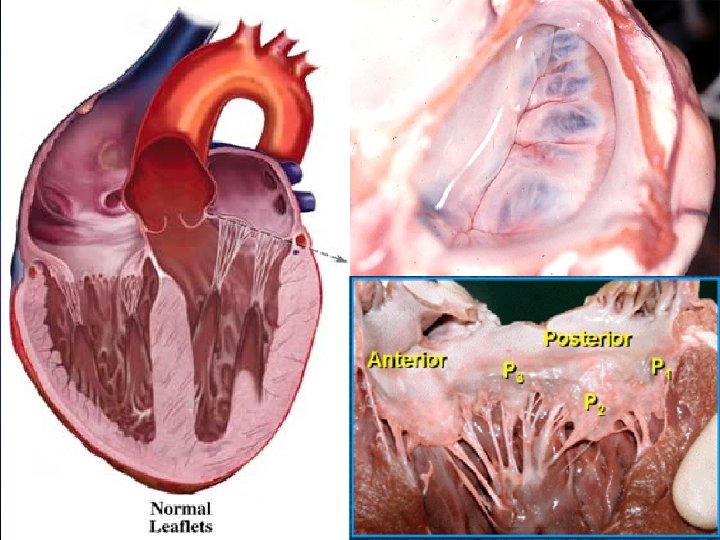
Anatomy and Pathology 1. Crucial to understand the anatomy of the mitral valve in order or perform valve repair/surgery 2. Mitral valve is composed of five separate components: a. valvular leaflets b. annulus c. chordae tendinae d. papillary muscles e. left ventricular wall
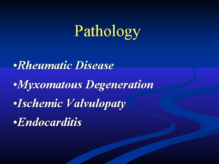
Pathology • Rheumatic Disease • Myxomatous Degeneration • Ischemic Valvulopaty • Endocarditis
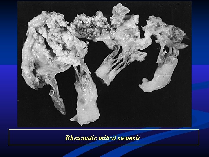
Rheumatic mitral stenosis
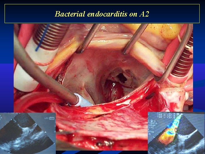
Bacterial endocarditis on A 2
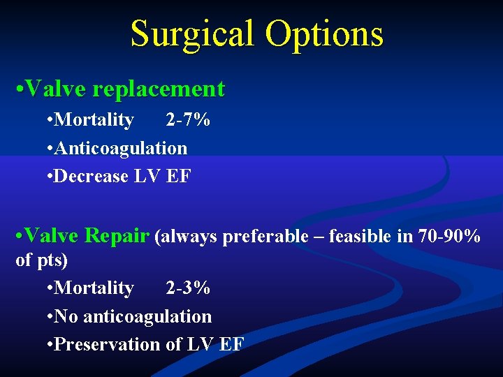
Surgical Options • Valve replacement • Mortality 2 -7% • Anticoagulation • Decrease LV EF • Valve Repair (always preferable – feasible in 70 -90% of pts) • Mortality 2 -3% • No anticoagulation • Preservation of LV EF
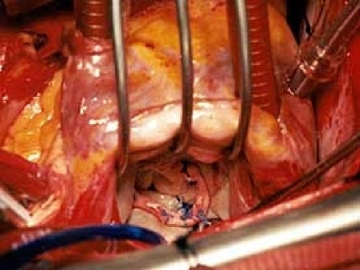
Tecniche Riparative Mitraliche • Riparazioni Anatomiche • Annulus • Lembi • Corde Tendinee • Muscoli Papillari • Riparazioni Funzionali
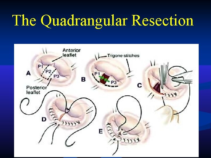
The Quadrangular Resection
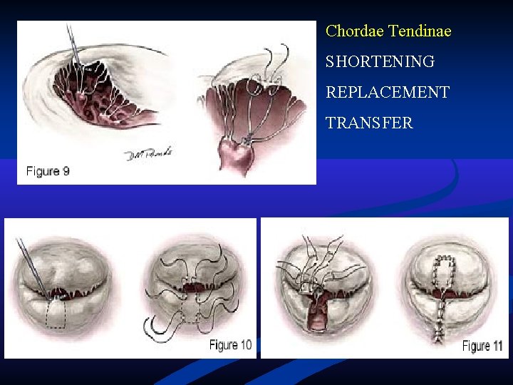
Chordae Tendinae SHORTENING REPLACEMENT TRANSFER
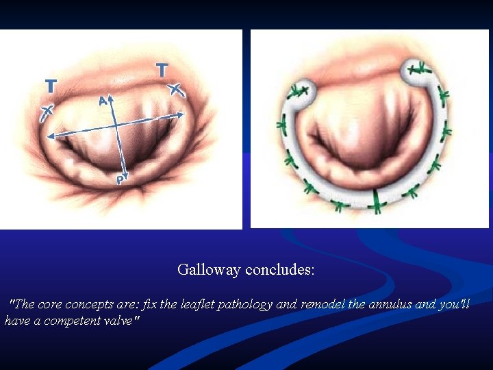
Galloway concludes: "The core concepts are: fix the leaflet pathology and remodel the annulus and you'll have a competent valve"
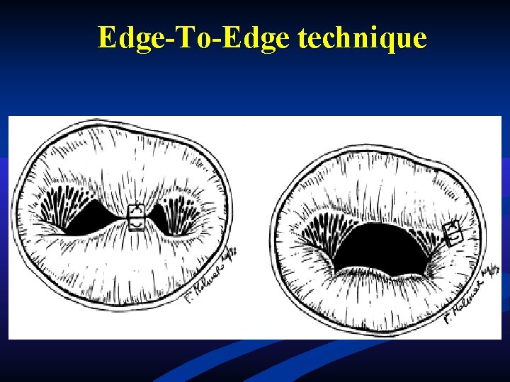
Edge-To-Edge technique
Valvulopatia Aortica
Anatomy and Pathology 1. Crucial to understand the anatomy of the aortic valve in order or perform valve repair/surgery 2. Aortic valve is composed of five separate components: a. valvular cusps b. annulus c. Valsalva’s sinus d. sinus-tubular junction e. Aortic root
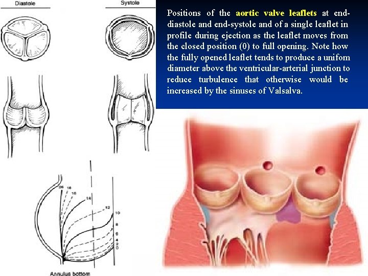
Positions of the aortic valve leaflets at enddiastole and end-systole and of a single leaflet in profile during ejection as the leaflet moves from the closed position (0) to full opening. Note how the fully opened leaflet tends to produce a unifom diameter above the ventricular-arterial junction to reduce turbulence that otherwise would be increased by the sinuses of Valsalva.
Pathology • Congenital Malformation • Rheumatic Disease • Degeneration • Endocarditis
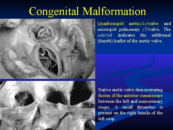
Congenital Malformation Quadricuspid aortic(Ao)valve and unicuspid pulmonary (P)valve. The asterisk indicates the additional (fourth) leaflet of the aortic valve. Native aortic valve demonstrating fusion of the anterior commissure between the left and noncoronary cusps. A small thrombus is present on the right lunula of the left cusp.
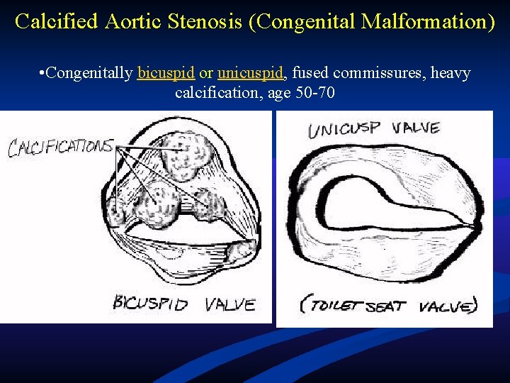
Calcified Aortic Stenosis (Congenital Malformation) • Congenitally bicuspid or unicuspid, fused commissures, heavy calcification, age 50 -70
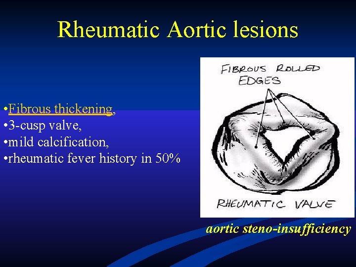
Rheumatic Aortic lesions • Fibrous thickening, • 3 -cusp valve, • mild calcification, • rheumatic fever history in 50% aortic steno-insufficiency
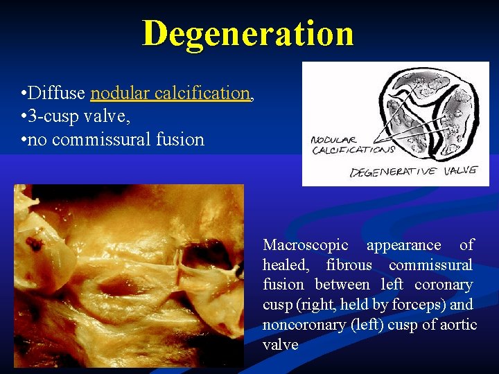
Degeneration • Diffuse nodular calcification, • 3 -cusp valve, • no commissural fusion Macroscopic appearance of healed, fibrous commissural fusion between left coronary cusp (right, held by forceps) and noncoronary (left) cusp of aortic valve
Endocarditis Infective endocarditis is defined as an infection of the endocardial surface of the heart, which may include one or more heart valves, the mural endocardium, or a septal defect. Endocarditis can be broken down into the following categories: • Native valve (acute and subacute) endocarditis • Prosthetic valve (early and late) endocarditis • Endocarditis related to intravenous drug use
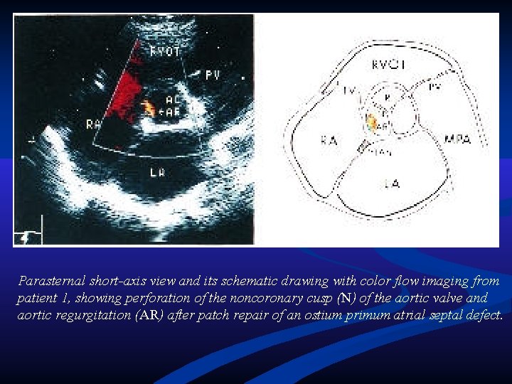
Parasternal short-axis view and its schematic drawing with color flow imaging from patient 1, showing perforation of the noncoronary cusp (N) of the aortic valve and aortic regurgitation (AR) after patch repair of an ostium primum atrial septal defect.
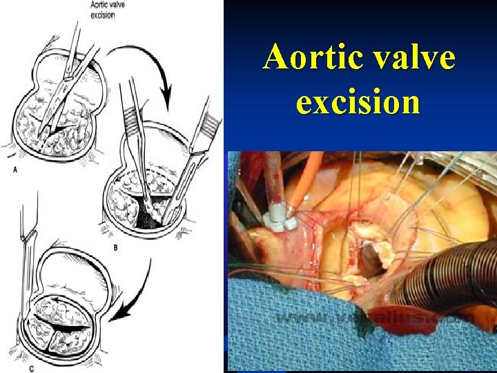
Aortic valve excision
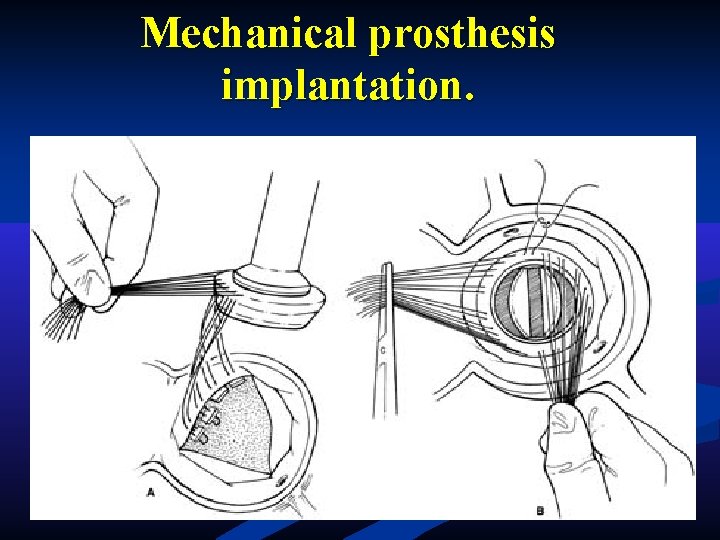
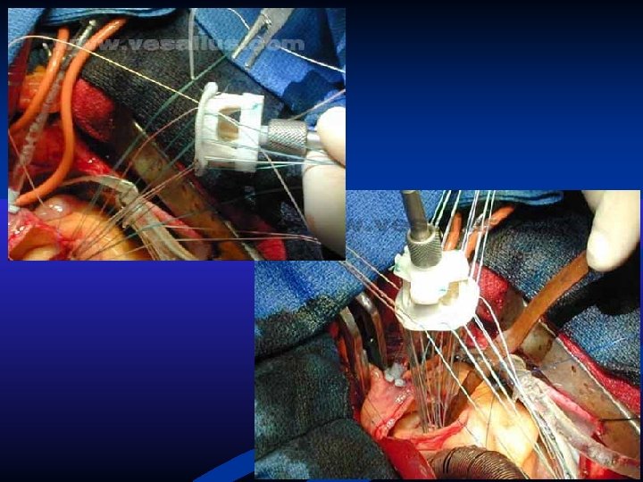
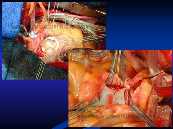
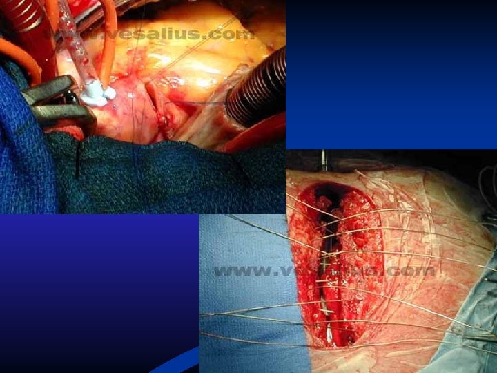
Mechanical prosthesis implantation.
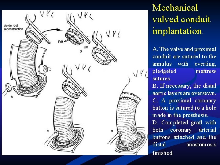
Mechanical valved conduit implantation. A. The valve and proximal conduit are sutured to the annulus with everting, pledgeted mattress sutures. B. If necessary, the distal aortic layers are oversewn. C. A proximal coronary button is sutured to a hole made in the prosthesis. D. Completed graft with both coronary arterial buttons attached and the distal anastomosis finished.
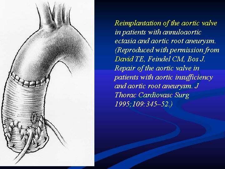
Reimplantation of the aortic valve in patients with annuloaortic ectasia and aortic root aneurysm. (Reproduced with permission from David TE, Feindel CM, Bos J. Repair of the aortic valve in patients with aortic insufficiency and aortic root aneurysm. J Thorac Cardiovasc Surg 1995; 109: 345– 52. )
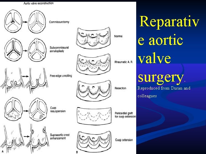
Reparativ e aortic valve surgery. Reproduced from Duran and colleagues
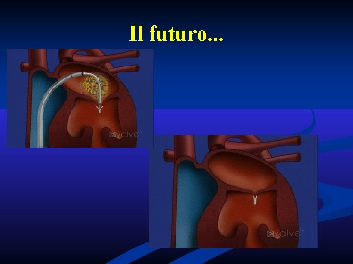
Il futuro. . .