The Cardiovascular System The major organs of the


 from the lungs to")


 Value • Mitral valve: separates the left Mitral valve: atrium from the")


 of the")


 • serves as the")
 • The impulse that originates from the SA node")


- Slides: 75
The Cardiovascular System The major organs of the cardiovascular system The heart structure and function
After today you should be able to: For more help: Chapter 13 pp. 329364 1. Name the organs of the cardiovascular system and discuss their functions. 2. Name and describe the locations and functions of the major parts of the heart. 3. Trace the pathway of the blood through the heart and the vessels of the coronary circulation.
Major organs of the cardiovascular system • The heart – located in the pericardial cavity, slightly to the left, close to the left lung, and rests on the diaphragm • Arteries – strong elastic vessels that are adapted for carrying blood away from the heart under high pressure
Major organs of the cardiovascular system • Arterioles – Arterioles smaller branches coming from the arteries • Capillaries smallest of the artery system, connect the smallest arterioles and the smallest venules
Major organs of the cardiovascular system • Venules – Venules the smallest vessels of the venous system, that continue from the capillaries and merge to form veins • Veins- carry blood back to the atria of the heart following pathways that are almost parallel to the arteries. Similar to arteries, but have thinner walls, and generally have flap like valves. Generally lower pressure than that of the arteries.
The Heart • In the course of a lifetime, a human heart can beat over two million times. • It can carry 7, 000 liters of blood throughout the body. • Composed of cardiac muscle tissue • The wall of the heart or tissue is made up of three levels: outer most: epicardium, middle: myocardium and inner: endocardium
The Heart • The heart is divided into four chambers: – The LEFT and RIGHT ATRIA – The LEFT and RIGHT VENTRICLES • A sophisticated valve system controls blood flow between the chambers. In fact, it is the latching (when they open and close) of the heart valves that creates the beating sound of the heart. • There are four distinct valves.
The Heart • The four chambers:
• The valves: The Heart
Superior Vena Cava The superior vena cava is one of the two main veins bringing de-oxygenated blood from the body to the heart. Veins from the head and upper body feed into the superior vena cava, which empties into the right atrium of the heart. Why is the blood here de-oxygenated?
Inferior Vena Cava • Inferior vena cava : is Inferior vena cava : one of the two main veins bringing de-oxygenated blood from the body to the heart. • Veins from the legs and lower torso feed into the inferior vena cava, which empties into the right atrium of the heart.
Right Atrium • The right atrium receives deoxygenated blood from the body through the superior vena cava and inferior vena cava.
Tricuspid Valve • Tricuspid valve: separates the right atrium from the Tricuspid valve: right ventricle. • It opens to allow the de-oxygenated blood from the right atrium to flow into the right ventricle. • It closes as the right ventricle contracts, preventing blood from returning to the right atrium. • Forces blood to exit into the pulmonary artery.
Right Ventricle • The right ventricle: receives de-oxygenated blood as the right atrium contracts. • This blood will move from the right ventricle to the pulmonary artery leading to the lungs.
Pulmonary Valve • Pulmonary valve: Pulmonary valve separates the right ventricle from the pulmonary artery. • As the ventricles contract, it opens to allow the deoxygenated blood from the right ventricle to flow to the lungs. • It closes as the ventricles relax, preventing blood from returning to the heart.
Pulmonary Artery • Pulmonary artery: is Pulmonary artery: the vessel transporting deoxygenated blood from the right ventricle to the lungs. • A common misconception is that all arteries carry oxygenrich blood. • It is more appropriate to classify arteries as vessels carrying blood away from the heart.
Summarize what we know so far: ANSWER THE FOLLOWING QUESTIONS: 1. 2. 3. 4. 5. Where does blood originate from? What in the heart does it enter first? Where does it go next? What are the roles of the 2 valves? Where does blood exit and go to from the right side of the heart? 6. Is it de-oxygenated (oxygen poor) or oxygenated (oxygen rich)?
Blood flow through the body
Cardio-respiratory connection
Cardio-respiratory connection
The Heart: Left side • Brings oxygenated (oxygen rich blood) from the lungs to the heart. • Begins with the Pulmonary Vein.
Pulmonary Vein • Pulmonary vein: is Pulmonary vein: the vessel transporting oxygenrich blood from the lungs to the left atrium. • A common misconception is that all veins carry deoxygenated blood. • It is more appropriate to classify veins as vessels carrying blood to the heart.
Left Atrium • Left atrium: receives Left atrium: oxygenated blood from the lungs through the pulmonary vein. • As the heart contracts (triggered by the sinoatrial node) Blood travels through the atria. • It passes through the mitral valve into the left ventricle.
Mitral (Bicuspid) Value • Mitral valve: separates the left Mitral valve: atrium from the left ventricle. • It opens to allow the oxygenated blood in the left atrium to flow into the left ventricle. • It closes as the left ventricle contracts, preventing blood from returning to the left atrium. • Forcing it to exit through the aortic valve into the aorta.
Left Ventricle • Left ventricle: receives Left ventricle: oxygenated blood as the left atrium contracts. • The walls of the left ventricle are thicker than the walls of the right ventricle, so that they can generate enough force to push the blood from the left ventricle into the aorta.
Aortic Valve • Aortic valve: separates the Aortic valve: left ventricle from the aorta. • As the ventricles contract, it opens to allow the oxygenated blood collected in the left ventricle to flow throughout the body. • It closes as the ventricles relax, preventing blood from returning to the heart.
Aorta • Aorta: is the largest single blood vessel in the body. • It is approximately the diameter of your thumb. • This vessel carries oxygenrich blood from the left ventricle to the various parts of the body.
Papillary Muscles • Papillary muscles: Papillary muscles attach to the lower portion of the interior wall of the ventricles. • They connect to the chordae tendineae on tendineae the valves, • The contraction of the papillary muscles opens the valves. When the papillary muscles relax, the valves close.
Papillary Muscles
Chordae Tendineae • Chordae tendineae are tendons linking the papillary Chordae tendineae muscles to the tricuspid valve in the right ventricle and the mitral valve in the left ventricle. • The chordae tendineae are string-like in appearance and are sometimes referred to as "heart strings. "
Ventricular Septum • Ventricular Septum: wall separating the lower chambers (the ventricles) of the heart from one another.
The Heart
The Heart
The Heart
As a group of 4: • With the construction paper: Create the heart. • Show the major organs that we discussed (arteries, veins, valves, chambers, etc) • Show the pathway that blood takes • Make sure the red and blue construction paper are in the correct location for oxygenated and deoxygenated. • Use the white and black construction paper to show where CO 2 is and goes and where O 2 is and goes. • The shape needs to look similar to the actual heart.
Act out the blood flow through the circulatory and respiratory system card activity
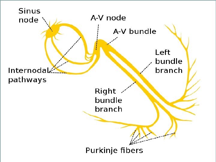
Sinoatrial Node (often called the SA node or sinus node) • serves as the natural pacemaker for the heart. • Nestled in the upper area of the right atrium, it sends the electrical impulse that triggers each heartbeat. • The impulse spreads through the atria, coordinated wave-like manner.
Atrioventricular node (or AV node) • The impulse that originates from the SA node strikes AV node • situated in the lower portion of the right atrium. • The AV node in turn sends an impulse through the nerve network to the ventricles to contract.
Right and Left Bundle Branches. • electrical network serving the upper ventricles • These nerve fibers send impulses that cause the cardiac muscle tissue to contract.
Purkinje Fibers • electrical network serving the lower ventricles • These nerve fibers send impulses that cause the cardiac muscle tissue to contract.
Electrical Conduction Pathway:
Electrical Conduction Pathway: • The SA Node to the AV Node to the left and right Bundle Branches - to the Purkinje Fibers = THE HEART BEAT and CONTRACTIONS
BLOOD • Blood is a mixture of Cells and Plasma • The heart pumps blood through arteries • Blood carries oxygen to the body and wastes away from the body.
Blood Cells: Contains 3 types of Cells: • RED BLOOD CELLS • WHITE BLOOD CELLS • PLATELETS
Blood Cells: Identify the components: white blood cell platelets plasma
Red Blood Cells: Erythrocytes • Biconcave discs that allows it to transport gases • Hemoglobin binded to oxygen gives it the red color. • When they are mature they lack nuclei. • RBC count for adults is: 4, 600, 000 -6, 200, 000 cells per mm 3 • 120 day life span • Made in the bone marrow
White Blood Cells: Leukocytes • Protect against disease • Part of the Immune system • Twice the size of red blood cells. • WBC count: 4, 000 -10, 000 normally • During an infection this number increases rapidly. After the infection goes back to normal.
White Blood Cells: Leukocytes • Six different types: * Neutrophils – 58% * Basophils – 1% • * Monocytes - 4 % *Eosinophils - 2 % *Bands - 3 % * Lymphocytes - 4 %
Platelets: Platelets Thrombocytes • Only fragments of cells that have broken off from Megakaryocytes in the bone marrow. • Their main function is in blood clotting. • Because of their function they contain: chemicals such as epinephrine, cytokines, and others that aid in blood clotting. • Ten day life span • VERY SMALL! • Platelet cell count: 130, 000 -360, 000
Plasma: Plasma • Clear yellowish fluid • Milky color when diet has a lot of lipids and fats. • 90% is made of water • 10% salts, minerals and nutrients dissolved in the plasma needed by your cells. • Also contains, CO 2, waste material, hormones, proteins, and sugars • Transports the cells.
Blood Typing: • Four main types of blood: _______ ______ • Different proteins found on the RBC and determine the blood type. • You can also be Negative or Positive • Blood type is a Genetic Factor.
Blood Type is Genetic: • Each of us has two ABO blood type alleles, because we each inherit one blood type allele from our biological mother and one from our biological father. • A description of the pair of alleles in our DNA is called the genotype.
Blood Type is Genetic and the Rh Factor! • A and B alleles are dominant. • O is recessive. To be type O blood you must have OO or two O alleles. • Someone who is "Rh positive" or "Rh+" has at least one Rh+ allele, but could have two. Their genotype could be either Rh+/Rh+ or Rh+/Rh-. Someone who Rhhas a genotype of Rh-/Rh-.
Finding blood types: • If mom is blood type A and dad is blood type B- with your table hypothesize what possible types the children could be. Now all you MOM Possible types Dad possible types have to do is AO BO genetics: Punnett Squares!! AA BB
Four options: Alleles B O A B O B B A A O Alleles B B Alleles A A A O
Blood Type and Genetics Practice: Work out the following problems: show your work on the back of this sheet: 1. What are the possible blood types of children if Mom is Type AA, and Dad is Type AB? 2. What Blood type(s) could mom be if Dad is Type B and their children are either Type O or Type B? 3. What are the possible blood Alleles A B types of children if mom is Type AB and dad is Type A? O 4. Finish this punnett square and tell me the possible blood types: O
How do you know who can donate to who? • By the antigens and antibodies located on the Red blood cell and in the plasma
Blood Transfusions: • The transfusion will work if a person who is going to receive blood has a blood group that doesn't have any antibodies against the donor blood's antigens But if a person who is going to receive blood has antibodies matching the donor blood's antigens, the red blood cells in the donated blood will clump
Blood type A Blood type B Blood type AB Blood type O A Antigens B Antigens AB Antigens No Antigens Antibodies in the plasma
What about the Negative or Positive factor? • That is called the Rh Factor. You are either Rh – or Rh + • This works the same way as the antigens and antibodies. • If you are Rh + you have the Rh antigen on your red blood cells. (which means you do not have the antibody in your plasma) • If you are Rh- you do not have the Rh antigen on your RBCs, BUT you DO have the Rh antibody in your plasma.
So what does all this mean? 1. When a certain blood type donates to another blood type, the antibodies and antigens can NOT aggulate (or clump together) 2. If they clump together these two types CAN NOT donate to each other! 3. The blood clumps and makes it difficult to pass through the blood vessels forming blood clots!
Blood Typing activity • Lets try and see who can donate and receive blood from whom.
Here is what happens: • If Type B gives to Type A: • Why did type A blood clump together? Because Type B has anti-A antibodies and they fit with the antigens on type A causing it to clump!
Blood Mixing Lab • With a partner work through the blood typing lab. Alleles & Antibodies O anti-A anti-B B anti-A AB None O None A Clump None B Clump None AB Clump None
Lets review Blood Types With your partner answer the following questions: 1. Who can Type A donate to? 2. Can Type B donate to Type AB? Why? 3. Which Type is considered the Universal Donor (Can donate to everyone)? 4. Which Type is considered the Universal Recipient (Can receive from everyone)?
Blood Donators and Receivers? Red blood cell Antigens TYPE A Blood Antigen A TYPE B Blood Antigen B TYPE AB Blood Antigens A and B Type O Blood No Antigens Plasma Antibodies None Blood Recipient (Receiving the blood) Blood Donor (Donating blood) Type A Type O Type AB Type O Type B Type AB Type O Type AB Type A Type B Type AB Type O
Blood Diseases: Problems of RBC Anemia Low iron or hemoglobin Person feels tired, weak & short of breath Cure: Add iron to diet
Sickle Cell Anemia Genetic disease RBC are sickle shaped RBC can’t pass through capillaries well Blood clots, lack of oxygen to cells. Can be deadly
Blood Diseases: Problems with White Blood cells: • Normal White blood cells help to ____________. • When there is an injury or invasion of bateria/virsus the number of WBC’s _______ in that area. • WBC’s numbers go back to normal after fighting off the infection.
WBC disease: Leukemia • • • Blood cancer WBC count increases abnormally Usually increases to 73 k or above #’s don’t decrease after time Leukemia WBC’s don’t fight infections Bone marrow is busy making “bad” WBC instead of RBC, which leads to a lack of oxygen
Blood Diseases: Problems of Platelets Bruises • • Platelet number is low • Blood clots can’t form • Genetic disease • Small black and blue • Platelets don’t marks contain a chemical that starts clotting • Trouble clotting blood when injured
Blood Vessels disease: • Atherosclerosis • Plaque builds up in the arteries that supply O 2 to the heart. • Can cause a heart attack because blood flow is blocked from getting to the heart. • Caused mainly by ______.
Counting Blood cells to determine disease packet/lab.
Monday 11/22/10 Review Tuesday 11/23/10 TEST!! • Review activities: – Vocab card activity for heart blood flow. (the order blood flows through the heart and the structure of the heart) – Draw the heart and label all of the components on red and blue paper in a group. – Blood typing review with cut outs – Draw a diagram showing how respiratory works with Cardio Sam’s white boards.