Spinal dysraphism Dr Demet Demirciolu Formation and separation


 the blastocyst is formed, • During")

, or localized failure of primary neurulation, that can")


 protruding above skin surface due")


: initial screening test")


 flush with skin surface.")


 lies outside of spinal canal")


 • Type II")
tethering cord proximal to • conus")

 • Directly from conus medullaris or")


- Slides: 56
Spinal dysraphism Dr. Demet Demircioğlu
• Formation and separation of the germ layers • Dorsal and ventral induction phases, and • Phases of neurogenesis, • Migration, • Organization and • Myelination.
• During week 1 (stages 2– 4) the blastocyst is formed, • During week 2 (stages 5 and 6) implantation occurs and the primitive streak is formed, • Formation of the notochordal process and the beginning of neurulation (stages 7– 10). • Somites first appear at stage 9. The neural folds begin to fuse at stage 10, • Rostral and caudal neuropores close at stages 11
• Three processes are responsible for further development of the CCM • Condensation • Canalisation • Retrogressive differentiation • Final derivatives • Distal sacral nerve roots, conus medullaris, terminal ventricle, filum terminale and sacrococcygeal remnant • Secondary neurulation continues till day 52
NTDs • neural tube defects (NTDs), or localized failure of primary neurulation, that can arise through one of two mechanisms. • The “nonclosure theory” proposes that NTDs represent primary failure of neural tube closure. • The “overdistention theory, ” introduced in 1769 by Morgagni and popularized by Gardner proposes that NTDs arise through overdistention and rupture of a previously closed neural tube.
Embryological classification Of Cong. dis
Spinal Dysraphism • Spinal dysraphism refers to a spectrum of disorders in which there is defective midline closure of neural, bony, or other mesenchymal tissues. • The “open” dysraphic states include myelocele, myelomeningocele, hydromyelia, Chiari II malformations, hemimyelocele, and myeloschisis. • The closed dysraphic states include entities such as dermal sinus, lipomyelomeningocele, tight filum terminale, meningocele, myelocystocele, diastemato- myelia, neurenteric cyst, slit notochord, and developmental tumors such as spinal lipomas
Classification of Dysraphism Myelomeningocele Open Hemi. MY/MYM E Myelocele Myelocys tocele Lipomyelomeningocele Meningocele Closed With S/C Mass Lipomyelocele Terminal Myelocystocele Spinal Dysraphism Closed without S/C Mass Simple Dysraphic state Dermal Sinus Intradural Lipoma Complex Dysraphic State Neuroenteric cyst Caudal Agenesis Segmental Vertebral
Myelomeningoceles • Disorder resulting from defective primary neurulation • 98% of all Spinal dysraphism • Incidence • 0. 4 per 1000 live births • Racially variable • 85% caudal thoraco lumbar spine, 10 % in the torax and the rest cervical • 80 -90 % associated with hydrocephalus and Chiari • Trisomy 13 and trisomy 18
Associated defects • Brain stem defect includes • Medullary kinking, tectal beaking, and intrinsic nuclei abnormalities • Supratentorial abnormalities include • partial or complete dysgenesis of the corpus callosum, • polymicrogyria, a large massa intermedia, and gray matter heterotopia. • Mesodermal development of the skull • • small posterior fossa, short clivus, low-lying tentorium and torcular Herophili, wide incisura, and enlarged foramen magnum. craniolacunia (scalloping of the skull bones)
Cause of neuro-deterioration • symptomatic hydrocephalus, • • syringomyelia Retethering of cord. Chiari II malformation, Risk factors mostly due to shunt malfunction resulting in hydrocephalus
Pathoanatomy • Failure of neural tube closure results in an exposed neural placode. The groove in the center of the placode is the remnant of the central canal. • The spinal roots exit from the anterior surface of the placode such that the ventral roots lie medially and the dorsal roots lie laterally. • The dura fuses with the defect in the fascia laterally. Functional neural tissue + either caudal to the placode or in the nerve roots exiting from the placode.
1. Axial schematic of myelomeningocele shows neural placode (star) protruding above skin surface due to expansion of underlying subarachnoid space (arrow). 2. Axial T 2 -weighted MR image in 1 -day-old boy shows neural placode (black arrow) extending above skin surface due to expansion of underlying subarachnoid space (white arrow), which is characteristic of myelomeningocele.
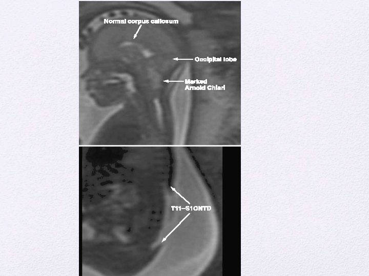
MR Image of myelomeningocele • Sagittal T 2 -weighted MR image from same patient with myelomeningocele shows neural placode (white arrow) protruding above skin surface due to expansion of underlying subarachnoid space (black arrow).
Clinical Examination • Signs of myelopathy, including hyperreflexia and clonus, because they often have an incomplete functional spinal cord transection in 2/3 rd • sensory level by stimulus - distally to proximally until the infant grimaces. • A stimulus is applied above the sensory level, and the distal-most voluntary motion seen for motor level determination • With an L 1 -3 level, the infant has hip flexion with extended knees and clubfeet. • The presence of intact hip adduction, hip flexion, and knee extension with inverted feet is indicative of an L 2 -4 level. • With an L 5 -S 2 level, the infant has hip adduction, knee extension, and knee flexion with dorsiflexed feet. • Infants with a sacral level may appear intact except for weakness of plantar flexion and rocker-bottom feet. • A flaccid pelvic floor and patulous anus are often present
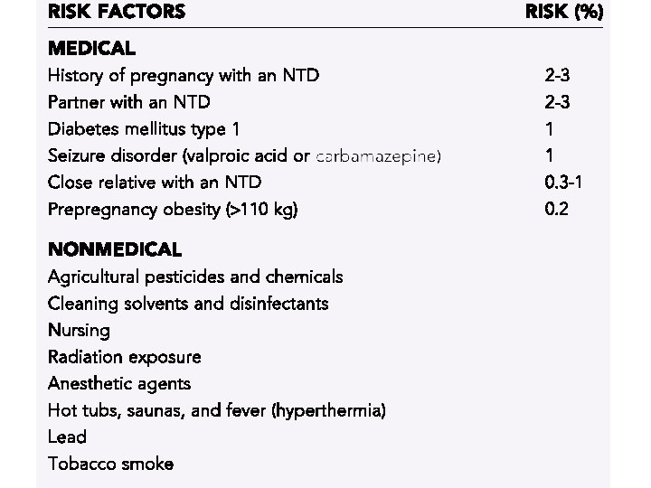
• Prenatal diagnosis • Maternal serum Alpha feto protein (MSAFP): initial screening test • High resolution fetal ultrasonography (USG) • Can also demonstrate hydrocephalus and Chiari II abnormality • Amniocentesis : if MSAFP and USG are suggestive • Ach esterase levels along with AFP • AFP can increase in other developmental anomalies of the gut and kidneys.
“lemon sign” Normal fetal head
D/D • At least 22 other fetal abnormalities myelomeningocele increase MSAFP levels. besides • Abdominal abnormalities such as omphalocele, cloacal exstrophy, esophageal atresia, annular pan- creas, duodenal atresia, and gastroschisis and • urologic abnormalities such as congenital nephrosis, polycystic kidneys, urinary tract obstruction, and renal • sacrococcygeal cystic teratoma
Preop evaluation- Clinical • General • Repaired within 72 hrs • Enteral feeding avoided to prevent fecal soiling of placode • Prone position , saline dressings • Neurosurgical • Sensory level determined • Motor evaluation –distal most voluntary motion evaluated. Limb abnormalities documented. • Anal tone and anal reflex evaluated
• Ventricular size documented with preop USG and NCCT head. • Imaging study shows that differentiating feature between a myelomeningocele and myelocele is the position of the neural placode relative to the skin surface • The neural placode protrudes above the skin surface with a myelomeningocele and is flush with the skin surface with a myelocele • Observe for symptoms of Chiari II • Renal evaluation • 90 % have neurogenic bladder. • All should have preop Renal ultrasound for detecting severe anomalies. • CIC if fails to void.
Myelocele 1. Axial schematic of myelocele shows neural placode (arrow) flush with skin surface. 2. Axial T 2 -weighted MR image in 1 -day-old girl shows exposed neural placode (arrow) that is flush with skin surface, consistent with myelocele. There is no expansion of underlying
Repair • Timing of repair: • Myelomeningocele repair can be performed safely up to 72 hours after birth • Delayed repair – Increases chance of ventriculitis by 5 times, • shunt infection developed in about 75%, and the mortality was 13% • In case Delay • 1) Cultures from the neural placode – No growth –n repair • If infection +, -external ventricular drainage and appropriate antibiotics until the infection clears – Then repair
• Shunt before repair – High chance of shunt inf. / Meningitits – IQ impairment (due to inf) • PREPARATION • Intraoperatively avoid hypothermia, hypovolemia, and hypoglycemia. • A doughnut-shaped sponge - to protect the myelomeningocele while intubation. • If severe Hydrocephalus - CSF diversion before closure of the myelomeningocele - to minimize pressure on the myelomeningocele dural closure • entire back and flanks are prepared and draped to facilitate extensive closure if needed. • Contact between povidone- iodine solution and the neural placode should be avoided
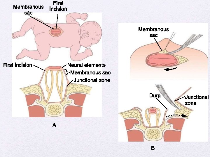
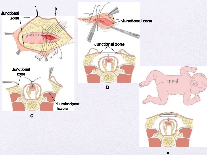
• purposes of myelomeningocele repair are to protect the functional spinal cord tissue, prevent loss of CSF, and minimize the risk for meningitis by reconstructing the neural tube and its coverings. • The margin between the arachnoid of the neural placode and the dystrophic epidermis, or the junctional zone, is the site of the initial incision. • The goal is to free the neural placode from the surrounding junctional zone circumferentially. • Duraplasty with thoracolumbar fascia or another dural substitute is performed when necessary to prevent leakage of CSF.
Postop Care • Post op antibiotics • Prevention of fecal contamination of wound • Nurse in Trendlenberg’s • Observe for Hydrocephalus – shunt if HCP present • Complications • Superficial wound dehiscence • Meningitis • Symptomatic chiari • Ensure functioning shunt • Hindbrain decompression
Occult spinal dysraphism • Aka Closed dysraphism • Of 2 types ; with/without s/c mass • Congenital spinal defects covered by intact skin • Causative lesions • • • Fatty filum terminale Lipomyelomeningocele Split cord malformations type I and II Inclusion lesions (dermoid, dermal sinus tract) Neurenteric cyst Myelocystocele
Closed / Occult type • • Closed With S/C Mass • Lipomyelomeningocele • Meningocele • Lipomyelocele • Myelocystocele • Terminal Myelocystocele Closed without S/C Mass • • Simple Dysraphic state • Dermal Sinus • Intradural Lipoma Complex Dysraphic State • Diastometamyelia • Neuroenteric cyst • Caudal Agenesis
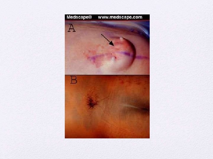
Manifestations of occult spinal dysraphism Cutaneous stigmata Orthopedic deformities Urologic problems Asymmetric gluteal cleft Foot or leg deformities Neurogenic bladder Capillary hemangioma Scoliosis UTIs Subcutaneous lipomas Sacral agenesis Incontinence Hypertrichosis Dermal sinus tract Cutis aplasia Delay in toilet training
Neurological signs and symptoms in different age groups Infants Toddler Older children Young adults Decreased spontaneous leg movements Delayed walking Asymmetric motor/ sensory development Back pain Absent reflexes Abnormal gait Back/leg pain Leg cramping/pain Leg atrophy UMN signs Spasticity Foot asymmetry Painless ulceration Hyperreflexia Decreased urinary stream Bowel/bladder incontinence.
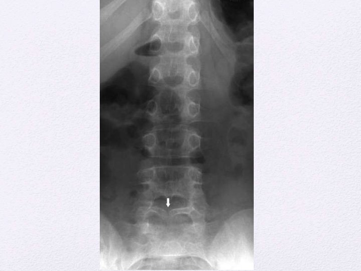
Plain radiological findings Structure Findings Lamina Fusion defects, midline defects, abnormal spinous processes Vertebral bodies Hemivertebrae, Butterfly vertebrae, Block vertebrae, Midline cleft defects, canal stenosis Disk space Congenital narrowing Pedicles Flattening, thinning Widening of spinal canal Interpedicular widening, scalloping of posterior border, Midline bony spur. Failure of development Reduced number of vertebral bodies, Absence of parts of vertebrae, sacral dysjunction Spinal curvature Scoliosis, kyphosis, lordosis.
Lipomyelomeningoceles • Lipoma tethering the cord to the subcutaneous tissue • Fascial, spinous and dural defect • Lipoma cord interface distracted out of the spinal canal by traction created by tethering
LMMC Axial schematic of lipomyelomeningocele shows placode–lipoma interface (arrow) lies outside of spinal canal due to expansion of subarachnoid space. Axial T 1 -weighted MR image in 18 -month-old boy shows lipomyelomeningocele (arrow) that is differentiated from lipomyelocele by location of placode–lipoma interface outside of spinal canal due to expansion of subarachnoid space.
LMC- Lipomyelocele • Axial schematic of lipomyelocele shows placode–lipoma interface lies within spinal canal. • Axial T 2 -weighted MR image in 3 -year-old girl shows placode–lipoma interface within spinal canal, characteristic for lipomyelocele.
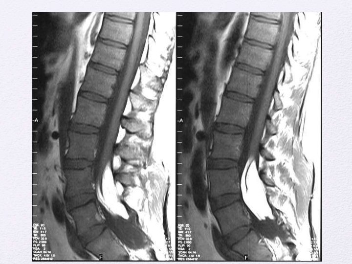
Lipomyelocele Sagittal T 1 -weighted MR image in 3 year-old girl with lipomyelocele shows subcutaneous fatty mass (black arrow) and placode–lipoma interface (white arrow) within spinal canal.
Chapman’s classification of LMM • Classification • Type I (dorsal lipoma) • Type II (transitional lipoma) • Type III ( terminal lipoma)
• • Dorsal lipoma (Type Fibrolipomatous stalk. I)tethering cord proximal to • conus • Usually at middle lumbar to lumbo sacral level • Dorsal spinal cord dysraphic at site of attachment of lipoma • Site of attachment medial to the dorsal root entry • Normal spinal cord distal to myeloschisis. • Roots lie within the subarachnoid space.
Dorsal lipoma
• Caudal or terminal lipomas (type III) • Directly from conus medullaris or filum terminale • Largely or wholly intradural • Nerve roots entangled in the lipoma • Lipoma cord interface caudal to the dorsal root entry zone. • Filum may be fatty, thickened and sometimes attached to subcuatneous tissue ( sacral dimple).
Terminal lipomas
• Transitional lipomas • Share the charcteristics of both Type 1 and type 2. • No normal spinal cord distal to lipoma attachment • Initially dorsal roots may be separate but caudally become enmeshed into the lipoma. • Frequently assymmetric attachment to cord.
Transitional lipomas
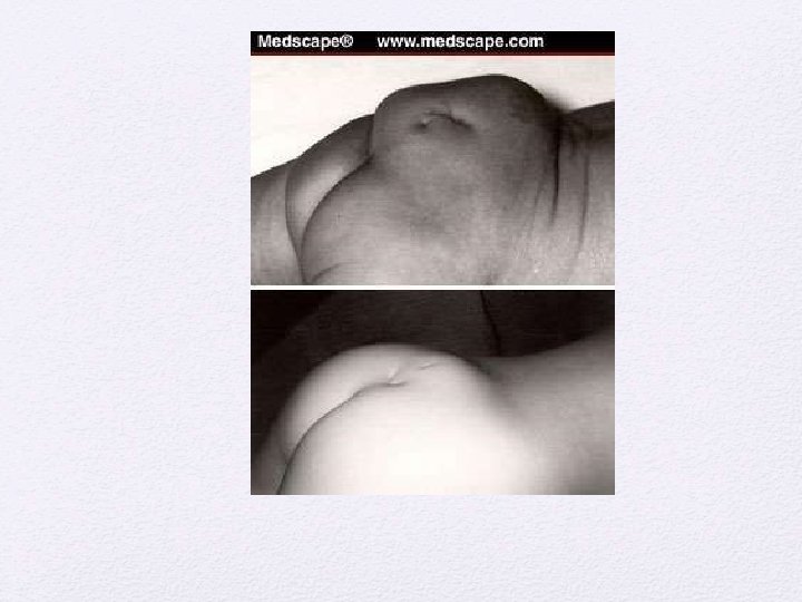
Clinical features • Subcutaneous masses over the back • Stigmata of occult dysraphism • • • Hypertrichosis Hemangioma Hypo/ hyperpigmented patch Dermal pit or sinus Atretic meningocele Assymmetric gluteal cleft
• Inexorable symptomatic progression - in untreated cases • Risk of precipitous neurologic deterioration • Orthopedic syndrome • Limb length discrepancy, high pedal arches, hammer toes, calcaneovarus/ valgus foot deformity. • Urologic syndrome • Urinary incontinence, post void dribbling, urgency, frequency • Intractable pain in the legs, back, pelvis or perineum.
Indication for surgical repair • Asymptomatic infant older than 2 months • Presence of orthopedic, pain or urologic syndrome • Neurological symptoms • Prior to corrective spinal surgery.
Principal Goal of surgery • Detethering of spinal cord • Decompression of the cord by removing as much lipoma as possible • Reconstruct the spinal cord and dural sac • Preservation of the functional tissue • Surgical principles • Relationship between the lipoma-cord interface and dorsal roots to be established • Conservative excision of the lipoma to avoid injury to the cord/ exiting roots.
• Complications of surgical repair • Early – CSF leak/ pseudo meningoceles & New Neurological deficit • Late – retethering of the cord • Mostly presenting between 3 -8, 11 -22 months after surgery • Upto 20% cases may demonstrate retethering • Diagnosis primarily clinical. • • Aseptic meningitis from host-graft inflammation meningitis, Intradural abscess, wound infection, and wound breakdown
Lipoma of the terminal filum Less severe form of Occult SD More than 2 mm thickness of the filum on MR imaging Frequently assosciated with sacral/gluteal cleft dimples. May be associated with VATER association, imperforate anus, cloacal extrophy and other urogenital abnormalities. Sometimes a/w sacral agenesis Reflects defective secondary neurulation
Orthopedic Clinical presentation Urologic Pain • All asymptomatic infants and symptomatic adults are surgical candidates. • Surgical procedure is the exposure of filum through lumbosacral laminectomy or interlaminar approach The filum identified separated from nerve roots and cut.