BRONCHOHYGIENIC THERAPY Dr Demet Demirciolu Bronchial Hygiene Therapy

BRONCHOHYGIENIC THERAPY Dr. Demet Demircioğlu

• Bronchial Hygiene Therapy involves the use of noninvasive airway clearance techniques designed to help mobilize and remove secretions and improve gas exchange.

Bronchial Hygiene Therapy • accepted as part of the care of critically ill patients, largely due to risks of ETT obstruction. • Short term, aim to remove obstructive secretions from the airways thereby – – – reducing work of breathing; improving delivery of mechanical ventilation; improving gaseous exchange; preventing and resolving respiratory complications; facilitating early weaning from the ventilator • Main et al, 2004; Ntoumenopoulos et al, 2002; Wallis and Prasad, 1999; Ciesla, 1996. • Longer term, aim to – Prevent postural deformities – Improve exercise tolerance – Return to optimal function

Indications for Bronchial Hygiene Therapy • “indications or contraindications for or against Bronchial Hygiene Therapy should never be formulated on the basis of diagnostic entities but should rather stem from a detailed analysis of the prevailing individual pathophysiology. ” – Oberwaldner (2000) Eur Respir J

Indications Components for a patient to receive bronchial hygiene regimes are – Excessive sputum production. Most authors state that more than 25 -30 ml/day ( 1/4 cup or 12 teaspoons) is excessive. Examples of common pathologies include: *cystic fibrosis *bronchitis *and bronchiectasis.

The second component required for bronchial hygiene therapy is an ineffective cough. Examples of causes for an ineffective cough are • weakness, • pain, and • placement of an artificial airway.

• PROPHYLACTIC - Pre-operative high risk surgical patient - Post-operative patient who is unable to mobilize secretions - Neurological patient who is unable to cough effectively - Patient receiving mechanical ventilation who has a tendency to retain secretions - Patients with pulmonary disease, who needs to improve bronchial hygiene

• THERAPEUTIC - Atelectasis due to secretions - Retained secretions - abnormal breathing pattern due to primary or secondary pulmonary dysfunction - COPD and resultant decreased exercise tolerance - Musculoskeletal deformity that makes breathing pattern and cough ineffective

Minimal to no benefit – Acute asthma • Asher et al, Pediatr pulmonol 1990 – Bronchiolitis • Webb et al (1985) Arch Dis Child • Nicholas et al (1999) Physiotherapy • Cochrane Systematic Review (Perrotta et al 2005) – Respiratory failure without atelectasis – Prevention of post-extubation atelectasis in neonates – Hyaline membrane disease • Schechter (2007) Resp Care – Prevention of atelectasis following surgery • Reines et al, 1982 – Undrained pleural collections

Contraindications Specific contraindications for bronchial hygiene therapy are: elevated intracranial pressure acute, unstable head, neck or spine injury increased risk of aspiration cardiac instability Other medical conditions that would be of concern when considering bronchial hygiene therapy are: pulmonary embolism and pulmonary edema associated with congestive heart failure.

Precautions • • • Untreated tension pneumothorax Abnormal coagulation profile Status epileptics or status asthmatics Immediately following intra cranial surgery Head injury with raised ICP Osteoporotic bones Recent acute myocardial infarction, unstable vitals Immediately after tube feedings Sutures and ICD’s

Complications • • • hypoxia increased metabolic demand O 2 consumption cardiac arrythmias changes in blood pressure raised intracranial pressure and decreased cerebral oxygenation gastro-oesophageal reflux pneumothoraces atelectasis and death. • Chalumeau et al, 2002; Krause and Hoehn, 2000; Wallis and Prasad, 1999; Harding et al, 1998; Button et al, 1997; Cross et al, 1992; Reines et al, 1982.

Goals • • • Prevent accumulation of secretions Improve mobilization and drainage of secretions Promote relaxation to improve breathing patterns Promote improved respiratory function Improve cardio-pulmonary exercise tolerance Teach bronchial hygiene programs to patients with chronic respiratory dysfunction

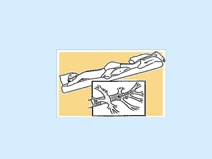
Traditional Forms Of Bronchopulmonary Hygiene Therapy The three traditional methods of BHT are: • Directed cough • Postural drainage • External manipulation of the thorax.

Techniques • • • Positioning Chest tapotement techniques Manual hyperinflation Airway suctioning Coughing techniques Breathing exercises Neuro physiological facilitation Controlled mobilization Patient education

Positioning • POSITIONING is the use of body position as a specific treatment technique • (it has a marked influence on gas exchange because of the unevenly damaged lungs- Tobin et al, 1994)

Physiological effects of Positioning • Optimizes oxygen transport by improving V/Q mismatch • Increases lung volumes • Reduces the work of breathing • Minimizes the work of heart • Enhances mucociliary clearance (postural drainage)

• Directed Cough is one of the simplest techniques to employ when the patient's own spontaneous cough is not adequate in clearing secretions.

Coughing Techniques • Coughing: It is a forced expiratory technique performed with a closed glottis. • Huffing: It is a forced expiratory technique performed with a open glottis. • Sniffing: Its an respiratory maneuver performed after a full inspiration or expiration.

Effects of Coughing • Cough removes secretions from the larger airways • Huff mobilizes the secretions from the distal airways. • Sniff augments collateral ventilation thereby preventing distal airway collapse.

Limitations: • Patients who are uncooperative , or comatose • Patients with an artificial airway, effective closure of the glottis is not possible • Extremely thick, tenacious secretion may require other modes of therapy If the patient has incisional pain, Splinting with a pillow or towel may be beneficial.

Breathing Exercises Breathing exercise is a technique which concentrates on ventilation to specific areas of lungs.

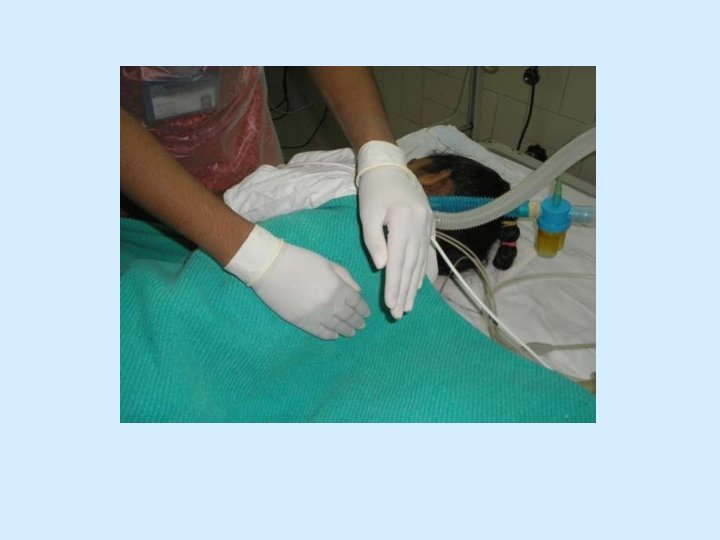
External Manipulation of the Thorax Commonly known as percussion and vibration. The patient is placed in the appropriate position. The therapist then either manually "claps" over the affected areas for 3 to 5 minutes. The force applied with the clapping or percussor varies greatly primarily due to the patient's tolerance.

Clapping/Chest Percussion • Percussion consists of rhythmic clapping on the chest with loose wrist & cupped hand. • Effect : Dislodges & loosens secretions from the lung

Chest Vibration • Vibrations consists of a fine oscillation of the hands directed inwards against the chest, performed on exhalation after deep inhalation. • Effects: Helpful in moving loosened mucous plugs towards larger airway
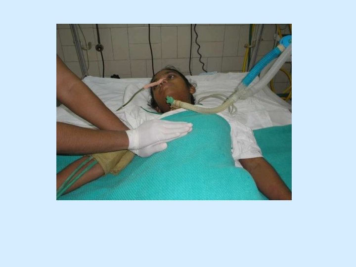

Rib Springing/Shaking • Shaking is a coarser movement in which the chest wall is rhythmically compressed. • Effects : Direct secretions towards larger airways & Stimulates cough.

Manual Hyperinflation • Was originally defined as inflating the lungs with oxygen and manual compression to a tidal volume of 1 liter requiring a peak inspiratory pressure of between 20 and 40 cm H 2 O (Med j Aust, 1972).
 • Improves oxygen saturation and lung")
Advantages of MH • Reverses atelectasis (Lumb 2000) • Improves oxygen saturation and lung compliance (Patman et al. , 1999) • Improves sputum clearance (Hodgson et al. , 2000)

Disadvantages of MH • Haemodynamic and metabolic upset (Stone, 1991 & Singer et al. , 1994) • Risk of barotrauma • Discomfort and anxiety

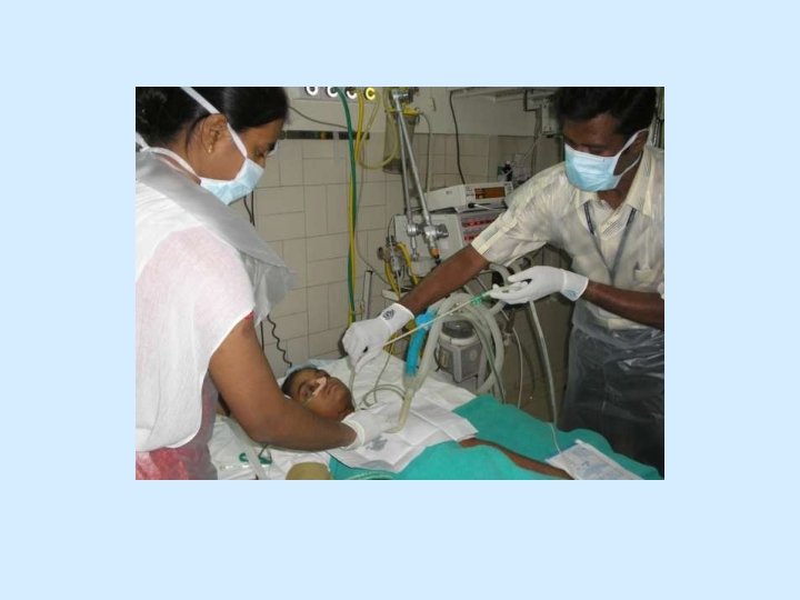
Suctioning • Suctioning is the mechanical aspiration of pulmonary secretions from a patient with an artificial airway in place.

criteria for suctioning: • Position client in fowlers for those with intact gag reflex. • Side lying for unconscious to prevent aspiration. • Set the pressure

• Apply suction for 5 to 10 seconds – - maximum of 15 seconds • Over suctioning can cause hypoxia and vagal stimulation. • Hyperventilate • Allow 20 to 30 second interval.
 adjuncts are used to mobilize")
Positive Airway Pressure Adjuncts • Positive airway pressure (PAP) adjuncts are used to mobilize secretions and treat atelectasis. • Types of PAP Adjuncts – Continuous positive airway pressure (CPAP) – Expiratory positive airway pressure (EPAP) – Positive expiratory pressure (PEP)

Indications of PAP Adjuncts – To reduce air trapping in asthma and COPD – To aid in mobilization of retained secretions (in cystic fibrosis and chronic bronchitis) – To prevent or reverse atelectasis – To optimize delivery of bronchodilators in patients receiving bronchial hygiene therapy
 • It is a method to deliver high")
High Frequency Chest Wall Compression (HFCC) • It is a method to deliver high frequency vibration over the chest wall to cause transient increases in airflow and improve mucus movement.
 It is a two-part system: the first, a")
High Frequency Chest Wall Oscillation (HFCWO) It is a two-part system: the first, a variable air-pulse generator, and the second, an unstretchable, inflatable vest that covers the patient creating an oscillatory motion against the patient’s thorax. HFCWO increases airflow velocity, which creates repetitive cough-like shear forces and decreases the viscosity of secretions. Therapy is usually performed in 30 -minute sessions at varying oscillatory frequencies ( 5– 25 Hz ). Depending on need, one to six therapy sessions may occur per day.
")
High Frequency Chest Wall Oscillation (HFCWO)

Flutter Valve Therapy • The Flutter Valve combines the technique of PEP with high frequency oscillations at the airway opening. •

• The effect is threefold: First, to vibrate the airways and thus, facilitate movement of mucus; Second, to increase endobronchial pressure to avoid air trapping and Third, to accelerate expiratory airflow to facilitate the upward movement of mucus
 • promoting or hastening the response of neuro muscular mechanism")
Neuro Physiological Facilitation (NPF) • promoting or hastening the response of neuro muscular mechanism through proprioceptors (dorothy voss et al, 1985). • Cutaneous and proprioceptive stimulation reflexly increases the depth of breathing (Jones, 1998). INDICATIONS: • Non alert patients such as those who are drowsy postoperatively. • Those with neurological conditions. • Partially breathing patient on ventilator, especially if they are unable to turn.
. • Perioral")
Techniques of NPF • Stimulation of diaphragm (Dorothy voss et al, 1985). • Perioral technique • Intercostal stretch • Co- contraction of abdominal muscles • Vertebral pressure (D. D. Bethune, 1975)

Mobilization and Exercise • Immobility is a major factor contributing to retention of secretions • Early mobilization and frequent position changes are preventive interventions for atelectasis. • Exercise also improves overall aeration and ventilation perfusion matching. • Exercise can improve a patients general fitness, self esteem and quality of life.
- Slides: 46