Physiology of Respiration Dr Demet Demirciolu Physiological Anatomyof


 - simple elasticity of the lungs DECREASES")


 - normal")
 = TV + IRV (MAXIMUM volume of")


 (Normal) AVR")
 As simple solution (3% i. e. 0.")
 Increased")
 leads")

 1. Partial Pressure Gradients & Solubilities")

 � 1. Oxygen: blood (104 mm)")


 �Decreased CO 2")


 exercise sufficient oxygen is supplied to muscles due to increased oxygen")
 increase during exercise? 1. During light exercise (walking)? By")


- Slides: 43
Physiology of Respiration Dr. Demet Demircioğlu
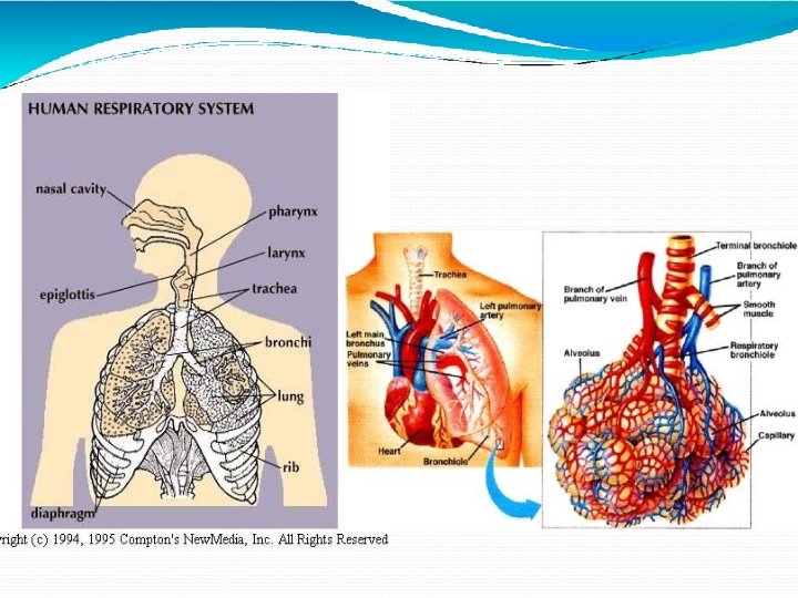
Physiological. Anatomyof Respiratory. System �The respiratory system consists of the nasal cavity, pharynx, larynx, trachea, bronchi, and lungs, which is whole together called as the Respiratory Tract. . �Upper respiratory tract refers to: Nasal cavity, pharynx, and associated structures. �Lower respiratory tract refers to: Larynx, trachea, bronchi, and lungs. The Lungs & Alveoli
Respiratory Unit Respiratory unit is the terminal portion of the Respiratory Tract. It includes: � 1. Respiratory Bronchioles � 2. Alveolar Ducts � 3. Antrum � 4. Alveolar Sacs � 5. Alveoli (Human beings have a thin layer of about 700 million alveoli within their lungs. Which is crucial for respiration, exchanging O 2 & CO 2 with the surrounding blood capillaries. ) Microscopic Structure of Alveoli
Respiratory Functions: � 1. Pulmonary Ventilation � 2. Diffusion of O 2 and CO 2 between the alveoli and the blood. � 3. Transportation of O 2 & CO 2 in the blood & body fluids to & from the body’s tissue cells. � 4. Regulation of Ventilation. �Non- Respiratory Functions: � 1. Olfaction, Vocalization � 2. Defense Mechanism � 3. Anticoagulant Function � 4. Regulation of Body Temperature & Acid Base Balance � 5. Maintenance of Water balance � 6. Secretion of ACE Angiotensin Converting Enzyme
Mechanism of Respiration Muscles of Respiration i. Primary Inspiratory: Diaphragm ii. Accessory Inspiratory: Sternomastoid, Scaleni, Anterior Serrati, Elevators of Scapula & Pectorals iii. Primary Expiratory: Internal Intercostal muscles iv. Accessory expiratory: Abdominal muscles Inspiration
Inspiration � 1. Diaphragm muscle contracts, increasing thoracic cavity size in the superior-inferior dimension � 2. External intercostal muscles contract, expanding lateral & anterior-posterior dimension � 3. INCREASED volume (about 0. 5 liter), DECREASED pulmonary pressure (-1 mm Hg), air rushes into lungs to fill alveoli �Deep/forced inspirations – as during exercise and pulmonary disease * scalenes, sternocleidomastoid, pectorals are used for more volume expansion of thorax.
Expiration � 1. Quiet expiration (exhalation) - simple elasticity of the lungs DECREASES volume INCREASED pulmonary pressure -> movement of air out of the lungs � 2. Forced expiration - contraction of abdominal wall muscles (i. e. obliques & transversus abdominus) further DECREASES volume beyond relaxed point ----> further INCREASE in pulmonary pressure ---> more air moves out.
Movements of Thoracic Cage
Movements of Lungs Factors holding lungs AGAINST the thorax wall: i. Surface tension holding the "visceral" and "parietal" pleura together. ii. Intrapulmonary pressure is ALWAYS slightly greater than intrapleural pressure by 4 mm Hg. iii. Atmospheric pressure acting on the lungs. a)Atelectasis (collapsed lung) - hole in pleural "balloon" causes equalization of pressure and collapse of the lung. b)Pneumothorax - abnormal air in the intrapleural space, can lead to collapsed lung.
Factors facilitating lung movement AWAY from thorax wall i. Elasticity of lungs allows them to assume smallest shape for given pressure conditions. ii. Fluid film on alveoli allows them to assume smallest shape for given pressure conditions.
Pulmonary. Volumes, Capacities&Function Tests A. Respiratory Volumes � 1. Tidal volume (TV) - normal volume moving in/out (0. 5 L). � 2. Inspiratory reserve volume (IRV) - volume inhaled AFTER normal tidal volume when asked to take deepest possible breath (2. 1 -3. 2 L). � 3. Expiratory reserve volume (ERV) - volume exhaled AFTER normal tidal volume when asked to force out all air possible (1. - 2. 0 L). � 4. Residual volume (RV) - air that remains in lungs even after totally forced exhalation (1. 2 L).
B. Respiratory. Capacities 1. Inspiratory capacity (IC) = TV + IRV (MAXIMUM volume of air that can be inhaled). � 2. Functional residual capacity (FRC) ERV + RV (all non-tidal volume expiration). � 3. Vital capacity (VC) = TV + IRV + ERV (TOTAL volume of air that can be moved). � 4. Total lung capacity (TLC) = TV + IRV + ERV + RV (the SUM of all volumes; about 6. 0 L).
C. Dead Space � 1. Anatomical dead space – all areas where gas exchange does not occur � 2. Alveolar dead space – non-functional alveoli. � 3. Total dead space – Anatomical + Alveolar.
D. Pulmonary Function. Tests � 1. Spirometer - measures volume changes during breathing. a. Obstructive pulmonary disease - increased resistance to air flow (bronchitis or asthma). b. Restrictive disorders - decrease in Total Lung Capacity (TB or polio). � 2. Minute respiratory volume (MRV) - total volume flowing in & out in 1 minute (resting rate = 6 L per minute). � 3. Forced vital capacity (FVC) - total volume exhaled after forceful exhalation of a deep breath. � 4. Forced expiratory volume (FEV) - FEV volume measured in 1 second intervals (FEV 1. . . ).
E. Alveolar Retention Rate AVR =Breath Rate X (TV - Dead space) (Normal) AVR = 12 /minute X (500 ml – 150 ml) (Normal) AVR = 4. 2 L/min Spirometer
Transportof Gases �Transport of O 2: i) As simple solution (3% i. e. 0. 3 ml/100 ml ) ii) In Combination with Hb (97%) �Transport of CO 2: i) As dissolved form (7%) ii) As carbonic Acid (Negligible) iii) As Bicarbonate (63%) iv) As Carbamino Compounds (30%)
A. Effects of Temperature 1. HIGHER Temperature 2. LOWER Temperature Decreased Affinity (right) Increased Affinity (left) B. Effects of p. H (Acidity) 1. HIGHER p. H Increased Affinity (left) 2. LOWER p. H Decreased Affinity (right) "Bohr Effect“ C. Effects of Diphosphoglycerate (DPG) 1. DPG - produced by anaerobic processes in RBCs 2. HIGHER DPG > Decreased Affinity (right) 3. Thyroxine, testosterone, epinephrine, NE - increase RBC metabolism and DPG production, cause RIGHT shift.
�Carbon Dioxide Dissociation Curve �Bohr Effect - Formation of Bicarbonate (through Carbonic Acid) leads to LOWER p. H (H+ increase), and more unloading of Ox to tissues. Since Hb "buffers" to H+, the actual p. H of blood does not change much.
Chloride Shift - Chloride ions move in opposite direction of the entering/leaving Bicarbonate, to prevent osmotic problems with RBCs
Exchangeof. Gases A. External Respiration (Air & Lungs) 1. Partial Pressure Gradients & Solubilities a. Oxygen: alveolar (104 mm) ---> blood (40 mm) b. Carbon Dioxide: blood (45 mm) ----> alveolar (40 mm) (carbon dioxide much more soluble than oxygen) 2. Alveolar Membrane Thickness (0. 5 -1. 0 micron) very easy for gas to diffuse across alveoli Edema - increases thickness, decreases diffusion b. Total Alveolar Surface Area for Exchange 3. a. Total surface area healthy lung = 145 sq. Meters b. Emphysema - decreases total alveolar surface area
4. Ventilation-Blood Flow Coupling Low O 2 in alveolus vasoconstriction High O 2 in alveolus vasodilation High CO 2 in alveolus dilate bronchioles Low CO 2 in alveolus constrict bronchioles
B. Internal respiration ( Blood & Tissues ) � 1. Oxygen: blood (104 mm) tissues (40 mm) � 2. Carbon Dioxide: tissues (>45 mm) blood (40 mm)
Regulationof Respiration Nervous Mechanism A. Medullary Respiratory �Inspiratory Center (Dorsal Resp Group - rhythmic breathing �Phrenic nerve, Intercostal nerves , diaphragm + external intercostals �Expiratory Center (Ventral Resp Group - forced expiration) �Phrenic nerve, Intercostal nerves, Internal intercostals + abdominals (expiration) 1. Eupnea - normal resting breath rate (12/minute) 2. Drug overdose - causes suppression of Inspiratory Center
B. Pons Respiratory Centre � 1. Pneumotaxic center - slightly inhibits medulla, causes shorter, shallower, quicker breaths � 2. Apneustic center – stimulates the medulla, causes longer, deeper, slower breaths
C. Control of Rate & Depth of Breathing � 1. Breathing rate - stimulation/inhibition of medulla. � 2. Breathing depth - activation of inspiration muscles. � 3. Hering-Breuer Reflex - stretch of visceral pleura that lungs have expanded (vagal nerve). D. Hypothalamic Control - emotion + pain to the medulla E. Cortex Controls (Voluntary Breathing) - can override medulla as during singing and talking
Chemical Mechanism A. Chemoreceptors � 1. Central chemoreceptors - located in the medulla � 2. Peripheral chemoreceptors - large vessels of neck
B. Overview of Chemical Effects Chemical �Increased CO 2 (more H+) �Decreased CO 2 (less H+) �Slight decrease in O 2 �Large decrease in O 2 �Decreased p. H (more H+) �Increased p. H (less H+) Breathing Effect Increase Decrease Effects CO 2 system Increases ventilation Increase Decrease
EXERCISE EFFECT ON THE RESPIRATORY SYSTEM
Exercise is the most influential physiological stress on breathing How is increased ventilation accomplished? Exercise increases the breaths/minute Exercise increases the amount of air in each breath (tidal volume)
During light exercise Ventilation increases linearly with oxygen uptake and carbon dioxide production This increase in ventilation is accomplished more by increased tidal volume (breathing deeper in and out)
During higher exercise levels Ventilation is increased more by increased breathing frequency This will keep the blood saturated with oxygen because the blood is in the alveoli capillaries long enough for complete diffusion of gases
Steady rate (moderate) exercise sufficient oxygen is supplied to muscles due to increased oxygen up take, there is little, or no, build up of lactic acid in the muscles some lactate will be produced and removed by the blood stream lactic acid is neutralized in the blood (this reaction produces carbon dioxide as a by-product) increased carbon dioxide in the blood will stimulate increased ventilation Increased ventilation is accomplished by both increased tidal volume and frequency
How does pulmonary ventilation (breathing) increase during exercise? 1. During light exercise (walking)? By increasing the tidal volume (breathing deeper) 2. During intense exercise (sprinting)? By increasing the frequency of breathing 3. During steady state exercise (jogging)? By increasing both the tidal volume and the frequency of breathing
The Energy Cost of Breathing
At rest and with light exercise the energy cost of breathing is minimal (4% of energy)
During intense exercise the energy use may increase from 1020% of total energy expenditure
In a person with respiratory disease the work of breathing itself during exercise may be exhausting
The Effects of Cigarette Smoking Cigarette smoking has little long term effect on athletes in the initial years Cigarettes have an immediate effect of airway resistance (as much as 3 times) that lasts about 35 minutes after only 15 puffs This could be significant in vigorous exercise
Ventilation Adaptations with Training
Minimal exercise ventilation during exercise improves with training (due to increased alveolar ventilation and increased tissue oxygen uptake)
Sub-maximal exercise The percentage of energy used in breathing is reduced after training only 4 weeks Tidal volume increases Breathing frequency is reduced This results in increased amounts of oxygen extracted from inspired air Untrained expired air contains 18% oxygen Trained expired air contains 14 -15% oxygen
This is important because It reduces the fatiguing of the ventilation musculature The freed up oxygen (not used by the respiratory muscles) can be available for the exercising muscle