Poultry Pathology Part III H L Shivaprasad CAHFS


 – duck.")
 – Ostrich.")
, duck - myocardial")
 – Chicken")
 and TEM")
 - turkeys Monensin toxicity – Clinical signs, turkeys. Skeletal muscle degeneration.")


 and zinc phosphide (grey) toxicity – pea fowl. Liver – hemorrhage. Crop")


 üHepatic Lipidosis üHemorrhagic fatty liver")
 üAortic rupture üCoronary artery aneurysm (turkeys) üRound")


 Øgout is a metabolic condition where abnormal accumulation of white chalky")
 Øthese")
 Onset Visceral urate deposit Usually acute")


 – visceral urate deposit")

: is a disease of")

 Type IIIB in Emus üMPS type IIIB, also called sanfilippo B syndrome")

")

, normal aorta (middle), rupture of coeliac artery (right)")


 v. Common condition in broiler chickens and turkeys and ducks vabnormal")
")
. Normal cartilaginous cores")


 Human errors: –omission of")


 ØVitamin B 6 (Pyridoxine) ØPantothenic acid/Biotin ØNiacin,")
 üPolyneuritis (B 1) and B 6")


")
 Microscopic pathology üSquamous metaplasia with or with out keratin formation")
 üBursa of Fabricius üFeather follicles üCornea üNasal")
")

")
 and parabronchiole (left)")


")

")


. Clinical signs - turkey poults and chickens")
, cerebellar hemorrhage - turkey poults")
 - turkey poults")
. Normal brain, malacia and hyaline thrombi")

 - chicken")
 – Brown pelican Skeletal muscle and heart")
– Brown pelican. Histopath: Skeletal muscles")
")
")


 and enlarged (right)")
. lymphocytic infiltration demyelination (right)")


 ØMost common")


 and hemangiosarcoma (lungs) - chickens")

 – Processed chicken, skin")


- Slides: 120
Poultry Pathology – Part III H. L. Shivaprasad CAHFS – Tulare University of California, Davis
Avian Toxicosis üMycotoxins üHeavy metals üIonophores üGases; PTFE, Ammonia, CO üSelenium, salt, calcium üVitamins üAntibiotics üRodenticides üPlants üOthers
Ergot toxicity – toes, quail
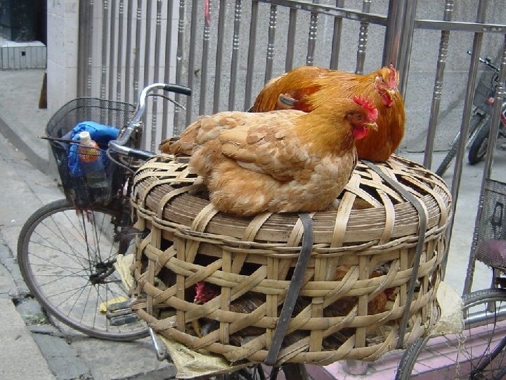
Gizzard contents (roofing nails, zinc toxicity) – duck.
Gizzard contents (Pennies, zinc toxicity) – Ostrich.
Lead toxicity, duck - lead pellets in gizzard Lead toxicity (gizzard), duck - myocardial degeneration
Gizzard contents (22 caliber, lead toxicity) – Chicken
Lead inclusions – kidney, psittacine. Histopathology ( H & E) and TEM
Ionophore toxicity (monensin) - turkeys Monensin toxicity – Clinical signs, turkeys. Skeletal muscle degeneration.
Salt toxicity–symmetrical encephalomalacia, turkey poult
Ammonia toxicity – chicken Corneal erosion/ulceration
Diphacinone (green) and zinc phosphide (grey) toxicity – pea fowl. Liver – hemorrhage. Crop contents
Aminoglycoside/tetracycline toxicity – nephrosis, chickens
Cotton defoliant toxicity Trachea - turkeys
Salt toxicity–environmental induced, Ruddy ducks
Avian Metabolic Diseases üHemochromatosis üAmyloidosis üGout (visceral and articular) üHepatic Lipidosis üHemorrhagic fatty liver syndrome (chickens) üAtherosclerosis üDiabetes mellitus
Avian Metabolic Diseases üMucopolysaccharidosis type IIIB (emu) üAortic rupture üCoronary artery aneurysm (turkeys) üRound heart disease (turkeys) üAscites syndrome (chickens) üTibial dyschondroplasia üDeep pectoral myopathy
Amyloidosis Øa condition where in amorphous eosinophilic substance, amyloid is deposited, primarily extracellularly Øtwo types of amyloid; primary and secondary Øprimary: associated with plasma cell neoplasia secondary: associated with chronic infections others: based on origin of amyloid, chemical composition (17 proteins), localized, endocrine (insulin), aging(senile), etc.
Amyloidosis – common in waterfowl, finches and canaries
Amyloidosis, ‘water belly’ in ducks Hepatic amyloidosis - ducks
Amyloidosis, liver, duck. Gross and histopathology
Amyloidosis, spleens - duck.
Amyloid arthropathy – Brown Leghorn chicken. Mycoplasma synoviae Amyloid arthropathy – Brown Leghorn chicken. Enterococcus faecalis
Amyloid arthropathy – Brown Leghorn chicken. Congo Red and green birefringence
GOUT (urate deposit) Øgout is a metabolic condition where abnormal accumulation of white chalky or white semi-fluid -like urates in soft tissues of various organs in the body Øuric acid is the endproduct of protein and purine metabolism (uricotelic) in birds, where as in mammals urea is the endproduct (ureotelic) Øbirds lack the enzyme, carbamylphosphate synthetase to dispose of ammonia and the enzyme uricase to decarboxylate uric acid to allontoin
Gout ØGout occurs as two distinct syndromes; visceral and articular urate deposits (gout) Øthese two syndromes differ in age of onset, frequency, sex predilection, gross and microscopic lesions, pathogenesis and causes Øgreat deal of confusion exists between the two syndromes because urate deposition takes place in joints in visceral gout also Øterm “visceral gout” should be replaced with the term “visceral urate deposits”
Differences between Visceral and Articular urate deposits (Gout) Onset Visceral urate deposit Usually acute Articular urate deposit Chronic Frequency Very common Rare Age of onset One day and above 4 - 5 months and above Sex Both sexes Mostly males
Visceral ‘vs’ Articular urate deposits Gross Lesions Kidney Visceral urate deposits Articular urate deposits Always involved; enlarged, pale with white precipitates Not involved. Can be involved secondary to dehydration. Soft tissues White precipitates on serosal surfaces; visceral organs, pleura, air sacs, pericardium, etc. Synovium. Comb, wattles, trachea in chronic conditions Joints Synovium and tendon Joints always involved; sheaths in severe cases enlarged
Visceral ‘vs’ Articular urate deposits Visceral urate deposits Articular urate deposits Microscopic lesions Nephrosis/nephritis – Granulomatous tophi synovitis Pathogenesis Failure to excrete urates Dehydration Toxicities Infectious agents Nutritional Neoplasia Anomalies Others Causes Metabolic defect in secretion Genetics High protein in diet Others?
Visceral gout – joints, chicken Visceral urate deposits – joints and viscera, 5 day-old chick
Visceral urate deposits – liver, heart, and heart/valves
Visceral urate – joint, chicken Visceral urate deposits – kidneys
Tophus (Nephritis/nephrosis) – visceral urate deposit
Articular urate deposits – joints, chicken
Hemorrhagic Fatty Liver Syndrome/Hepatic Lipidosis ØHemorrhagic Fatty Liver Syndrome (HFLS): is a disease of obese chicken layers in cages characterized by extremely fatty liver, drop in egg production and increased mortality due to ruptured liver ØObesity with fatty livers is common in pet birds; amazons, budgerigars, rose-breasted cockatoos and others
Hemorrhagic fatty liver syndrome - chicken
Mucopolysaccharidosis (MPS) Type IIIB in Emus üMPS type IIIB, also called sanfilippo B syndrome üfirst description in any animal üdeficiency of N-acetyl-a-D-glucosaminidase üprobably inherited as a autosomal recessive trait üsudden death, neurological signs in birds between 3 weeks and 6 months of age üruptured liver or subcutaneous hemorrhage üaccumulation of membrane bound substance in neurons of nervous tissue and visceral organs
MPS IIIB – Emu. Clinical signs MPS IIIB – emu brain, swollen neurons
MPS IIIB – emu brain, retina and TEM (brain)
Metabolic diseases – cont. v. Aortic rupture –most common in male turkeys, also in ostrich, emu –longitudinal slit/tear in aorta at the origin of coeliac –medial degeneration and loss of elastic fibers, plaque –genetics, hypertension, low copper, vasa vasorum defect ? v. Coronary artery rupture – 15 -16 weeks-old male turkeys, 1. 5 -3. 5% mortality –hemopericardium, hemorrhage at base of heart, medial degeneration of coronary artery, rupture –genetics, hypertension, low copper, increased body weights?
Aortic Rupture, turkey – hemorrhage (left), normal aorta (middle), rupture of coeliac artery (right)
Aortic Rupture, turkeys – rupture of coeliac artery
Aortic rupture – ostrich and emu Histopathology - aorta
Coronary artery aneurysm – Turkeys Hemopericardium, hemorrhage, heart
Coronary artery aneurysm–turkey. H & E and elastic stains
Metabolic diseases – cont. q. Round heart disease of turkeys: –also called spontaneous cardiomyopathy –common condition in young commercial turkeys –dilated ventricles, chronic passive congestion of liver • cause is not known, genetics ? q. Ascites syndrome of chickens: –common condition in broiler chickens –right heart hypertrophy, dilation, passive congestion of liver and ascites –rapid growth coupled with insufficient pulmonary capillary capacity aggravates pulmonary hypertension leading to right heart failure
Round Heart disease - turkeys
Ascites syndrome – broiler chicken
Tibial Dyschondroplasia (TD) v. Common condition in broiler chickens and turkeys and ducks vabnormal masses of cartilage below the growth plate primarily in the proximal tibiotarsus but occurs also in the tarsometatarsus üpresence of pre-hypertrophic cartilage with no vascular channels vetiology: multifactorial; nutrition, genetics, mycotoxin, etc. vsimilar picture as TD can be seen grossly in the long bones of ratites but it is NOT TD nor it is pathological üit is normal embryonic cartilage which gets resorbed by 68 weeks of age
Tibial Dyschondroplasia – chicken and turkey (right)
Tibiotarsus – ostrich chicks (1 - 3 weeks-old). Normal cartilaginous cores
Metabolic diseases – cont. q. Deep Pectoral Myopathy: –seen in well muscled broilers and turkeys –green discoloration of primarily supracoides muscle due to ischemic necrosis q. Perirenal hemorrhage: –seen in rapidly growing turkeys, 8 -14 weeks of age –hemorrhage over portion or entire kidney –underlying vascular and cardiac problems ? q. Xanthomatosis: –common in psittacines and occasionally in chickens –yellow subcutaneous swelling or nodules in the body cavity • giant cells, lymphocytes, macrophages with cholesterol clefts
Deep pectoral myopathy – broiler chicken
Diseases of Malnutrition in Birds
Nutrition Øprocess of furnishing cells inside the animal with that portion of external environment for optimum functioning of the many metabolic chemical reactions Øinvolves procurement, ingestion, digestion and absorption
Nutrition ØNutrients: essential for normal growth, development, livability, work and reproduction Øshould be in the diets in proper concentration and balance Øenergy, amino acids, carbohydrates, fats, vitamins, inorganic chemical elements, water and oxygen
Nutrient Requirements üWell worked out for growing chicks, poults and laying-type chickens üNot so well established for broiler and turkey breeders üEmpirical in pet and exotic birds and ratites üMore is being learned about wild birds in captivity
Malnutrition v. Cab be absolute ‘VS’ marginal deficiency ümarginal deficiency is probably more common and difficult to diagnose ØCan manifest as generalized or specific disease ØSuppresses immune system ØDecreases reproductive performance ØDecreases weight gain, feather problems ØDecreased response to therapeutic agents
Factors Influencing Malnutrition ØDiet composition (deficiency or absence of nutrients) Human errors: –omission of ingredient(s), equipment failure –improper mixing and/or storage –miscomputation in feed formulations –feeding to wrong species, sex or age Poor Nutritive value of ingredients Nutrients, vitamins and mineral interactions Poor shelf life
Factors Influencing Malnutrition ØInsufficient feed intake –inanition or starvation –anorexia –dysphagia or aphagia ØMaldigestion and malabsorption Ø Diseases affecting the GI tract ØDecreased storage or utilization ØIncreased excretion or secretion ØIncreased requirement or inhibition ØLack of knowledge on nutrition for some species
Diseases of Malnutrition Diagnoses ØHistory, clinical signs, gross and microscopic lesions ØAnalysis of feed ØAnalysis of liver, serum ØHematology, serum chemistry, radiography ØFeeding trials ØPeroxide (for rancidity) level in feed ØTreatment
Diseases of Malnutrition ØVitamin A ØVitamin D 3 ØCalcium/phosphorus ØVitamin E/Selenium Xerophthalmia Rickets/osteomalacia Encephalomalacia Exudative diathesis/ muscular dystrophy ØVitamin B 2(Riboflavin) Curled-toe paralysis
Diseases of Malnutrition ØVitamin B 1 (Thiamin) ØVitamin B 6 (Pyridoxine) ØPantothenic acid/Biotin ØNiacin, Folic acid ØCholine/Manganese ØIodine ØOthers: Zinc, Copper, Salt, Methionine, Vitamins K and B 12 Polyneuritis Perosis/nervous Perosis, dermatitis Perosis and others Perosis/slipped tendon Goiter
Diseases of Malnutrition Most common üXerophthalmia (Vitamin A) üPolyneuritis (B 1) and B 6 Deficiency üRickets/osteomalacia üEncephalomalacia üMuscular dystrophy/exudative diathesis (not since 1980’s) üCurled-toe paralysis (Vitamin B 2) üPerosis/slipped tendon
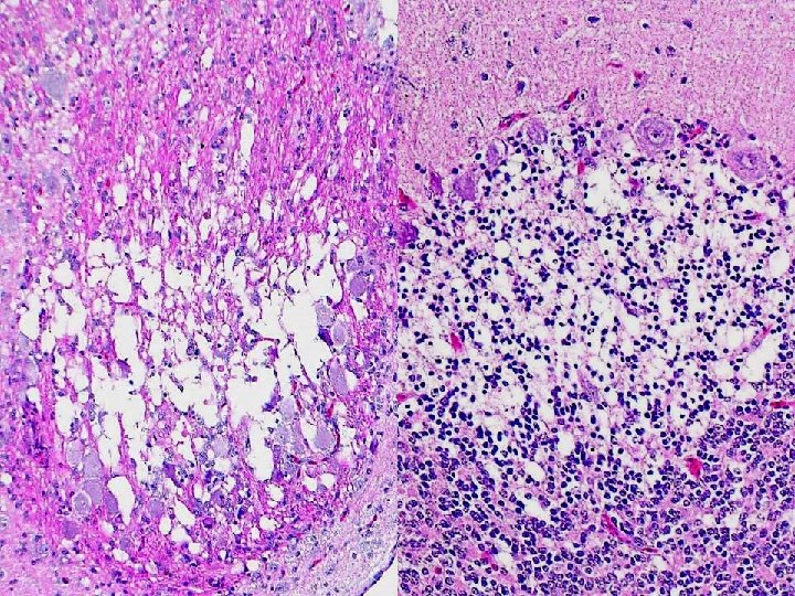
Diseases of Malnutrition ØXerophthalmia: –in non commercial poultry, psittacines, etc. Etiology: vitamin A deficiency Lesions: pustule-like nodules in upper GI tract, exudate in conjunctiva, nasal cavity, nephrosis, opaque dry cornea, hyperkeratosis of plantar surfaces • micro; squamous metaplasia of epithelium, hyperkeratosis
Vitamin A deficiency – chicken and Golden pheasant. Conjunctivitis and keratitis
Vitamin A deficiency – chickens. Oral cavity and esophagus
Vitamin A deficiency – turkey. Naso lachrymal duct and trachea
Vitamin A deficiency – turkey. Oral cavity, upper esophagus and bursa of Fabricius (right)
Vitamin A deficiency (turkeys) Microscopic pathology üSquamous metaplasia with or with out keratin formation üMucosa and submucosal glands üProximal esophagus, conjunctiva, and (third eye-lid? ) consistently affected üSalivary glands, tongue, pharynx, proventriculus üSinuses, turbinates, larynx, trachea, bronchi and parabronchi
Vitamin A deficiency Microscopic pathology (Cont. ) üBursa of Fabricius üFeather follicles üCornea üNasal and lacrimal glands and gland of Harder üGlands of the external ear üKidneys not affected
Vitamin A deficiency – chicken. Squamous metaplasia, esophagus and trachea (Dr. D. Swayne)
Vitamin A deficiency, chicken. Squamous metaplasia - esophagus and proventriculus
Vitamin A deficiency, chicken. Squamous metaplasia - conjunctiva and Bursa (right)
Vitamin A deficiency, turkey. Squamous metaplasia secondary bronchus (right) and parabronchiole (left)
Diseases of Malnutrition ØRickets/Osteomalacia: rickets in younger birds and osteomalacia in older birds –in poultry, ratites, psittacines, zoo birds, etc. Etiology: deficiency or imbalance of calcium, phosphorus and vitamin D 3 Lesions: soft and pliable beak, claws and keel, beading of ribs, enlarged epiphysis, fractures of long bones, enlarged parathyroid glands
Rickets – pheasant poults and a turkey poult
Rickets – pliable beak, chicks
Rickets – broiler chicken. Crooked keel and beaded ribs
Osteomalacia – adult chicken. Beaded ribs and hemorrhage (Path. fract. )
Rickets – broiler chicken. Parathyroids enlarged
Ostrich Rheas Rickets – tarsometatarsus, ostrich and rhea (Path. fract. )
Growth plate, tibiotarsus – chicken. Normal, rickets and TD. Courtesey: Dr. David Swayne
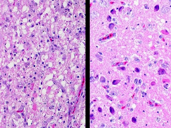
Diseases of Malnutrition ØEncephalomalacia: –in young chickens, turkeys, pheasants, geese, ducks, etc. Etiology: vitamin E deficiency Lesions: soft and enlarged cerebellum, petechiae, hemorrhages in turkey poults micro: malacia, hemorrhages, hyaline thrombosis, demyelination in brain occasionally spinal cord involved Yellow fat disease in wild birds; herons, etc. –fat is brownish yellow, steatitis
Encephalomalacia (Vit. E def. ). Clinical signs - turkey poults and chickens
Encephalomalacia (Vit. E def. ), cerebellar hemorrhage - turkey poults
Encephalomalacia (Vitamin E def. ) - turkey poults
Encephalomalacia (Vit. E def. ). Normal brain, malacia and hyaline thrombi
Diseases of Malnutrition ØMuscular dystrophy/Exudative diathesis –in chickens, quail, turkeys, ducks, etc. Etiology: vitamin E/Selenium deficiency Lesions: subcutaneous green-tinged fluid pale streaks in skeletal muscles, gizzard, heart –degeneration of skeletal muscle, heart, pancreatic acinar necrosis
White muscle disease (Vit. E/Sel. def. ) - chicken
White muscle disease (Vit. E/Sel. def. ) – Brown pelican Skeletal muscle and heart
White muscle disease (Vita. E/Sel. def. )– Brown pelican. Histopath: Skeletal muscles
Myocardial degeneration, hearts - Ducks (Selenium/vitamin E deficiency)
Gizzard myopathy - quail Histopath: Pancreatic acinar necrosis – chick (Selenium/Vitamin E deficiency)
Diseases of Malnutrition ØCurled-toe paralysis –seen in chicks, turkey poults, ducklings –one of the most common diseases –chicks most sensitive to B 2 deficiency ØEtiology: vitamin B 2 (riboflavin) Clinical signs: paralysis, curled toe/s (may not be in acute stages) Lesions: swelling (edema) of peripheral nerves, loss of striations, axon and myelin degeneration, lymphocytic infiltration, Schwann cell proliferation, etc.
Curled toe paralysis – chicks. Clinical signs, nerve
Curled toe paralysis – chicks. Nerves – normal (left) and enlarged (right)
Curled toe paralysis – Nerves. Normal (top). lymphocytic infiltration demyelination (right)
Diseases of Malnutrition ØPerosis/Slipped tendon: in chicks, poults Etiology: Manganese and Choline deficiency Lesions: deformity of hocks, enlargement of condyle, bowing of tarsometatarsus, slipping of gastrocnemius tendon –deficiency of pantothenic acid, biotin, folic acid, niacin cause similar lesions –in addition dermatitis in birds deficient in biotin and pantothenic acid and poor feathering and anemia in birds deficient in niacin
Perosis - slipped tendon ‘vs’ normal. Chicken
Diseases of Malnutrition ØGoiter: –common in psittacines especially in budgerigars –also seen in pheasants, geese, chickens, etc. Etiology: Iodine deficiency Lesions: enlarged thyroid glands, some times cystic Micro: severe hyperplasia of follicular epithelium with papillary projections, pale or lack of colloid, hemorrhage and severe effacement of the gland in extreme cases
Goiter – goose and pheasant
Goiter. Normal thyroid and severe follicular hyperplasia, goose.
Avian Neoplasia ØHistory and significance: Rou’s sarcoma, oncogenes, vaccine for Marek’s (lymphoma) ØMost common in chickens and psittacines especially budgerigars among birds –birds in the order Passeriformes have the lowest incidence –in chickens most are caused by viruses such as retroviruses and herpesvirus (Marek’s Disease) –Others of unknown etiology: Squamous cell carcinoma (keratoacanthoma) of the skin, adenocarcinoma of the ovary and/or oviduct (carcinomatosis), leiomyoma of the mesosalpinx , etc.
Avian Neoplasia ØEtiology is not known in other species of birds ØIncidence in budgerigars can range from 16 to 24 % ØSome of the common tumors in psittacines include renal tumors, fibrosarcoma, lymphoma, papilloma, tumors of gonads, gastric carcinoma, leiomyosarcoma, pituitary adenoma, etc.
Neuroma – beak, chicken
Hemangioma (trachea) and hemangiosarcoma (lungs) - chickens
Fibroma – chicken leg
Squamous cell carcinoma (keratoacanthoma) – Processed chicken, skin
Leiomyoma, mesosalpinx chicken Teratoma - duck
Carcinomatosis – chicken, abdominal cavity
Carcinomatosis – chicken abdominal cavity Carcinoma, ovary - chicken
Thank you