PATHOGENIC FUNGI Medical mycology field of medicine concerned
















- Slides: 124
PATHOGENIC FUNGI
Medical mycology • field of medicine concerned with the diagnosis, managements and prevention of mycoses
MYCOSES • diseases caused by fungi • Mode of infection: – Inhalation – Trauma – Ingestion – Rarely from person to person • Most mycoses are not contagious
Clinical Manifestations of Mycoses • Fungal infections (most common) • Toxicoses (poisoning) • Allergies (hypersensitivity reactions)
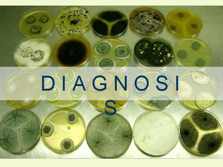
DIAGNOSI S
DIAGNOSIS • Sabourad dextrose agar – the medium where fungi is cultured • Potassium hydroxide (KOH) preparations – dissolve keratin in skin scrapings or biopsy specimens, leaving only fungal cells for examination • Gomori methenamine silver (GMS) stain – used on tissue to stain fungal cells black • Direct immunofluorescence - labeling of antibodies or antigens with fluorescent dyes.
Mycoses are also among the most difficult diseases to heal.
WHY? • Fungi possess the biochemical ability to resist the oxidative damage perpetuated by T cells during cell-mediated immune response • Fungi are eukaryotic and thus biochemically similar to human cells, which means most fungicides are toxic to human tissues
The Gold Standard!!! • Amphotericin B – considered the best drug for treating systemic mycoses and other fungal infections that do not respond to other drugs
The downside…. . It is still toxic to humans
The alternative. . • Azole drugs – which only inhibit the fungi (fungistatic) rather than kill it – less toxic to humans – e. g. ketoconazole, itraconazole and fluconazole
Categories of Fungal Agents • True fungal pathogens • Opportunistic fungi
True pathogens • Have the ability to actively attack and invade tissues • Can infect any host, regardless of its immune status
Opportunistic Fungi • Lack genes for proteins that aid in colonizing the body • Do not cause disease in healthy individuals • Can infect only susceptible individuals, those whose body’s defenses are low
The only fungi known to cause disease in humans: • • Blastomyces dermatiditis Coccidiodes immitis Histoplasma capsulatum Paracoccidiodes brasiliensis The remaining fungi that cause disease are considered opportunistic fungi.
The following slides may contain scenes not suitable for very young audiences. Those with weak constitutions are asked to continue paying attention no matter how utterly repulsive you may find it to be. VIEWER’S DISCRETION IS ADVISED.
SYSTEMIC MYCOSIS caused by TRUE PATHOGENS
SYSTEMIC MYCOSES • those fungal infections that spread throughout the body • result of infections by one of the four pathogenic, dimorphic fungi of the division Ascomycota. • Ascomycota: Blastomyces, Coccidioides, Histoplasma, and Paracocidioides
BLASTOMYCOSI S
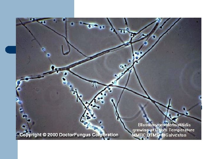
BLASTOMYCOSIS • Pathogen: Blastomyces dermatitidis. • B. dermatitidis normally grows in soil rich in organic material, such as decaying vegetation and animal wastes, where cool, damp conditions favor growth and sporulation.
BLASTOMYCOSIS • Mode of infection: Inhalation of dust disturbed from the environmental sources carries fungal spores and pieces of hyphae into the lungs.
BLASTOMYCOSIS • Pulmonary Blastomycosis – the most common manifestation of Blastomyces infection in humans. • Initial pulmonary lesions are asymptomatic. • If symptoms do develop, they are vague and include cough, fever, malaise, and weight loss.
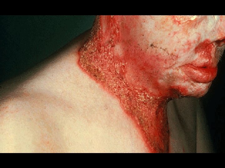
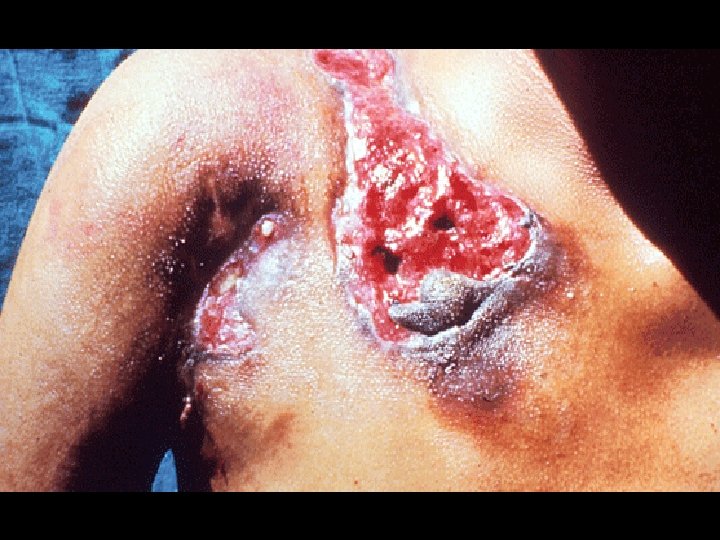
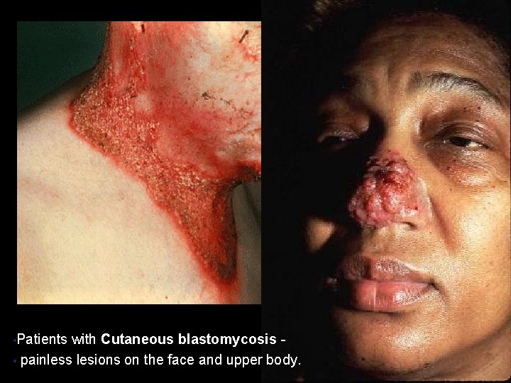
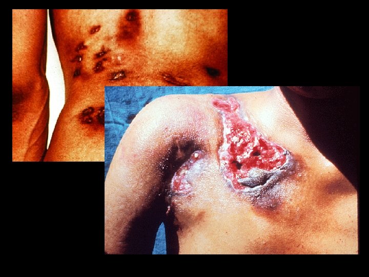
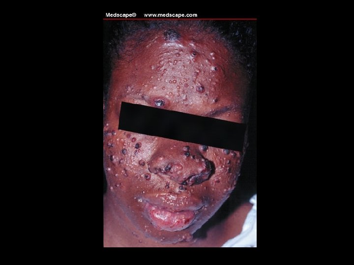
BLASTOMYCOSIS • Cutaneous blastomycosis – consists of generally painless lesions on the face and upper body. • Can be raised and wartlike, craterlike due to the death of tissue.
BLASTOMYCOSIS • Osteoarticular blastomycosis – a condition where fungus spreads to the spine, pelvis, cranium, ribs, long bones, or subcutaneous tissues surrounding joints.
• Patients with Cutaneous blastomycosis • painless lesions on the face and upper body.
BLASTOMYCOSIS • Diagnosis – relies on identification of B. dermatitis following culture, or direct examination of various samples such as sputum, bronchial washings, biopsies, cerebrospinal fluid, or skin scrapings. • Treatment – amphotericin for 10 weeks.
COCCIDIOIDOMYCOSI S
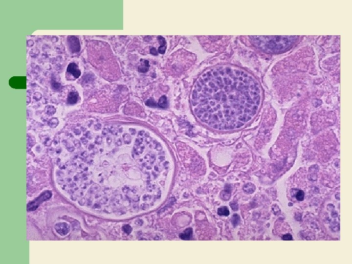
COCCIDIOIDOMYCOSIS Pathogen: Coccidioides immitis • Can be recovered from desert soil, rodent burrows, archaeological remains, and mines. • Once inhaled in dust, anthrospores become trapped in the alveolar spaces of the lungs.
COCCIDIOIDOMYCOSIS • Major manifestation - pulmonary • Invasion of the CNS may result in meningitis, headache, nausea, and emotional disturbance
COCCIDIOIDOMYCOSIS • Diagnosis – based on the identification of spherules in KOH- or GMS- treated samples collected from patients • Amphotericin B – the drug of choice
HISTOPLASMOSI S
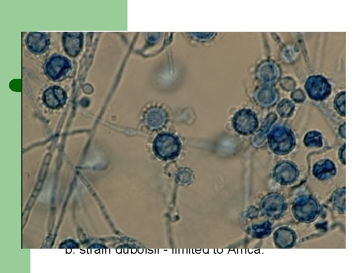
HISTOPLASMOSIS • Pathogen: Histoplasma capsulatum • most common fungal pathogen affecting humans. • Two strains recognized: a. strain capsulatum - intracellular parasite, first attacks alveolar macrophages in lungs b. strain duboisii - limited to Africa.
HISTOPLASMOSIS • Diagnosis – based on the identification of the distinctive budding yeast in KOH- or GMS - prepared samples of skin scrapings, sputum, cerebrospinal fluid, or various tissues.
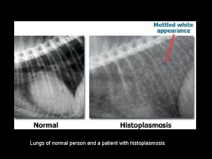
Lungs of normal person and a patient with histoplasmosis
HISTOPLASMOSIS TREATMENT: • Amphotericin B - prescribed when symptoms do not resolve. • Ketoconazole – used to treat mild infections.
PARACOCCIDIOIDOMYCOSI S
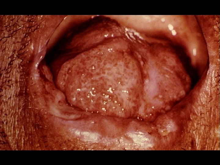
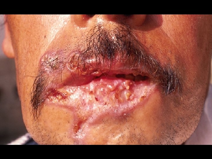
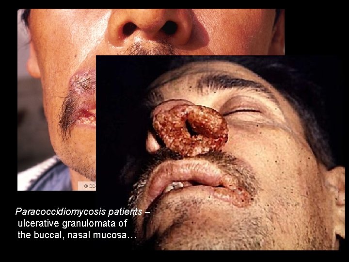
PARACOCCIDIOIDOMYCOSIS • Pathogen: Paracoccidioides brasiliensis • chronic granulomatous disease that characteristically produces a primary pulmonary infection, and then disseminates to form ulcerative granulomata of the buccal, nasal and occasionally the gastrointestinal mucosa.
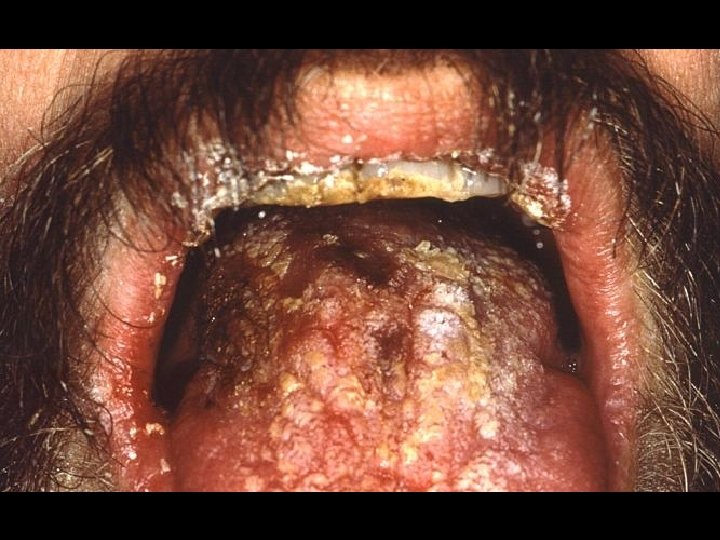
Paracoccidiomycosis patients – ulcerative granulomata of the buccal, nasal mucosa…
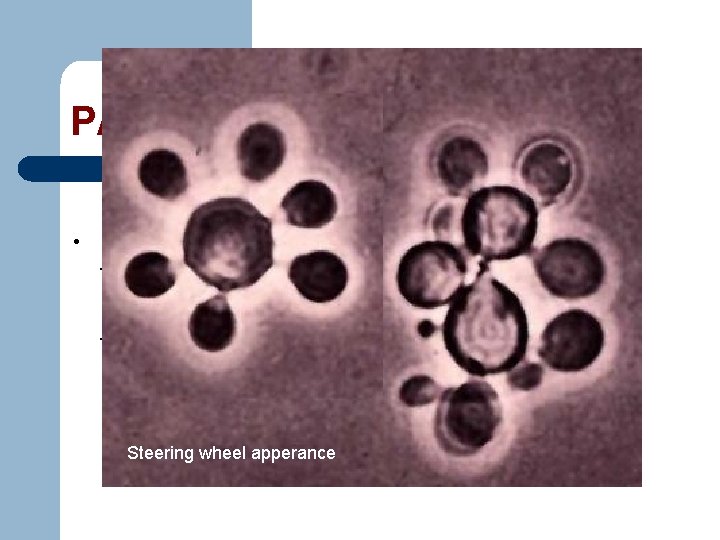
PARACOCCIDIOIDOMYCOSIS • Diagnosis - KOH or GMS preparations of tissue samples revealing yeast cells with multiple buds in a “steering wheel” formation. Steering wheel apperance
PARACOCCIDIOIDOMYCOSIS • Treatment - Amphotericin B or Ketoconazole
End of slideshow, click to exit. Aww… sorry, wrong number. .
Systemic Mycoses caused by OPPORTUNISTIC FUNGI
Systemic Mycoses Caused by Opportunistic Fungi • Opportunistic mycoses do not typically affect health humans. • One of the more significant causes of human disease and death and appear only when their hosts are weakened. • considered “classical” opportunists because they are routinely encountered.
• 5 genera of opportunistic fungi that cause systemic mycoses: – Aspergillus – Candida – Cryptococcus – Pneumocystis – and Mucor
ASPERGILLOSI S
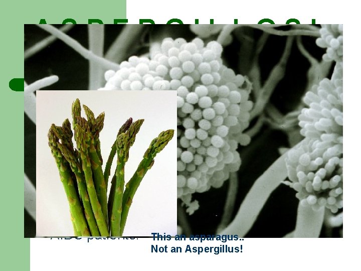
ASPERGILLOSI S • Pathogens: Aspergillus fumigatus Aspergillus niger and Aspergillus flavus • a term for several diseases caused by the inhalation of spores of fungi • causes allergies and a growing problem for AIDS patients. This an asparagus. . Not an Aspergillus!
ASPERGILLOSI S • 3 clinical manifestations: -Hypertensitivity aspergillosismanifests as asthma -Noninvasive aspergillomas- ballmasses of fungal hyphae like
ASPERGILLOSI S - Acute invasive pulmonary aspergillosismore serious. Signs and symptoms, which include fever, cough, and pain, may present as mild pneumonia.
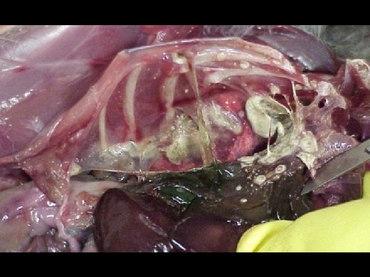
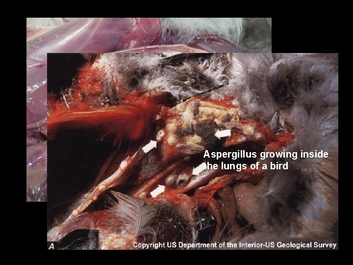
Aspergillus growing inside the lungs of a bird
ASPERGILLOSI S • TREATMENT of hypersensitivity - either the use of various allergy medications or desensitization to the allergen. • Invasive disease – treated by surgical removal of aspergillomas and surrounding tissue, plus high-dose, intravenous administration of amphotericin B.
CANDIDIASI S
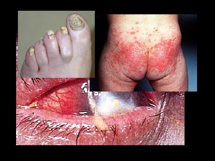
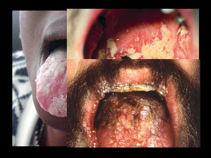
CANDIDIASIS • Pathogen: Candida albicans. • Common members of the microbiota of the skin and mucous membranes.
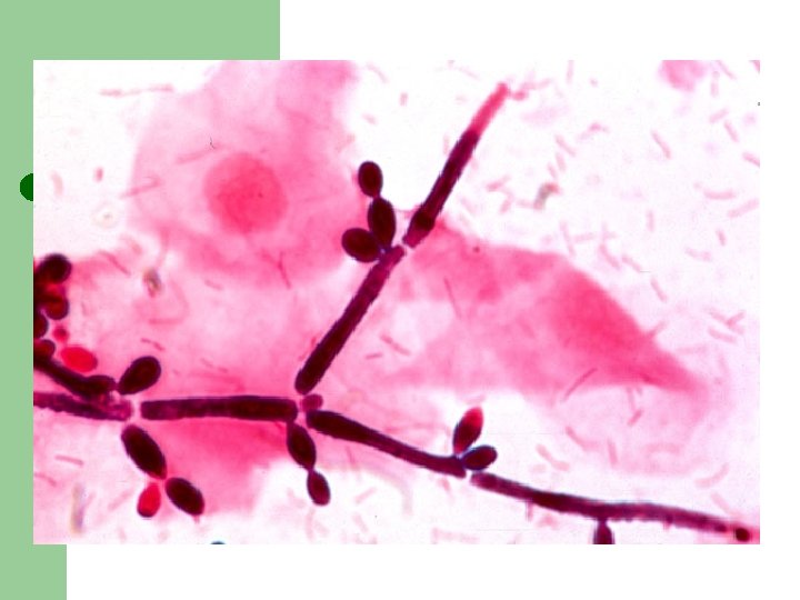
CANDIDIASIS • Diagnosis – KOH and GMS preparations of skin samples, nail scrapings, cerebrospinal fluid, serum, urine, sputum, or tissue biopsies can reveal clusters of budding yeast and branching pseudohyphae.
CANDIDIASIS l TREATMENT – Nystatin for oral candidiasis in infants. l Amphotericin B-treatment for invasive candidiasis.
C R Y P T O C C OS I S
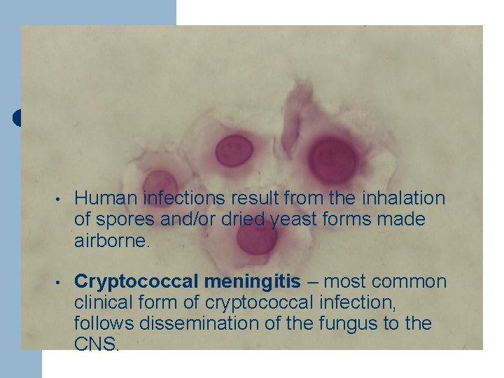
CRYPTOCOCCOSIS • Pathogen: Cryptococcus neoformans • Human infections result from the inhalation of spores and/or dried yeast forms made airborne. • Cryptococcal meningitis – most common clinical form of cryptococcal infection, follows dissemination of the fungus to the CNS.
CRYPTOCOCCOSIS • Diagnosis – involves collecting specimen samples that correlate with the symptoms.
CRYPTOCOCCOSIS • Treatment – Amphotericin B and 5 fluorocytosine administered together for 6 -10 weeks.
PNEUMOCYSTIS PNEUMONIA
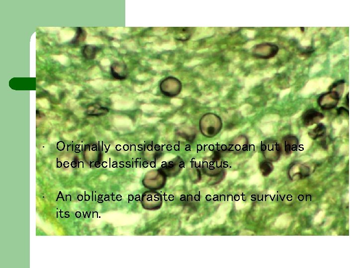
PNEUMOCYSTIS PNEUMONIA • Pathogen: Pneumocystis jiroveci • Originally considered a protozoan but has been reclassified as a fungus. • An obligate parasite and cannot survive on its own.
PNEUMOCYSTIS PNEUMONIA l Transmission occurs through inhalation of droplet nuclei containing fungus. l DIAGNOSIS – relies on clinical and microscopic findings. Chest X-rays usually reveal abnormal lung features. l Treatment- antiprotozoan drugs trimethoprim and sulfanilamide.
ZYGOMYCOSES
ZYGOMYCOSES • are opportunistic fungal infections caused by various genera of fungi, Mucor, Rhizopus, and Absisia. • Commonly seen in patients with uncontrolled diabetes, people who inject illegal drugs, in some cancer patients, and in some patients receiving antimicrobial agents. • Infections generally develop in the face and head are.
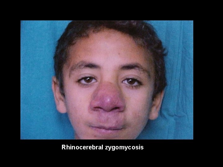
ZYGOMYCOSES l Conditions which resulted from the dissemination of the infections throughout the body in severely immunocompromised individuals: a. Rhinocerebral zygomycosis b. Pulmonary zygomycosis c. Gastrointestinal zygomycosis d. Cutaneous zygomycosis
Rhinocerebral zygomycosis
ZYGOMYCOSES • DIAGNOSIS – clinically based on patient history and microscopic findings in fungus. • TREATMENT- physical removal of infected tissues and management of predisposing factors. • Drug of choice- Amphotericin B administered intravenously for 8 -10 weeks.
End of slideshow, click to exit. Di pa-reportun si KATSUS! Nyahahaha! Aw. . Cge nlng….
Superficial, Cutaneous, and Subcutaneous Mycoses caused by OPPORTUNISTIC FUNGI
Superficial Mycoses • Usually acquired by direct contact with the fungus and are the most common infections. • Confined at the outer, dead layers of the skin, nails, or hair, all of which are composed of dead cells filled with a protein called keratin.
Superficial Mycoses l Piedra- a superficial infection that forms nodules on hair shafts - infections are normally associated with the scalp, but faial and pubic hairs can also be infected -transmission is via shared hair brushes and combs, and several members of a family are typically infected at the same time
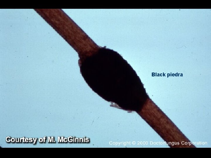
Superficial Mycoses l Black piedra- caused by Piedra Hortae - characterized as hard, black nodules encircling the hair shafts l White piedra- caused by Trichosporon beigelii - characterized as soft, gray to white nodules on the hair shafts
Black piedra
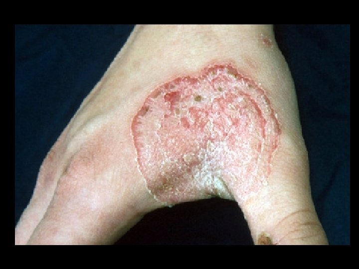
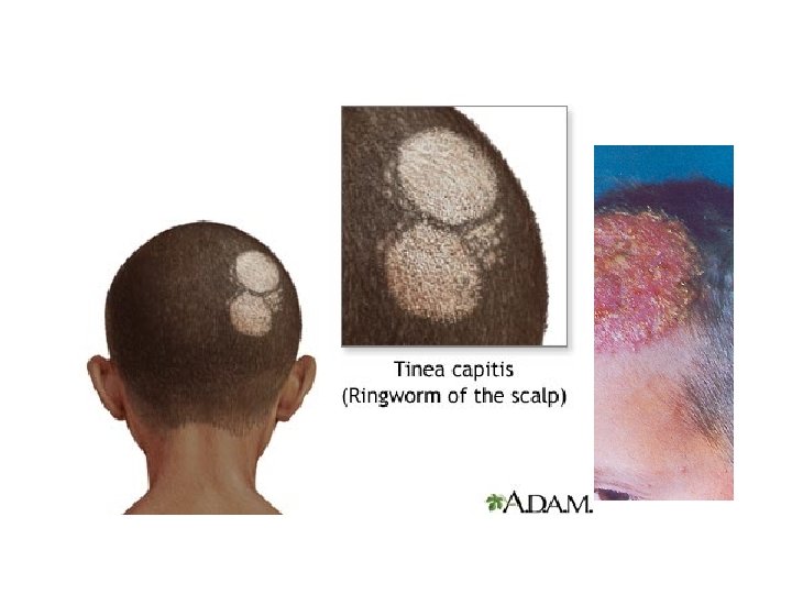
Dermatophytoses l Infections caused by dermatophytes. l Fungal infection of nails and skin. l Infections were previously called ringworms because they resemble a worm lying below the surface of the skin.
Dermatophytoses Result from the fungi that use keratin as a nutrient source and thus colonize only dead layers of the skin, nails, and hair. l No living tissues are infected. l Caused by various Epidermophyton, Microsporum, Trichophyton species. l
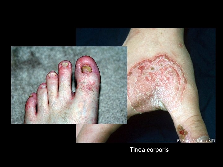
Types of Diseases Caused by Dermatophytes Tinea infections (ringworm) l - Athlete’s foot (Tinea Pedis) l -jock itch (Tinea cruris) l -yeast infections of the mouth and vagina often caused by Candida species. l
Tinea corporis
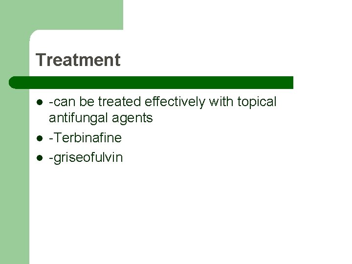
Treatment l l l -can be treated effectively with topical antifungal agents -Terbinafine -griseofulvin
MALASSEZIA
MALASSEZIA INFECTIONS • -Malassezia furfur is a fungus that infects the skin. Clinical manifestations include pityriasis, in which fungal growth disrupts melanin production to produce discolored patches, and the most common manifestation of M. furfur infectiondandruff.
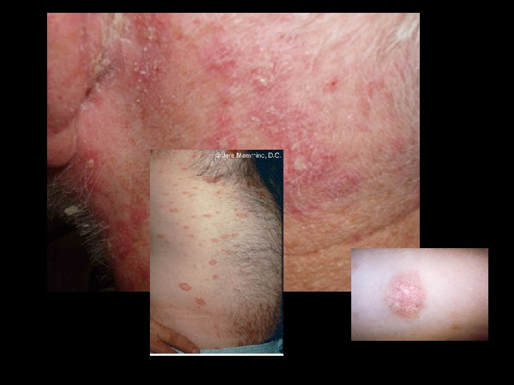
MALASSEZIA INFECTIONS • Folliculitis- caused by M. furfur - involves infection of the hair follicles on the back, chest, and upper arm, producing follicular papules or pustules
MALASSEZIA INFECTIONS • Diagnosis - examining the patient under ultraviolet illumination, though some other fungi also fluoresce. • Treatment - solutions of antifungal imidazoles, such as ketoconazole shampoos. - topical applications of zinc pyrithione -selenium sulfide lotions - propylene glycol
Cutaneous and Subcutaneous Mycoses
Cutaneous and Subcutaneous Mycoses • Infection requires traumatic introduction of fungal elements beneath the outer, dead layers of skin.
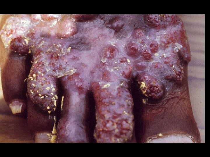
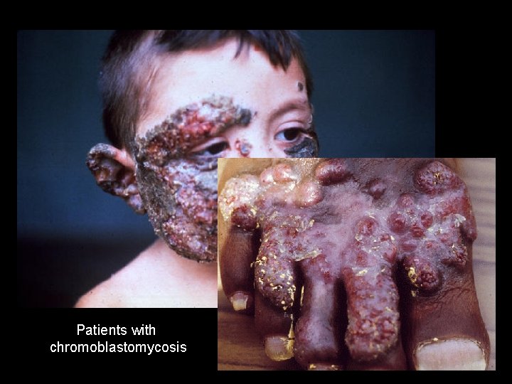
CHROMOBLASTOMYCOSIS AND PHAEOHYPHOMYCOSIS l Chromoblastomycosis- small, itchy but painless, scaly lesions on the skin surface resulting from fungal growth in subcutaneous tissues near the site of inoculation.
PHAEOHYPHOMYCOSIS Paranasal sinus phaeophomycosis – involves colonization of the nasal passages and sinuses; it occurs in allergy sufferers and AIDS patients l Cerebral phaeophomycosis – caused by Cladophiolophora bantiana - has a preference for neural cells and actively invades the brain
Patients with chromoblastomycosis
Treatment l Phaeohyphomycosis can be treated with intraconazole, but the disease is permanently destructive to tissues l Chromoblastomycosis requires surgical removal of infected and surrounding tissues followed by antifungal therapy.
MYCETOMAS
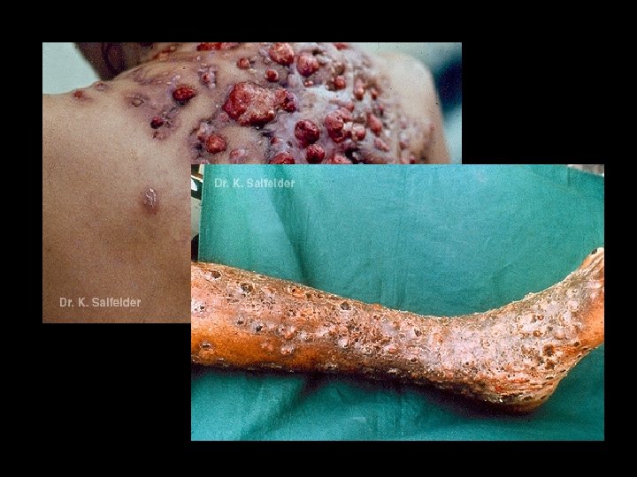
MYCETOMAS l - tumorlike infections of the skin, fascia (lining of muscles), and bones of the hands of feet caused by mycelial fungi l l This fungi live in soil and are introduced into humans via the prick wounds and scrapes caused by twigs, thorns, or leaves contaminated with fungi.
Treatment l l Surgical removal of the mycetoma Anti fungal therapy with ketoconazole
SPOROTRICHOSIS
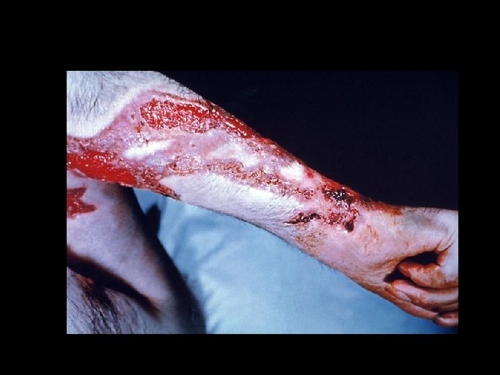
SPOROTRICHOSIS l Pathogen: Sporothrix schenckii l Also called “rose-gardener’s disease”, a subcutaneous infection limited to the arms and legs.
Treatment l l Topical applications of saturated potassium iodide for several months Itraconazole Terbinafine It can be prevented by wearing of gloves, long clothing and shoes to prevent inoculation.
Summing it all up…
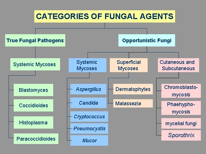
CATEGORIES OF FUNGAL AGENTS True Fungal Pathogens Opportunistic Fungi Systemic Mycoses Superficial Mycoses Blastomyces Aspergillus Dermatophytes Coccidioides Candida Systemic Mycoses Cryptococcus Histoplasma Pneumocystis Paracoccidioides Mucor Malassezia Cutaneous and Subcutaneous Chromoblastomycosis Phaehyphomycosis mycelial fungi Sporothrix
Going back. . .
Clinical Manifestations of Mycoses • Fungal infections (most common) • Toxicoses (poisoning) • Allergies (hypersensitivity reactions)
TOXICOSIS
Toxicosis • Mycotoxicosis – caused by eating food contaminated with fungal toxins only • Mycetismus - poisoning caused by eating the fungus
Mycotoxicosis • Mycotoxins – chemicals produced by fungi during metabolism, that are poisonous to humans • Aflatoxins – are the most well-known mycotoxins, produced by certain species of Aspergillus
Mycetismus • Amanita phalloides – “death cap mushroom” – Produces the deadliest mushroom toxin
Allergies to Fungi • Allergens – spores or other fungal elements that cause type I hypersensitivities, or more rarely, type III