Male Reproductive System Spermatogenesis Dr Surojit Das Ph


- Slides: 38

Male Reproductive System & Spermatogenesis Dr Surojit Das, Ph. D Assistant Professor Biomedical Laboratory Science & Management Vidyasagar University 1 1

Gametogenesis: Spermatogenesis + Oogenesis

SOME TERMS Gamete: egg or sperm Gametogenesis: production of eggs or sperm Oogenesis: production of eggs Spermatogenesis: production of sperm Spermiogenesis: differentiation of sperm morphology Follicle: where eggs mature in the ovary Ovulation: release of egg from follicle Polar body: nonfunctional product of meiotic divisions in oogenesis Zygote: Fertilized egg Oogonia: mitotically dividing cells in the ovary, will become Oocytes Primary oocyte: decision has been made to undergo meiosis, cell has grown. Cells are arrested at this stage until puberty. Secondary oocyte: has completed first meiotic division the division is unequal in terms of cytoplasm Ovum: Ovulated egg, ready to be fertilized. If fertilized, the second meitoic division will occur, another polar body will be given off.

Hypothalamic pituitary axis and the hormonal feedback system

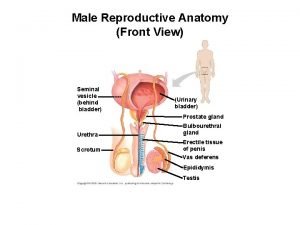
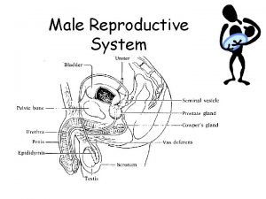
Male Reproductive System

Male Reproductive System Production of: 1. Semen 2. Hormones 6

7

Function of Male Reproductive System

Testis: Groups of Seminiferous Tubules 9

10

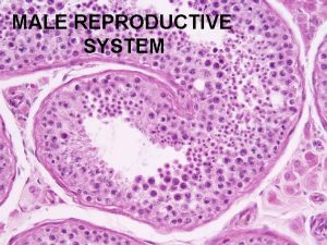
Histology of Testis 11

Cross Section through Testis Cells in spermatogenesis: 1. Spermatogenic cells 2. Sertoli cells 3. Leydig cells 12

Testis (cross-sectional view) 13 Stain: hematoxylin-eosin. Low magnification

Sertoli cells 14

-prominent euchromatic nucleus and prominent nucleolus - cell sends processes around developing germ cells making cell margins difficult to observe at LM level - inter-Sertoli cell tight junctions at the base of the tubule form blood testis barrier -synthesizes and secretes a fluid into the lumen containing: 1. androgen binding protein (ABP) that binds testosterone and creates increased (total) concentration of testosterone - secretion of ABP stimulated by FSH and testosterone 2. inhibin that suppresses FSH secretion from pituitary -provides environment for germ cell development (nurse cell) -phagocytose and digest shed residual bodies produced by developing germ cells

Interstitial cells of Leydig (Leydig cells): - steroid-synthesizing cell with enriched s. ER and mitochondria - produce and secrete testosterone - stimulated by LH and prolactin from pituitary 16

Spermatogenesis

Structure of seminiferous tubules

Spermatogenesis begins at puberty Spermatogonia primary spermatocytes mitotic division, occurring in basal compartment primary spermatocytes cross tight junction to adluminal compartment Primary spermatocyte secondary spermatocytes first meiotic division, occurring in adluminal compartment Secondary spermatocyte spermatids second meiotic division, adluminal compartment Spermatids spermatozoa no division, maturation process

Section of the germinal epithelium in the seminiferous tubule Sertoli cells divide the germinal epithelium into a basal and adluminal compartment, via the Sertoli cell. Spermatozoa are released into the lumen

Major events in the life of a sperm involving spermatogenesis, spermiogenesis, and spermiation during which the developing germ cells undergo mitotic and meiotic division to reduce the chromosome content

Cell Divisions during Spermatogenesis

Cell Divisions during Spermatogenesis

Cell Divisions during Spermatogenesis

Spermatogenic cycle The following is an example of how the number of spermatozoa is increased by repetitive mitotic divisions of spermatogonial cells followed by the two meiotic divisions. There actually more than 4 types of spermatogonia, so the actual number of mature spermatozoa originating from the initial division of a type A 1 spermatogonium is actually greater than 96. A 1 24 primary spermatocytes Mitosis Meiosis I 2 A 1 48 secondary spermatocytes Mitosis A 1 3 A 2 Reductional division Meiosis II Mitosis Equational division 96 spermatids Mitosis First meiotic division lasts several weeks in humans Second meiotic division takes about 8 hours in humans An entire spermatogenic cycle in humans takes about 64 days. 6 B 1 Mitosis Spermiogenesis = Spermateleosis = Spermatozoan metamorphosis 12 B 2 96 mature spermatozoa The maturing spermatids remain attached by cytoplasmic bridges as they mature => syncytium

Spermiogenesis 26

• The shape of a human sperm cell is adapted to its function Plasma membrane Middle piece Neck Head Tail Mitochondrion (spiral shape) Nucleus Acrosome 27

Cross Section through the Tail 28

29

Schematic representation of the development of a diploid undifferentiated germ cell into a fully functional haploid spermatozoon along the basal to the adluminal compartment and final release into the lumen. Different steps in the development of primary, secondary, and spermatid stages are also shown and the irreversible and reversible morphological abnormalities that may occur during various stages of spermatogenesis

Differentiation of a human diploid germ cell into a fully functional spermatozoon 31

Diagrammatic representation of the series of cellular and chromatin changes during the development of the germ cell into a spermatozoon and its subsequent release and storage into the epididymis and its journey into the female reproductive tract

Diagrammatic representation of the steps where the histones are replaced with the transition proteins and protamines in the round spermatid progresses into a condensed spermatid just before it is released into the lumen


Hormonal Regulation of Spermatogenesis 35

Semen 1. 5% Sperm 2. 65% Secretions of seminal vesicles: proteins, enzymes, mucus, fructose, prostaglandins 3. 30% secretions of prostate glands: milky alkaline fluid, high in basic sugars, p. H ~ 7. 3 -8 36

Semen Analysis – reference values Semen value Volume Normal range > 2. 0 ml Concentration > 20 million/ml Motility > 40% motile Morphology >14% 37

THANK YOU
 Function of cervix
 Function of male reproductive system
 Seminal tubules
 Exercise 42 anatomy of the reproductive system
 Lutalphase
 Function of fsh
 What is reproductive system
 Function of reproductive organs
 Reproductive physiology
 Reproduction in human
 Art-labeling activity: the male reproductive system, part 1
 Male reproductive system information
 Where is semen stored
 Male cow reproductive system diagram
 Function of prostate gland
 Male reproductive system plants
 Function of male reproductive system
 Male fish reproductive system
 Reproductive system of pila
 Mammary papilla pig
 Male plant reproductive system
 Figure 28-1 the male reproductive system
 Primary sex organ of the male reproductive system? *
 Lesson 20.2 the male reproductive system
 Boar reproductive system
 Figure 28-2 the female reproductive system
 Colon function in male reproductive system
 Figure 16-1 male reproductive system
 Similarities between male and female reproductive system
 Female reproductive system label
 Pathway of sperm in male reproductive system
 Drawing of the male and female reproductive system
 Male plant reproductive system
 Summary of reproductive system
 Female external genitalia
 Layers of scrotum anatomy
 Note on male reproductive system
 Oogenesis
 Male genital


