Liver function tests Liver function tests ITests that


")


: • In hepatic cirrhosis the albumin will")


: Clotting is the end result of a complex")


- Renal Oedema: total proteins are decreased, albumin is decreased and proteinuria.")
 & in these conditions proteinuria")

 • Anti-LKM")


 Liver cell injury causes transient increase in: • AST, ALT and")

 Alpha fetoprotein")


: • It occurs in")

: • It is due to")

- Slides: 51
Liver function tests
• Liver function tests: • I-Tests that detect injury to hepatocytes (serum enzyme tests): • Aminotransferases and alkaline phosphatase • II-Tests of the liver’s capacity to transport organic anions and to metabolize drugs • III-Tests of the Liver’s biosynthetic capacity: Serum proteins, albumin • IV-Tests that measure hepatic metabolic function • V-Tests for accurate etiological diagnosis.
• I-Tests that reflect injury to hepatocytes: • A. Enzymes that detect hepatocellular necrosis (aminotransferases or transaminases ): • Aspartate aminotransferase(AST, formerly serum glutamate oxaloacetic transaminase. SGOT) and alanine amino transferase( ALT, formerly serum glutamic pyruvate transaminase -SGPT). • ALT is primarily localized to the liver. • AST is present in a wide variety of tissues like the heart, skeletal muscle, kidney, brain and liver. • The AST and ALT levels are increased to some extent in almost all liver diseases.
• Elevations of aminotransferases • 1. Severe ( > 20 times, 1000 U/L) : The highest elevations occur in severe viral hepatitis, drug or toxin induced hepatic necrosis and circulatory shock • 2. Moderate (3 -20 times): acute hepatitis, neonatal hepatitis, chronic hepatitis, autoimmune hepatitis, drug induced hepatitis, alcoholic hepatitis and acute biliary tract obstructions • 3. Mild (1 -3 times): sepsis induced neonatal hepatitis, extrahepatic biliary atresia (EHBA), fatty liver, cirrhosis, non alcoholic steatohepatitis (NASH), drug toxicity, myositis, duchenne muscular dystrophy and even after vigorous exercise.
• Biliary atresia is a childhood disease of the liver in which one or more bile ducts are abnormally narrow, blocked, or absent. It can be congenital or acquired. • Myositis is inflammation or swelling of the muscles. • Duchenne muscular dystrophy is a severe type of muscular dystrophy. • Wilson disease a hereditary disease that is characterized by excessive accumulation of copper in the body (as in the liver or brain) due to abnormal copper metabolism. • AST: ALT ratio: The ratio of AST to ALT is of use in Wilson disease, chronic liver disease (CLD) and alcoholic liver disease and a ratio of more than 2 is usually observed • In NASH the ratio is less than one in the absence of fibrosis on liver biopsy. • The lack of ALT rise is probably due to pyridoxine deficiency
• In viral hepatitis the ratio is usually less than one • The ratio invariably rises to more than one as cirrhosis develops possibly because of reduced plasma clearance of AST secondary to impaired function of sinusoidal cells. • ALT exceeds AST in toxic hepatitis, viral hepatitis, chronic active hepatitis and cholestatic hepatitis. • Falsely low aminotransferase levels : in patients on long term hemodialysis probably secondary to either dialysate or pyridoxine deficiency. • Low levels have also been seen in uremia.
• B. Enzymes that detect cholestasis: • Cholestasis is a decrease in bile flow due to impaired secretion by hepatocytes or to obstruction of bile flow through intra-or extra-hepatic bile ducts. • 1 -Alkaline phosphatases(ALP) : are a family of zinc metaloenzyme. • In liver, alkaline phosphatase is found histochemically in the microvilli of bile canaliculi and on the sinusoidal surface of hepatocytes. • Fresh unhemolysed serum is the specimen of choice for estimation. • The test should not be done on plasma if citrate, oxalate or EDTA were used as anticoagulants, they form a complex with zinc and the alkaline phosphatase, causing irreversible enzyme inactivation.
• Highest levels of alkaline phosphatase occur in cholestatic disorders. Elevations occur as a result of both intra-hepatic and extra-hepatic obstruction to bile flow and the degree of elevation does not help to distinguish between the two. • ALP levels are likely to be very high in EHBA. • In acute viral hepatitis, alkaline phosphatase is usually either normal or moderately increased. • Hepatitis A may present a cholestatic picture with marked and prolonged itching and elevation of (ALP). • Hepatic and bony metastasis (cancer spreads), infiltrative liver diseases, abscesses, granulomatous liver disease and amyloidosis can also cause elevated levels of alkaline phosphatase.
• Low levels of alkaline phosphatase occur in hypothyroidism, pernicious anemia, zinc deficiency and congenital hypophosphatasia. • alkaline phosphatase in Wilson’s disease: • Wilson’s disease complicated by hemolysis and FHF(fulminant hepatic failure) , may also have very low levels of alkaline phosphatase. • Ratio of alkaline phosphatase and bilirubin is low in fulminant Wilson disease. • This might be the result of replacement of cofactor zinc by copper and subsequent inactivation of alkaline phosphtase.
• Drugs like cimetidine, frusemide, phenobarbitone and phenytoin may increase levels of alkaline phosphtase. • 2. γ glutamyl transpeptidase (GGT): is a membrane bound glycoprotein which catalyses the transfer of γ glutamyl group to other peptides, amino acids and water. • levels of ã glutamyl transpeptidase are high in neonates and infants up to 1 yr and also increase after 60 yr of life. Men have higher values. Children more than 4 yr old have serum values of normal adults. • The normal range is 0 -30 IU/L. • In acute viral hepatitis the levels of (GGT)may reach its peak in the second or third week of illness and in some patients they remain elevated for 6 weeks. • In EHBA GGT is markedly elevated.
• Other conditions causing elevated levels of γ glutamyl transpeptidase include: uncomplicated diabetes mellitus, acute pancreatitis and myocardial infarction. Drugs like phenobarbitone, phenytoin, paracetamol, tricyclic antidepressants may increase the levels of (GGT). • Non-hepatic causes of increased levels of the enzyme include anorexia nervosa, Gullian barre syndrome(GBS) , hyperthyroidism, obesity and dystrophica myotonica. • Anorexia nervosa is an eating disorder, characterized by low weight, food restriction, fear of gaining weight, and a strong desire to be thin. (GBS) is a rapid-onset muscle weakness caused by the immune system damaging the peripheral nervous system
Myotonic dystrophy is a long-term genetic disorder that affects muscle function. It is a type of muscular dystrophy. 3. Other enzymes that detect cholestasis: 5 Nucleotidase and Leucine aminopeptidase (not routinely estimated). …………………………………………… Estimation of serum liver enzymes (Alanine transaminase): The enzyme (ALT) is widely distributed with high conc in liver. Elevated serum ALT is found in hepatitis, cirrhosis, obstructive jaundice, carcinoma of the liver, and chronic alcohol abuse. Assay Principle:
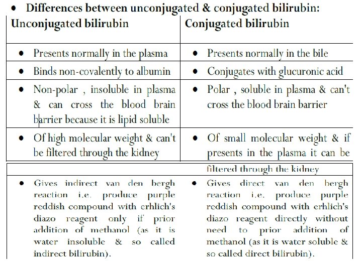
• ALT activity is measured by monitoring the conc of pyruvate hydrazone formed with 2, 4 dinitrophenylhydrazine. ……………………………………………………………………………………… • II-Tests of the liver’s capacity to transport organic anions and to metabolize drugs. • 1. Serum bilirubin: Bilirubin is an endogenous anion • • • derived from hemoglobin degradation from the RBC. The classification of bilirubin into direct and indirect bilirubin are based on the original van der Bergh method of measuring bilirubin. Serum and plasma samples must be kept in dark before measurements, because its photosensitive. Types of bilirubin: 1. Total bilirubin: Normal range is 0. 2 -0. 9 mg/dl. It is slightly higher by in males as compared to females. Helps to diagnose Gilbert syndrome in males easily.
• 2. Direct Bilirubin: Normal range 0. 3 mg/dl • gives estimation of conjugated bilirubin. • 3. Indirect bilirubin: the difference between total and direct bilirubin. • Hyperbilirubinemia: • 1 - Increased unconjugated bilirubin (Unconjugated hyperbilirubinemia > 80% of total bilirubin): results from • overproduction of bilirubin • Impaired uptake, conjugation
• 2 - Increased conjugated bilirubin (Conjugated hyperbilirubinemia > 20% of total bilirubin • Results from : • Impaired intrahepatic excretion. • Regurgitation of unconjugated or conjugated bilirubin from hepatocytes of bile ducts. • Classifed to: • Intrahepatic. eg , Neonatal hepatitis IHBA • Extrahepatic. Eg, EHBA Choledochal cyst. • Choledochal cysts are congenital bile duct anomalies
• Hyperbilirubinemia and Hemolysis: • 1. Blood Bilirubin: Bilirubin itself is not soluble in water and is bound to albumin and thus does not appear in urine. • Hemolysis of RBC with overproduction of bilirubin and concomitant reduced GFR cause decreased excretion and can lead to high bilirubin levels. • Severe parenchymal disease, septicemia and renal failure causes hyperbilirubinemia.
• 2. Urine bilirubin: urine bilirubin indicates hepatobiliary disease. • Unconjugated bilirubin is tightly bound to albumin and not filtered by the glomerulus and thus not present in urine. • Because the renal threshold for conjugated bilirubin is low and the laboratory methods can detect low levels of bilirubin in urine, so conjugated bilirubin may be found in urine when the serum bilirubin levels are normal. This is the case in early acute viral hepatitis.
• 3. Urobilinogen: An increase in the urobilinogen in urine is a sensitive indicator of hepatocellular dysfunction. • It is a good indication of alcoholic liver damage, well compensated cirrhosis or malignant disease of the liver. • In viral hepatitis it appears early in urine. • It is markedly increased in hemolysis. • Urobilinogen gives a purple reaction to Ehrlich’s aldehyde reagent.
• 4. Delta bilirubin: • Direct covalent albumin-bound bilirubin increases in prolonged jaundice. • Half-life is 18 days (the same as albumin), This accounts for the relatively slow regression of jaundice & presence of conjugated hyperbilirubinaemia without bilirubinuria as during recovery from acute hepatitis.
• III-Tests of the Liver’s biosynthetic capacity: • 1. Serum proteins: The liver is the major source of most the serum proteins. The parenchymal cells are responsible for synthesis of albumin, fibrinogen and other coagulation factors and most of the α and β globulins • 2. Albumin: Albumin is quantitatively the most important protein in plasma synthesized by the liver and is a useful indicator of hepatic function • Albumin synthesis is affected by liver disease nutritional status, hormonal balance and osmotic pressure • Serum levels are typically depressed in patients with cirrhosis and ascites.
• Albumin to globulin ratio (A/G): • In hepatic cirrhosis the albumin will be greatly decreased & (A/G) ratio will fall below one. • Normal serum values range from 3. 5 g/dl to 4. 5 g/dl. • Hypoalbuminemia is not specific for liver disease and may occur in protein malnutrition, nephrotic syndrome and chronic protein losing enteropathies
• 3. Prealbumin: • The serum prealbumin level is 0. 2 - 0. 3 g/L. • These levels fall in liver disease presumably due to reduced synthesis. • Because of its short half life, changes may precede alteration in serum albumin • Determination of prealbumin has bee considered particularly useful in drug-induced hepatotoxicity. • 4. Serum ceruloplasmin: It is synthesized in the liver and is an acute phase protein. • Normal plasma levels are 0. 2 -0. 4 g/L
• The plasma concentration rise in infections, rheumatoid arthiritis, pregnancy, non Wilson liver disease and obstructive jaundice. This is an important diagnostic marker in Wilson disease, in which the plasma level is usually low. • Low levels may also be seen in neonates, Menke’s disease, kwashiorkor, marasmus, protein losing enteropathy, copper deficiency and aceruloplasminemia. • Menke’s disease is an inherited disorder in which the body has a problem absorbing copper. • Kwashiorkor is a form of severe protein malnutrition characterized by edema and an enlarged liver with fatty infiltrates.
• Marasmus is a form of severe malnutrition characterized by energy deficiency. • Enteropathy is a disease of the intestine, especially the small intestine. • Aceruloplasminemia is a rare autosomal recessive disorder in which the liver can not synthesis the protein ceruloplasmin properly, which is needed to transport copper around the blood. • 5. α 1 Antitrypsin : is a glycoprotein synthesized by the liver and is an inhibitor of serine proteinases, especially elastase. • Its normal concentration is 1 - 1. 6 g/L. • it is an acute phase protein.
• serum levels increase with inflammatory disorders, pregnancy and after oral contraceptive pills. • Liver disease is usually seen with deficiency of α 1 antitrypsin, an inherited disorder • 6. α feto protein: It is increased in hepatocellular carcinoma (HCC) and more than 90% of such patients have raised levels. • Raised values are also found in other liver diseases like chronic hepatitis, in regeneration phase of acute hepatitis and in hepatic metastasis. • This is also raised in adenomas associated with tyrosinemia. • Tyrosinemia is an error of metabolism, usually inborn, in which the body cannot effectively break down the amino acid tyrosine
• 7. Prothrombin time (PT): Clotting is the end result of a complex series of • enzymatic reactions that involve at least 13 factors. The liver is the major site of synthesis of 11 blood coagulation proteins. • The standard method to assess is the one stage prothrombin time of quick, which evaluate the extrinsic coagulation pathway. • In acute and chronic hepatocellular disease the PT may serve as a prognostic indicator.
• Estimation of serum proteins using Biuret’s Reagent: • • • Total plasma proteins: -6 -8 gm/dl. Serum Albumin : - 3. 5 -5 gm/dl. A/G ratio : - 2 : 1. Serum proteins fractions by electrophoresis are : Albumin, α-1, α-2, β, γ-Globulins. Plasma proteins maintaine blood osmolarity & if decrease leads to oedema.
• Estimation of serum albumin: • Albumin is the major serum protein in normal individuals. It maintains the plasma colloidal osmotic pressure, binds and solubilizes many compounds such as calcium and bilirubin • Elevated serum albumin levels are usually the result of dehydration. • Hypoalbuminemia is very common in many diseases including malabsorption, liver diseases, Kidney diseases, severe burns, infections, cancer and some genetic abnormalities. In severe hypoalbuminemia (less than 2. 5 g/d. L), the low plasma oncotic pressure allows water to move out of the blood capillaries into the tissues causing edema.
• Oedema or edema: an excess of watery fluid collecting in the cavities or tissues of the body. • Types of Oedema: • 1)- Nutritional Oedema: total proteins decreased , albumin is normal and urine is normal. • Causes: 1 - decrease intake in diet as in Kwashiorkor in infants. • 2 - increase needs as in pregnancy • 3 -impaired absorption as in malabsorption syndrome. • 4 - increase loss as in protein losing entropathy & burns. • 5 - Dilutional as in excessive intravenous fluids.
• 2)- Renal Oedema: total proteins are decreased, albumin is decreased and proteinuria. • Causes: - 1 - Nephrotic syndrome. • 2 - urinary tract infection. 3 - renal failure. • 3 - Hepatic oedema: - total proteins are normal, albumin is decreased, and urine analysis is normal. • A/G ratio inverted. • Causes: - Liver cirrhosis. • Proteinuria: it may be physiological occurring in healthy individual due to prolonged standing (called postural proteinuria).
• May be occurring following exercise(called exercise proteinuria) & in these conditions proteinuria is transient. • Organic proteinuria due to renal glomerular or tubular disorders. Persistent Proteinuria. • Proteinuria are classified according to level of 24 hrs. proteins in urine into: • 1 - Mild proteinuria: - if it is less than 1 gm/day. • 2 - Moderate proteinuria: - if it is from 1 -3 gm per day. • 3 - Sever proteinuria: - if it is more than 3 gm/day.
• V- liver Tests for accurate etiological diagnosis: • A. Tests of Viral or toxic liver disease: • Depend on detection of viral Antigen or Antibodies. • (HCV Hepatitis C virus, HBV, HEV, HAV, CMV, EBV. . etc). • B. Auto-antibodies in autoimmune diseases: • Anti-mitochondrial antibodies (AMA) are autoantibodies, consisting of immunoglobulins formed against mitochondria. • AMA in the blood or serum may be indicative of the presence of, or the potential to develop, the autoimmune disease primary biliary cirrhosis (PBC).
• Antinuclear antibodies (ANAs, also known as antinuclear factor or ANF) • Anti-LKM antibody (anti–liver-kidney microsomal antibody or LKM antibody). • Anti-smooth muscle antibodies(ASM Ab) are antibodies (immunoglobulins) formed against smooth muscle. • ANA Ab, ALKM Ab and ASM Ab are detected in in autoimmune diseases.
• C. Tests for metabolic diseases: • Wilson’s disease: decreased ceruloplasmin level. • Haemochromatosis: transferrin level 90% saturated (N. 30%) • Haemochromatosis is an inherited (genetic) disorder causing the body to absorb too much iron from the diet. The excess iron causes damage to an organ in which it collects. especially liver, heart and pancreas • Transferrins are iron-binding blood plasma glycoproteins that control the level of free iron (Fe) in biological fluids. • Alpha 1 antitrypsin deficiency: alpha 1 antitrypsin decreases.
D. Electrophoretic pattern of serum proteins: Ig A rise in Alcoholic cirrhosis. Ig. M rise in primary biliary cirrhosis (PBC). Ig. G rise in chronic and autoimmune hepatitis. A single liver function test is of little value in liver disease. • The use of battery of liver function tests, however constitutes a highly sensitive procedure. • • •
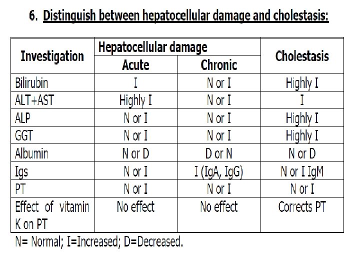
• Laboratory findings in different liver diseases: • 1. Cirrhosis: it may be alcoholic, postnecrotic or biliary cirrhosis. • Laboratory findings in cirrhosis include: • A) Metabolic abnormalities which lead to: • Decreased albumin. • Increased immunoglobulins • Decreased cholesterol. • Prolonged prothrombin time • B) Cholestasis is responsible for transient increase in: • Bilirubin. • GGT. • Alkaline phosphatase
• C) Liver cell injury causes transient increase in: • AST, ALT and LD. H. • D) In post necrotic cirrhosis the increase in bilirubin is higher and increases in AST and ALT are usually more persistent. • E) In biliary cirrhosis serum ALP is the earliest abnormal result. The bilirubin, AST and ALT are only slightly elevated. • 2. Hepatic failure: It may be the result of chronic liver disease or it may be of sudden onset caused by massive cell destruction.
• Laboratory findings of hepatic failure: • Elevated plasma and urine bilirubin (more rise in unconjugated bilirubin). • ALP, AST and ALT will be elevated early in the disease. They return to normal levels either as the condition improves or as the liver cell injury become massive. • Elevated GGT. • Low level of albumin. • Low level of cholesterol. • Prolongation of prothrombin time which parallels the degree of liver failure.
• 3. Liver carcinoma: • Oncofetal tumor markers include: • A) Alpha fetoprotein (AFP): Marked elevations of AFP are almost always associated with advanced hepatocellular carcinoma (HCC). • Patients with chronic hepatitis or cirrhosis with positive serology for HCV or HBV should be screened with serum ALP. • B) Carcinoembryonic antigen (CEA): It is elevated in colon cancer, liver diseases including primary and secondary carcinoma and alcoholic liver disease. • C) Hepatic enzymes: elevations in ALP and GGT in(HCC).
• 4. Fatty liver: • ALT and AST are increased 2 -3 times, usually ALT more than AST in non-alcoholic fatty liver (NAFL). • ALP is normal or slightly increased in less than 50% of patients. • Increased serum ferritin in 60% of cases. • Negative hepatitis viral markers
• • 5. Chronic hepatitis: Definition : Hepatitis more than 6 months. Causes: Viruses (HBV, HDV, HCV, CMV, EBV) Metabolic as: wilson's disease Hemochromatosis. α 1 antitrypsin deficiency.
• 7. Ascites : an abnormal accumulation of fluid • • within the (peritoneal) cavity. It may be a transudate( protein content˂ 3 g/dl) Transudate is fluid pushed through the capillary due to high pressure within the capillary. Or an exudates (protein ˃3 g/dl). Exudate is fluid that leaks around the cells of the capillaries caused by inflammation. Causes of exudates: liver disease (commonest); congestive heart failure; hepatic vein thrombosis and nephrotic syndrome.
• Other causes of exudates: • GIT malignancy (increased alpha fetoprotein, CEA and positive cytology); • Chronic pancreatitis (increased amylase and lipase activity); • T. B (bacteriological examination may help Lymphocytes are predominant cells); • Bacterial peritonitis (bacteriological examination, neurophils are the predominant cells).
• Bilirubin & Jaundice • • • Serum & urinary levels of the bilirubin: Total serum bilirubin: < 1. 1 mg/dl. Serum indirect bilirubin: < 0. 7 mg/dl. Serum direct bilirubin: < 0. 4 mg/dl. Urinary urobilinogens: 0 -4 mg/day Increased in hemolytic anemia. Decreased in micro-obstruction as in hepatitis. Absent in obstructive jaundice. Urine normally contains no bilirubin but it present if micro-obstruction (hepatitis) or obstructive jaundice occurs.
• Hyperbilirubinemia & jaundice: • Hyperbilirubinemia: the presence of bilirubin in the blood at level more than 1 mg/dl. • It is due to: 1. Overproduction of bilirubin more than the liver capacity. • 2. Liver cell damage & failure of excretion of bilirubin into the bile. • 3. Obstruction of the bile pathways. • Jaundice "icterus": If the blood bilirubin exceed 2 mg/dl, it will binds loosely to the albumin (at the site of low affinity) so it diffuse to the tissues (skin, mucous membrane & sclera) staining them yellow.
• Types of jaundice • 1. Pre-hepatic (hemolytic jaundice): • It occurs in all types of hemolytic anemias due to excessive destruction of the R. B. Cs. • Characterized by : • Unconjugated hyperbilirubinemia as it exceed the liver capacity. • Increased urobilinogen with abscent bilirubin in urine & increased stercobilinogens (normally: 30 -300 mg/day). • Stercobilinogen (fecal urobilinogen) is a chemical created by bacteria in the gut.
• 2. Hepatic jaundice: • It is due to liver cell damage by hepatitis or cirrhosis & there is associated micro-obstruction of some bile canaliculi. • Characterized by : • Increased both conjugated & unconjugated bilirubin in the blood. • Decreased urobilinogens in urine & stercobilinogens in feces. • Presence of bilirubin in the urine.
• 3. Obstructive or cholestatic jaundice (posthepatic hyperbilirubinemia): • It is due to obstruction of the bile flow by: • Mechanically by stone in the common bile duct. • Carcinoma of the biliary tree. • Carcinoma of the head of the pancrease. • Characterized by: • Increased conjugated bilirubin in the blood. • Presence of bilirubin in urine. • Absent urobilinogens & absent stercobilinogens ( giving clay color of the stool).
• Delta bilirubin: • Appears in the plasma of patients with chronic obstructive jaundice. • In these patients, some conjugated bilirubin react covalently with albumin in the liver cells forming bilirubin –albumin complex, called bilirubin.