Introduction to Hemolytic Anemias Dr Nisha Assistant professor


- Slides: 25

Introduction to Hemolytic Anemias Dr. Nisha Assistant professor Dept: of practice of medicine

HEMOLYTIC � Definition ANEMIAS Introduction � Classification � Pathogenisis � General clinical features � Laboratory evaluation of hemolysis

Hemolytic Anemias Definition �A group of disorders leading to anemia caused by a reduction in red cell life span. � RBC’s normally survive 60 - 120 days. � Bone marrow has the capacity to increase erythropoiesis 6 - 8 times than normal. � Anemia is the result of premature destruction of red cells exceeding the erythropoietic capacity of the bone marrow.

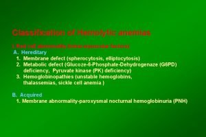
Hemolytic Anemias Classification � Hemolytic anemias may be classified as I- Hereditary or acquired or II- Intracorpuscular or Extracorpuscular

Hemolysis may occur in two compartments I- Intravascular or II- Extravascular ( eg: spleen )

CLASSIFICATION HERIDITARY ACQUIRED INTRA CORPUSCULAR EXTRA CORPUSCULAR • Haemoglobinopathies • Enzymopathies • Membrane cytoskeleton defect • Familial hemolytic uremic syndrome • Paroxysmal nocturnal haemoglobinuria(PNH) • Mechanical destruction(microangiop athic) • Toxic agents • Drugs • Infections • autoimmune

Red cell destruction Extravascular Hb Intravascular Hpt and Hpx RES Globin Haem+globin Hb- Hpt complex Plasma protein pool Plasma iron pool Protoporphyrin Expired CO Free plasma Hb Haemopexin-methem Excess Hb Liver Unconjugated bilirubin Liver Conjugated bilirubin GI tract Faeces methem methaemalbumin Kidney Hb Urine Urobilinogen Haemosiderin met. Hb

Clinical Manifestations in Summary � Onset may be acute or insidious � Symptoms and signs of anemia � Jaundice ◦ Acholuric ◦ Without pruritus � Symptoms and signs specific to the type of hemolytic anemia � Symptoms related to the underlying disease � Splenomegaly � Cholelithiasis (gall stones) symptoms � Leg ulcers (sickle cell, spherocytosis) � Skeletal abnormalities (thalassemia) � Crises (chronic hemolytic disease) ◦ Aplastic crises (HPV-B 19) ◦ Hemolytic ◦ Megaloblastic � Changes in urine color

LABORATORY FİNDİNGS I- Increased RBC destruction � Decreased RBC life span � Increased haem(heme) catabolism ◦ Increased serum unconjugated bilirubin* ◦ Increased endogenous CO production ◦ Increased urobilinogen excretion � Increased serum LDH* � Absence or decrease of serum haptoglobin* �> 1 g /dl /week fall in blood Hb level* � Reduced glycosylated Hb � Signs of intravascular hemolysis ◦ Hemoglobinemia* ◦ Hemoglobinuria* ◦ Hemosiderinuria* ◦ Methemalbuminemi a ◦ Reduced serum hemopexin level

LABORATORY FİNDİNGS II-Increased bone marrow activity and RBC production � Blood ◦ ◦ ◦ Reticulocytosis Macrocytosis Polychromatophilia Erythroblastosis Leukocytosis and thrombocytosis � Bone marrow ◦ Erythroid hyperplasia � Ferrokinetic ◦ Increased plasma iron turnover ◦ Increased RBC iron turnover � Biochemical ◦ Increased RBC creatine ◦ Increased activity of RBC enzymes eg: hexokinase, etc

Laboratory Evaluation of Hemolysis Extravascular Hematologic � Blood film Polychromatophilia � Reticulocyte Increased � Bone marrow Erythroid hyperplasia Plasma or serum � Bilirubin unconjugated � Haptoglobin , absent � Plasma free Hb N� LDH Urine � Bilirubin 0 � Hemosiderin 0 � Hemoglobin 0 � Urobilinogen Intravascular Polychromatophilia Increased Erythroid hyperplasia unconjugated absent 0 + + ( severe cases)

Laboratory tests useful in differential diagnosis � Examination of peripheral blood � Special Lab. examinations

CLASSIFICATION HERIDITARY ACQUIRED INTRA CORPUSCULAR EXTRA CORPUSCULAR • Haemoglobinopathies • Enzymopathies • Membrane cytoskeleton defect • Familial hemolytic uremic syndrome • Paroxysmal nocturnal haemoglobinuria(PNH) • Mechanical destruction(microangiop athic) • Toxic agents • Drugs • Infections • autoimmune

ACQUIRED HEMOLYTIC ANEMIAS

1. AUTOIMMUNE HEMOLYTIC ANEMIA § Auto antibodies(warm antibody- agglutinate the RBC at 37 C or cold antibody- agglutinate at lower temp, 0 to 4 C) develop against the erythrocytes. § Warm antibodies generally belong to Ig G class, whereas cold antibodies are immunoglobuline M (Ig M). § In the warm antibody type, hemolysis takes place extravascularly in the spleen, while in the cold antibody type hemolysis occurs intravascularly or in

ETIOLOGY CAUSES OF COLD AGGLUTININ DISEASES CAUSES OF WARM AGGLUTININ DISEASES VIRAL INFECTIONS IDIOPATHIC INFECTIOUS MYCOPLASMA DRUGS(penicillin) MULTIPLE MYLOMA SLE AND OTHER COLLAGEN DISORDERS KAPOSI, S SARCOMA ANTI PHOSPHOLIPID SYNDROME WALDENSTROM’S MACROGLOBULINEMIA LYMPHOMAS AND CLL VENOM-ARTHROPODES AND SNAKES INFECTIONSmaleria, bartenellosis

PATHOGENESIS � Antibody is usually of Ig. G class � Reacting with erythrocyte Rh antigen complex � Compliment activation may or may not occur � Antibody coated cells will be removed by the splenic macrophages � partially damaged erythrocytes may escape destruction � these repair themselves and appear as spherocytes.

SPHEROCYTES

CLINICAL FEATURES � Anemia � Jaundice � Splenomegaly � Acute Massive Hemolysis With Shock And Renal Failure

� Hemolytic anemia of the cold antibody type may give rise to two clinical syndromes on exposure to cold environment. 1. 2. Cold agglutinine syndrome Paroxysmal cold hemoglobinuria

1. � Ø Ø Cold agglutinine syndrome Occurs in older patients, it presents with acrocynosis Raynaud’s phenominon Gangrene of fingertips and mild chronic hemolytic anemia

2. Paroxysmal cold hemoglobinuria � First identified auto immune disease described by Karl landsteiner, a pathologist from Vienna. � Antibody reacts with erythrocytes in the cold. � In PCH sudden hemolysis develops and leads to hemoglobinuria.

LAB INVESTIGATIONS in peripheral blood film shows � Marked anisopoikilocytosis � Polychromasia � Spherocytosis � Increase reticulocytes. � Neutrophil leukocytosis � Rarely platelets may be reduced due to development of antibodies against them(Evan’s syndrome)

anisopoikilocytosis polychromasia

� The antibodies present on erythrocytes can be demonstrated by direct coomb’s test. � Free antibodies in the serum can be identified by the indirect coomb’s test.
 Promotion from assistant to associate professor
 Fok ping kwan
 Clasificacion morfologica de la anemia
 Megaloblastic anemia
 Vcm anemia ferropenica
 Clasificación morfológica de las anemias
 Classificacao das anemias
 Bilirrubina indireta
 Anemia valores normales en niños
 Titulos pequenos
 Anemias premedulares
 Site:slidetodoc.com
 Nisha patel family feud
 Name of technique
 Dr nisha patel
 Nisha nagarajan
 Nisha patel md
 Nisha patel md
 Nisha singh md
 Nisha patel md
 Dr nisha rughwani
 Hemolytic transfusion reaction
 Hemolysis treatment
 Intravascular hemolytic anemia
 Extrinsic hemolytic anemia
 Intracorpuscular hemolytic anemia


