Eosinophilic Esophagitis Nisha Patel MD Eugene Vortia MD
- Slides: 27
Eosinophilic Esophagitis Nisha Patel, MD Eugene Vortia, MD Jonathan Moses, MD Cleveland Clinic 2013 Resident Education Series Reviewed by Sandeep Gupta, MD of the Professional Education Committee
Definition • Chronic immune and antigen mediated esophageal disease characterized clinically by symptoms related to esophageal dysfunction and histologically by eosinophil-predominant inflammation* * J Allergy Clin Immunol 2011; 128: 3– 20
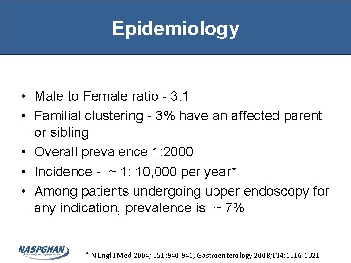
Epidemiology • Male to Female ratio - 3: 1 • Familial clustering - 3% have an affected parent or sibling • Overall prevalence 1: 2000 • Incidence - ~ 1: 10, 000 per year* • Among patients undergoing upper endoscopy for any indication, prevalence is ~ 7% * N Engl J Med 2004; 351: 940 -941, Gastroenterology 2008; 134: 1316 -1321
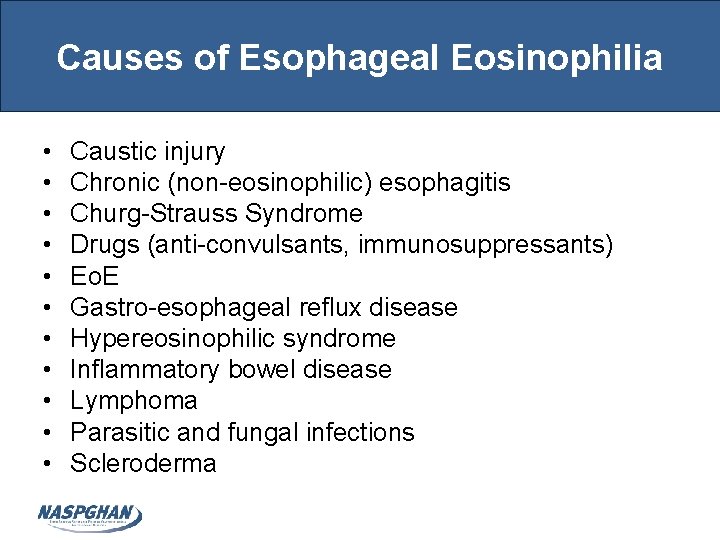
Causes of Esophageal Eosinophilia • • • Caustic injury Chronic (non-eosinophilic) esophagitis Churg-Strauss Syndrome Drugs (anti-convulsants, immunosuppressants) Eo. E Gastro-esophageal reflux disease Hypereosinophilic syndrome Inflammatory bowel disease Lymphoma Parasitic and fungal infections Scleroderma
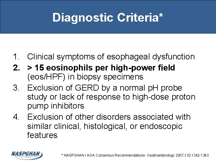
Diagnostic Criteria* 1. Clinical symptoms of esophageal dysfunction 2. > 15 eosinophils per high-power field (eos/HPF) in biopsy specimens 3. Exclusion of GERD by a normal p. H probe study or lack of response to high-dose proton pump inhibitors 4. Exclusion of other disorders associated with similar clinical, histological, or endoscopic features * NASPGHAN / AGA Consensus Recommendations- Gastroenterology 2007; 133: 1342 -1363
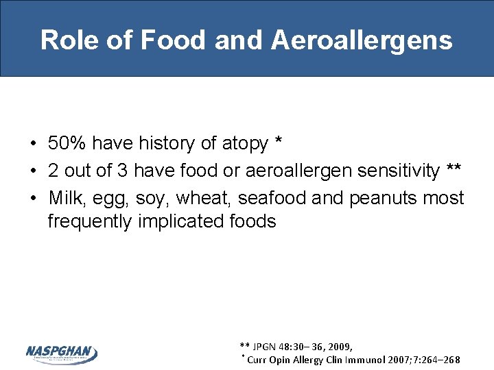
Role of Food and Aeroallergens • 50% have history of atopy * • 2 out of 3 have food or aeroallergen sensitivity ** • Milk, egg, soy, wheat, seafood and peanuts most frequently implicated foods ** JPGN 48: 30– 36, 2009, * Curr Opin Allergy Clin Immunol 2007; 7: 264– 268
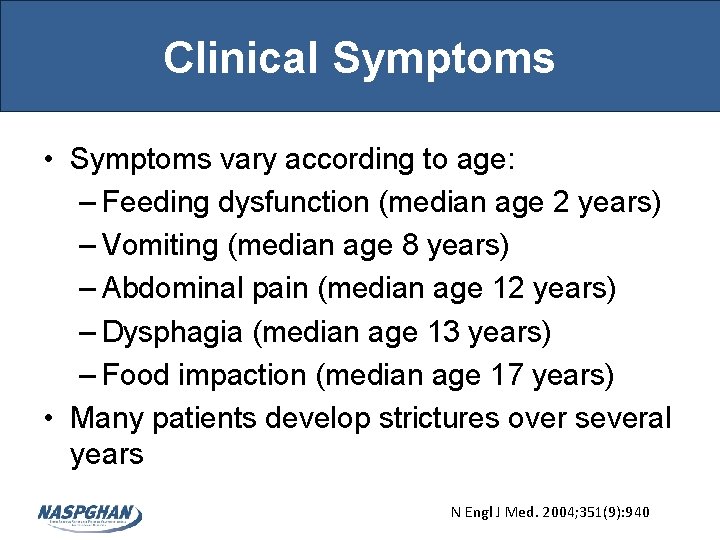
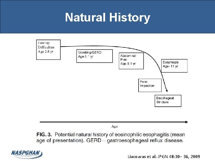
Clinical Symptoms • Symptoms vary according to age: – Feeding dysfunction (median age 2 years) – Vomiting (median age 8 years) – Abdominal pain (median age 12 years) – Dysphagia (median age 13 years) – Food impaction (median age 17 years) • Many patients develop strictures over several years N Engl J Med. 2004; 351(9): 940
Natural History Liacouras et al. JPGN 48: 30– 36, 2009
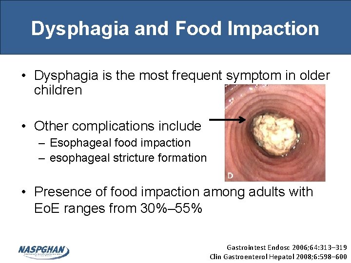
Dysphagia and Food Impaction • Dysphagia is the most frequent symptom in older children • Other complications include – Esophageal food impaction – esophageal stricture formation • Presence of food impaction among adults with Eo. E ranges from 30%– 55% Gastrointest Endosc 2006; 64: 313– 319 Clin Gastroenterol Hepatol 2008; 6: 598– 600
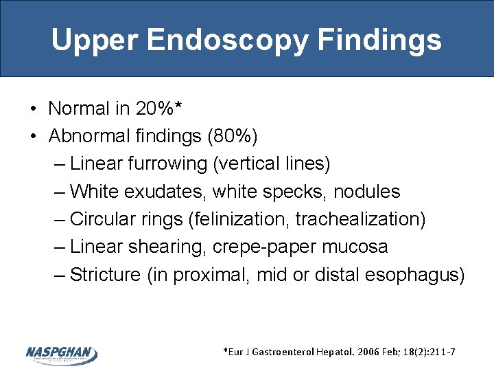
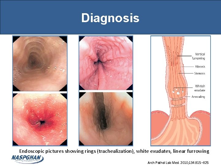
Upper Endoscopy Findings • Normal in 20%* • Abnormal findings (80%) – Linear furrowing (vertical lines) – White exudates, white specks, nodules – Circular rings (felinization, trachealization) – Linear shearing, crepe-paper mucosa – Stricture (in proximal, mid or distal esophagus) *Eur J Gastroenterol Hepatol. 2006 Feb; 18(2): 211 -7
Diagnosis Endoscopic pictures showing rings (trachealization), white exudates, linear furrowing Arch Pathol Lab Med. 2010; 134: 815– 825
Biopsy Procurement • Eo. E is a patchy disease and sensitivity of diagnosis varies based on number of biopsies • Recommendation is to obtain 2 -4 biopsies each from both mid and distal esophagus* • Using 15 eos/hpf as the threshold, the sensitivity of 2, 3 and 6 biopsies was found to be 84%, 97% and 100%, respectively** *J Allergy Clin Immunol 2011; 128: 3 -20 **Am J Gastroenterol 2009; 104: 716– 721
Histopathology • Consensus criteria – Peak eos count of >15 per 40 x eos/HPF • Other criteria – Eosinophilic micro-abscesses - cluster of > 4 eos – Superficial layering - preferential distribution of eos in the upper ½ to ⅓ of epithelium – Basal zone hyperplasia - > 20% of epithelium – Papillary elongation • Lack of correlation between symptoms and histologic findings
Histopathology Esophageal Eosinophilia Arch Pathol Lab Med. 2010; 134: 815– 825
Histopathology Schematic representation of the characteristic histology of Eo. E* *Arch Pathol Lab Med. 2010; 134: 815– 825
Treatment • Acid suppression • Dietary elimination • Elemental diet • • • Allergy evaluation Topical steroids Leukotriene inhibitors Systemic steroids and Biologics Esophageal dilatation
Acid Suppression • Useful part of diagnostic evaluation • GERD can coexist with Eo. E • May help with symptoms • Some patients have PPI-responsive esophageal eosinophilia and so Eo. E can be excluded in these patients* *J Allergy Clin Immunol 2011; 128: 3 -20
Food Elimination • Empiric 6 food elimination diet – Based on milk, soy, eggs, wheat, peanuts/tree nuts, and seafood (6 most common foods) – Foods are reintroduced with monitoring of symptoms and endoscopy/hisstopathology • Skin prick and atopy patch-directed elimination – Culprit foods identified and then eliminated • 6 -food elimination diet or targeted elimination leads to complete histologic remission in 50% of patients * *J Allergy Clin Immunol 2012 Aug; 130 (2): 461 – 467. e 500
Elemental diet • Elemental amino acid-based diet • Superior at inducing histologic remission compared with 6 -food elimination and skin test– directed diets. • Remission occurred in 96%, 81%, and 65% of patients on elemental, 6 -food elimination, and directed diets. J Allergy Clin Immunol 2012; 129: 1570 -8
Allergy evaluation and avoidance • Evaluation by an allergist and/or immunologist recommended for all patients • Skin prick testing (SPT) and atopy patch testing (APT) useful in effective avoidance/elimination strategies • Evidence does not currently support serum specific Ig. E-based food allergen testing J Allergy Clin Immunol 2012 Aug; 130 (2): 461 – 467. e 5
Topical Steroids • Provide short term resolution of clinical and pathologic features • Small risk of systemic side effects (1% absorbed) • Esophageal candidiasis occurs in 15 -20% after 3 months of therapy • Fluticasone (MDI) and Budesonide (viscous) are most commonly used
Fluticasone Propionate • Shown to be safe and effective in Pediatric randomized controlled trials ±* • Dose: 220 - 440μg MDI swallowed daily without the use of a spacer • No rinsing, eating or drinking for 30 minutes afterward, and then drink small amount of liquid *Gastroenterology 2006; 131: 1381– 1391 ± Clinical Gastroenterology and Hepatology 2008; 6: 165– 73
Oral viscous Budesonide • Good option in younger children (<4 yrs) • Dosing: 1 mg - 2 mg of 0. 5 mg/2 ml Budesonide nebulizer suspension mixed with 10 gm of sucralose (Splenda® – 1 gm/packet) orally at bedtime • Dosed according to height <5 ft (1 mg) or >5 ft (2 mg) • No eating, drinking or rinsing mouth for 30 minutes, and then drink small amount of liquid * Gastroenterology 2010; 139: 418 -429
Leukotriene Inhibitors • May help symptoms in some patients • Studies show no impact on esophageal eosinophilia • Symptoms usually recur on discontinuation * Gut 2003 Feb; 52(2): 181 -5
Oral Steroids and Biologics • Systemic corticosteroids may be used for emergent situations (severe dysphagia, hospitalization, weight loss)* • Chronic use of steroids in management of Eo. E results in systemic side effects, sometimes severe • Mepolizumab and Reslizumab (anti-IL 5) are biologics studied in trials for treatment of Eo. E • Safety and sustained efficacy of biologics not fully established J Allergy Clin Immunol 2011; 128: 3 -20
Esophageal Dilatation • Used in symptomatic patients who have strictures with critical esophageal narrowing • Increased risk of mucosal tearing and rarely, perforation during procedure • After dilation, strictures with dysphagia recurs in half of the patients after 2 years* * Gastroenterology 2003; 125: 1660 -1669
Summary • Eo. E is an increasingly recognized esophageal disorder characterized dysphagia and in severe cases food impaction; characteristic endoscopic findings; and intraepithelial eosinophilia not responsive to PPI therapy • Exact pathogenesis remains under investigation, but food allergens, aeroallergens, genetics and acid exposure have been implicated • Accepted treatments include allergy testing/dietary changes, topical steroids, dilation, and PPI. • Complications include esophageal strictures • Based on current evidence, there does not appear to be malignant potential