OVARIAN CANCER Prof Nisha Singh MD AIIMS FICOG





- Slides: 35
OVARIAN CANCER Prof. Nisha Singh MD (AIIMS), FICOG Dept. of Obstetrics & Gynecology King George Medical University Lucknow
INTRODUCTION • • • Fourth most common cause of death in women 30% of genital malignancies in the developed countries 5% of all gynecological cancers in India. Lifetime risk of having ovarian cancer 1. 7% Majority (70%) of cases are diagnosed in advanced stage.
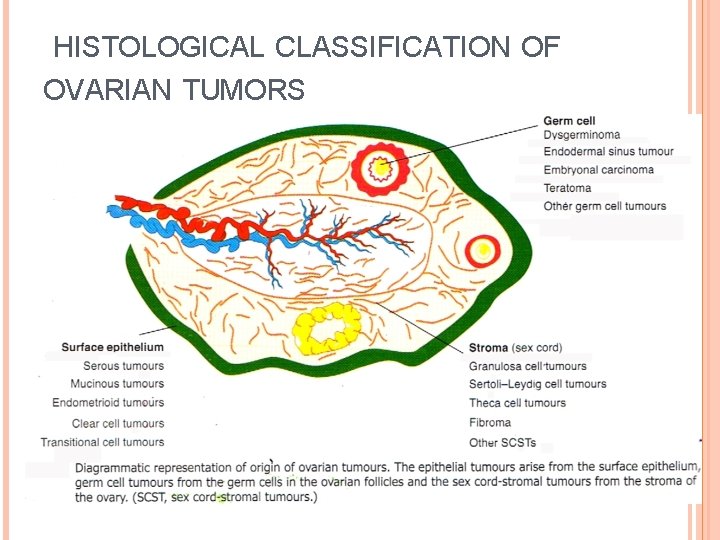
HISTOLOGICAL CLASSIFICATION OF OVARIAN TUMORS
EPITHELIAL CANCER Most common ovarian cancer- 90%. 80% are primary in ovary 20% -metastatic from breast , GIT , and colon Mean age at diagnosis - 60 years. Effect of menopausal status In menopausal women- 30% of ovarian neoplasm are malignant In premenopausal women- 7% are malignant.
ETIOLOGY Various theories Etiology not well known Hereditary or familial ovarian cancer 1) BRCA 1&2 mutations 2) Ras oncogenes, 3) p 53 mutations
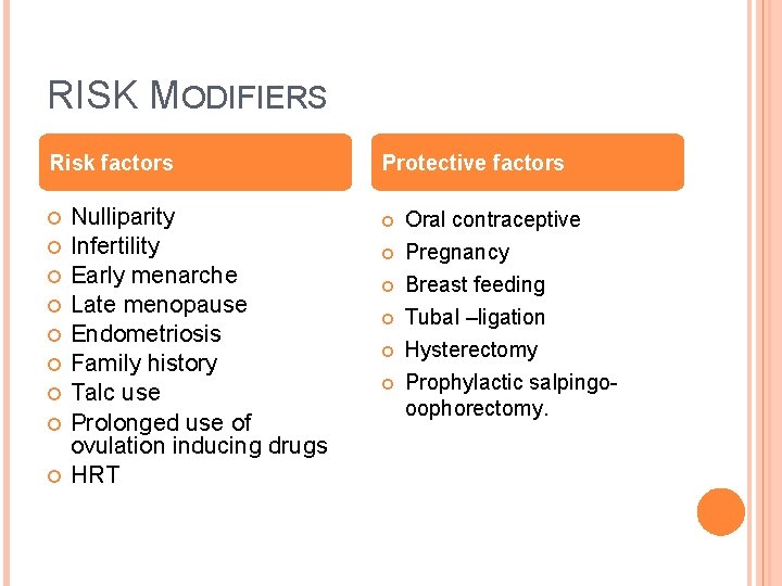
RISK MODIFIERS Risk factors Nulliparity Infertility Early menarche Late menopause Endometriosis Family history Talc use Prolonged use of ovulation inducing drugs HRT Protective factors Oral contraceptive Pregnancy Breast feeding Tubal –ligation Hysterectomy Prophylactic salpingooophorectomy.
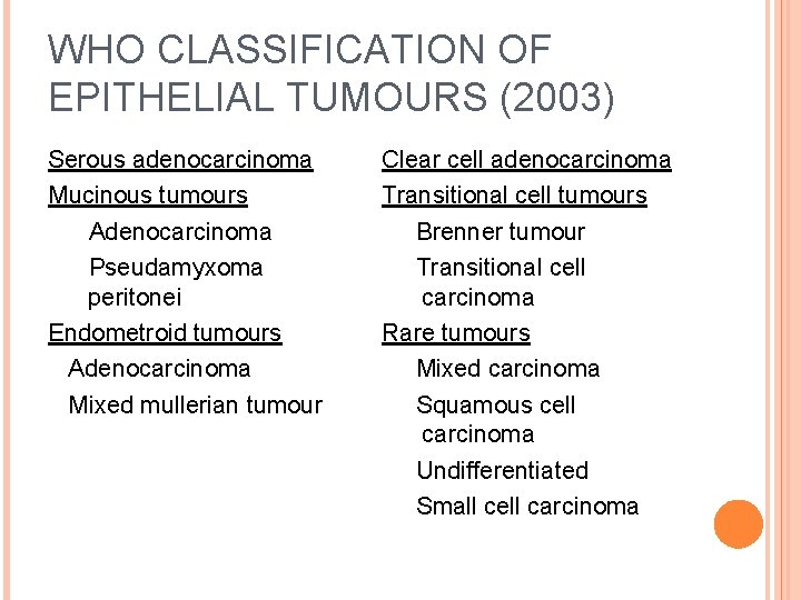
WHO CLASSIFICATION OF EPITHELIAL TUMOURS (2003) Serous adenocarcinoma Mucinous tumours Adenocarcinoma Pseudamyxoma peritonei Endometroid tumours Adenocarcinoma Mixed mullerian tumour Clear cell adenocarcinoma Transitional cell tumours Brenner tumour Transitional cell carcinoma Rare tumours Mixed carcinoma Squamous cell carcinoma Undifferentiated Small cell carcinoma
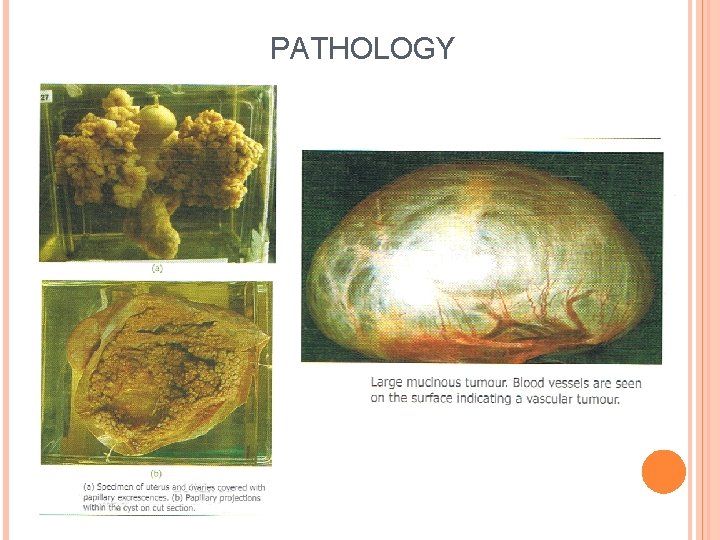
PATHOLOGY
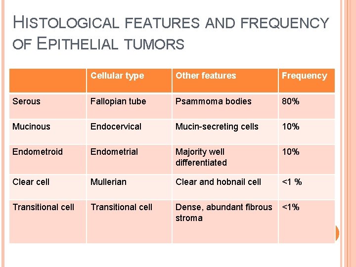
HISTOLOGICAL FEATURES AND FREQUENCY OF EPITHELIAL TUMORS Cellular type Other features Frequency Serous Fallopian tube Psammoma bodies 80% Mucinous Endocervical Mucin-secreting cells 10% Endometroid Endometrial Majority well differentiated 10% Clear cell Mullerian Clear and hobnail cell <1 % Transitional cell Dense, abundant fibrous stroma <1%
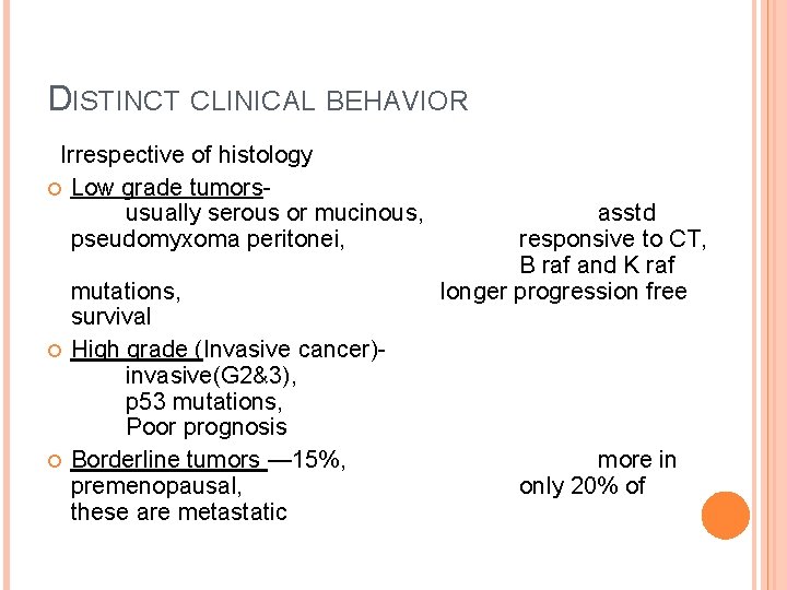
DISTINCT CLINICAL BEHAVIOR Irrespective of histology Low grade tumorsusually serous or mucinous, pseudomyxoma peritonei, mutations, survival High grade (Invasive cancer)invasive(G 2&3), p 53 mutations, Poor prognosis Borderline tumors — 15%, premenopausal, these are metastatic asstd responsive to CT, B raf and K raf longer progression free more in only 20% of
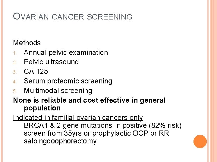
OVARIAN CANCER SCREENING Methods 1. Annual pelvic examination 2. Pelvic ultrasound 3. CA 125 4. Serum proteomic screening. 5. Multimodal screening None is reliable and cost effective in general population Indicated in familial ovarian cancers only BRCA 1 & 2 gene mutations- if positive (82% risk) screen from 35 yrs or prophylactic OCP or RR salpingooophorectomy
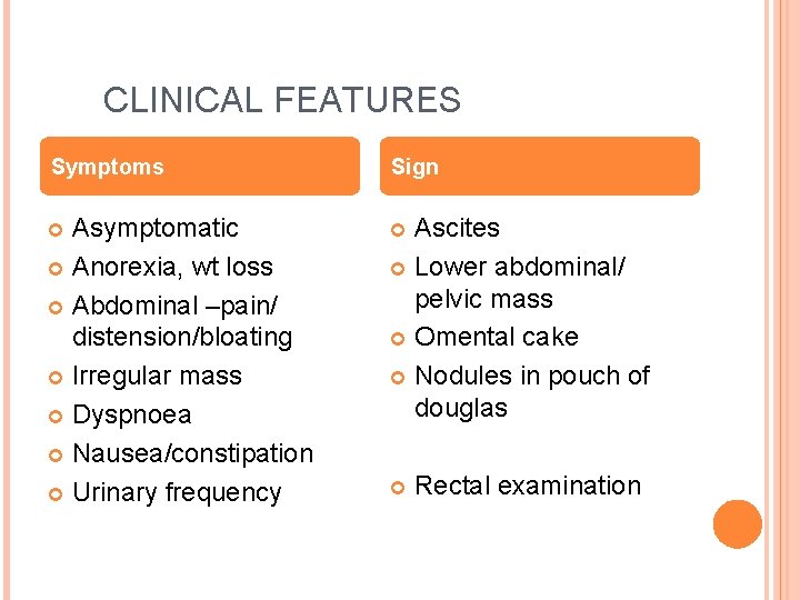
CLINICAL FEATURES Symptoms Sign Asymptomatic Anorexia, wt loss Abdominal –pain/ distension/bloating Irregular mass Dyspnoea Nausea/constipation Urinary frequency Ascites Lower abdominal/ pelvic mass Omental cake Nodules in pouch of douglas Rectal examination
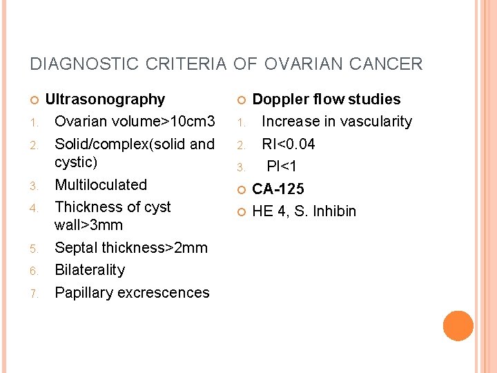
DIAGNOSTIC CRITERIA OF OVARIAN CANCER 1. 2. 3. 4. 5. 6. 7. Ultrasonography Ovarian volume>10 cm 3 Solid/complex(solid and cystic) Multiloculated Thickness of cyst wall>3 mm Septal thickness>2 mm Bilaterality Papillary excrescences 1. 2. 3. Doppler flow studies Increase in vascularity RI<0. 04 PI<1 CA-125 HE 4, S. Inhibin
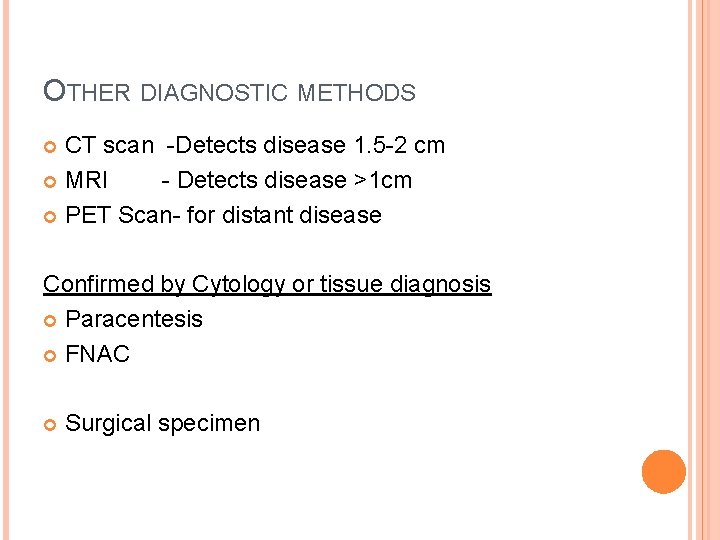
OTHER DIAGNOSTIC METHODS CT scan -Detects disease 1. 5 -2 cm MRI - Detects disease >1 cm PET Scan- for distant disease Confirmed by Cytology or tissue diagnosis Paracentesis FNAC Surgical specimen
ASSESSING RISK OF MALIGNANCY IN OVARIAN TUMOUR 1. 2. 3. 4. 5. Morphological index Risk of malignancy index Serum biomarker levels Colour flow doppler Serum proteomics.
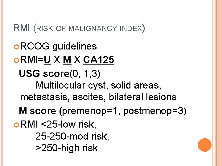
RMI (RISK OF MALIGNANCY INDEX) RCOG guidelines RMI=U X M X CA 125 USG score(0, 1, 3) Multilocular cyst, solid areas, metastasis, ascites, bilateral lesions M score (premenop=1, postmenop=3) RMI <25 -low risk, 25 -250 -mod risk, >250 -high risk
FIGO STAGING OF OVARIAN CANCER 1 ST JAN 2014 Stage I Growth limited to the ovaries IA Growth limited to one ovary, No tumor on surface, negative washings IB Growth in both ovaries else same as IA, IC Tumor limited to one or both ovaries IC 1 Surgical spill IC 2 Tumor rupture before surgery or tumor on surface IC 3 Malignant cells in ascites or peritoneal washings
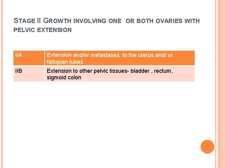
STAGE II GROWTH INVOLVING ONE OR BOTH OVARIES WITH PELVIC EXTENSION IIA Extension and/or metastases to the uterus and/ or fallopian tubes IIB Extension to other pelvic tissues- bladder , rectum, sigmoid colon
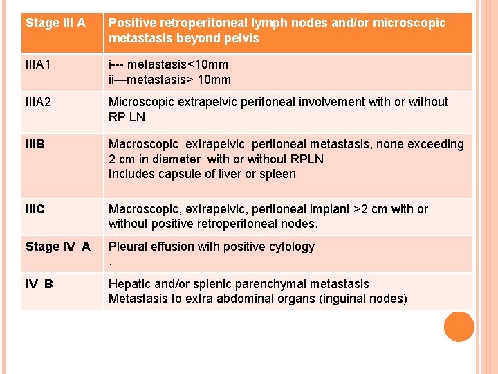
Stage III A Positive retroperitoneal lymph nodes and/or microscopic metastasis beyond pelvis IIIA 1 i--- metastasis<10 mm ii—metastasis> 10 mm IIIA 2 Microscopic extrapelvic peritoneal involvement with or without RP LN IIIB Macroscopic extrapelvic peritoneal metastasis, none exceeding 2 cm in diameter with or without RPLN Includes capsule of liver or spleen IIIC Macroscopic, extrapelvic, peritoneal implant >2 cm with or without positive retroperitoneal nodes. Stage IV A Pleural effusion with positive cytology. IV B Hepatic and/or splenic parenchymal metastasis Metastasis to extra abdominal organs (inguinal nodes)
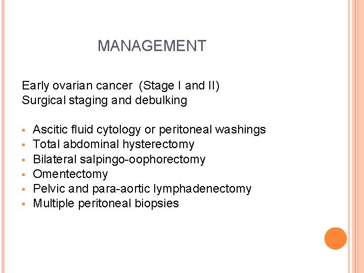
MANAGEMENT Early ovarian cancer (Stage I and II) Surgical staging and debulking § § § Ascitic fluid cytology or peritoneal washings Total abdominal hysterectomy Bilateral salpingo-oophorectomy Omentectomy Pelvic and para-aortic lymphadenectomy Multiple peritoneal biopsies
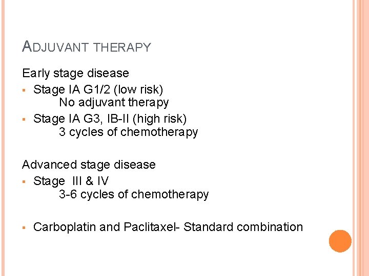
ADJUVANT THERAPY Early stage disease § Stage IA G 1/2 (low risk) No adjuvant therapy § Stage IA G 3, IB-II (high risk) 3 cycles of chemotherapy Advanced stage disease § Stage III & IV 3 -6 cycles of chemotherapy § Carboplatin and Paclitaxel- Standard combination
MANAGEMENT OF ADVANCED-STAGE EPITHELIAL OVARIAN CANCER § § Staging laparotomy and primary cytoreductive surgery followed by Postoperative adjuvant chemotherapy. Best results with Nil residual disease Debulk to microscopic level For inoperable tumors or high surgical risk cases Neoadjuvant Chemotherapy (NACT) followed by surgery and post operative chemotherapy. §
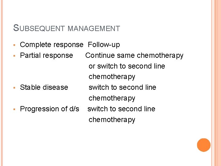
SUBSEQUENT MANAGEMENT § § Complete response Follow-up Partial response Continue same chemotherapy or switch to second line chemotherapy Stable disease switch to second line chemotherapy Progression of d/s switch to second line chemotherapy
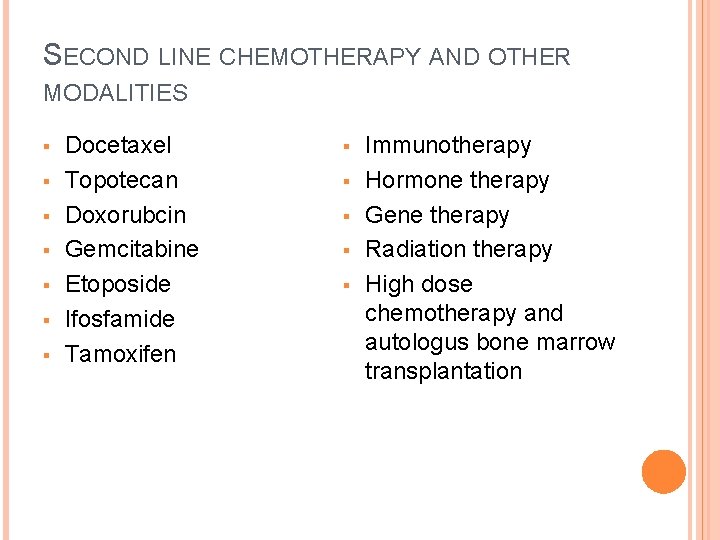
SECOND LINE CHEMOTHERAPY AND OTHER MODALITIES § § § § Docetaxel Topotecan Doxorubcin Gemcitabine Etoposide Ifosfamide Tamoxifen § § § Immunotherapy Hormone therapy Gene therapy Radiation therapy High dose chemotherapy and autologus bone marrow transplantation
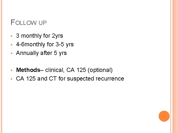
FOLLOW UP § § § 3 monthly for 2 yrs 4 -6 monthly for 3 -5 yrs Annually after 5 yrs Methods– clinical, CA 125 (optional) CA 125 and CT for suspected recurrence
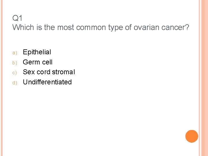
Q 1 Which is the most common type of ovarian cancer? a) b) c) d) Epithelial Germ cell Sex cord stromal Undifferentiated
Q 2 All are risk factors for ovarian cancer EXCEPT Nulliparity Early menarche Endometriosis Tubal ligation
Q 3 Screening for ovarian cancer is recommended for a) b) c) d) Women above 40 years Women above 60 years All women Women at high risk for familial ovarian cancer
Q 4 FIGO stage III of ovarian cancer includes a) b) c) d) Tumor limited to one ovary Tumor involving both ovaries Tumor extending to pelvic organs Tumor extending to abdominal cavity
Q 5 Ovarian cancer is primarily managed by a) b) c) d) Chemotherapy Radiotherapy Immunotherapy Staging Laparotomy
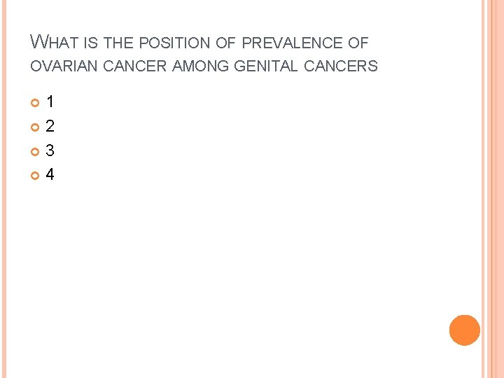
WHAT IS THE POSITION OF PREVALENCE OF OVARIAN CANCER AMONG GENITAL CANCERS 1 2 3 4
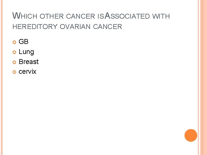
WHICH OTHER CANCER IS ASSOCIATED WITH HEREDITORY OVARIAN CANCER GB Lung Breast cervix
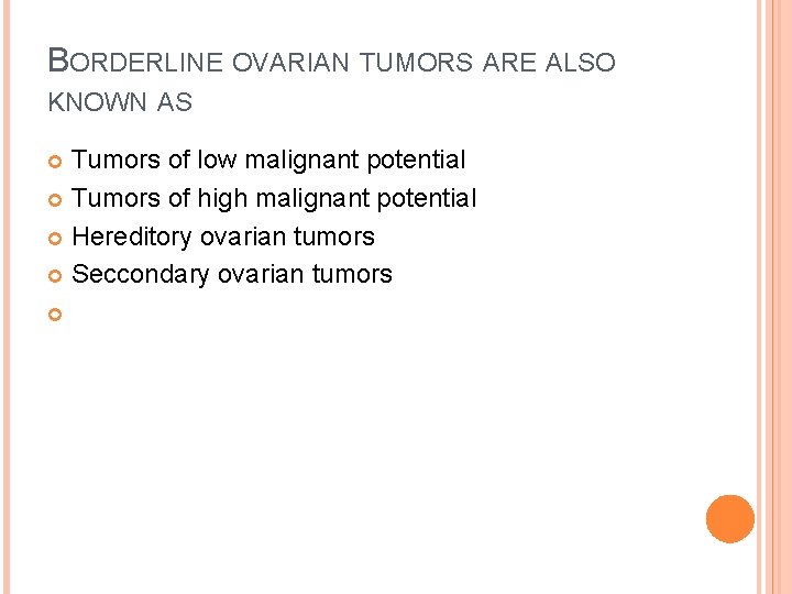
BORDERLINE OVARIAN TUMORS ARE ALSO KNOWN AS Tumors of low malignant potential Tumors of high malignant potential Hereditory ovarian tumors Seccondary ovarian tumors
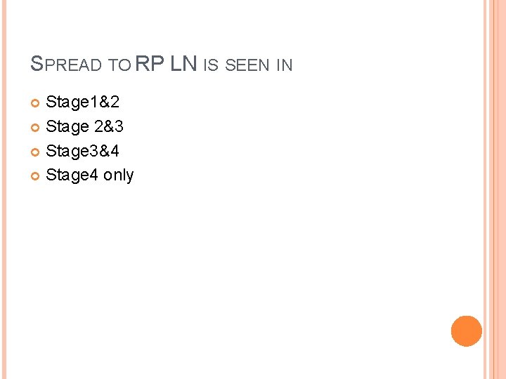
SPREAD TO RP LN IS SEEN IN Stage 1&2 Stage 2&3 Stage 3&4 Stage 4 only
5 YR SURVIVAL RATE S OF TAGEI IS 30% 50% 70% 90% OVARIANCA