International Conference Diagnosis Treatment of Inner Ear Disorders


 – – – most")


- Slides: 50
International Conference Diagnosis & Treatment of Inner Ear Disorders Genetics of deafness Lech Korniszewski The Medical University of Warsaw Institute of Physiology and Pathology of Hearing
Hearing loss – incidence: 6 -8% of population – when all causes are combined hearing loss – most common birth defect 1 in 1000 newborns are deaf 1 in 300 children are affected with congenital hearing loss of a lesser degree additional 1 in 1000 become profoundly hearing impaired before adulthood
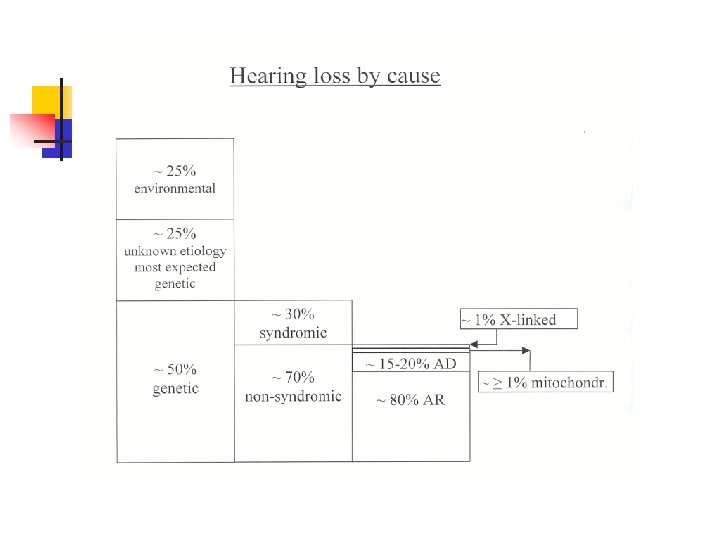
Genetic hearing loss approximately 1% of all human genes are involved in the hearing process inheritance: autosomal recessive autosomal dominant X-linked mitochondrial ® ® ® allelic mutatione in some genes can cause recessive and dominant hearing loss mutations in the same gene may cause syndromic or nonsyndromic hearing loss recessive hearing loss may be caused by a combination of two mutations in differrent genes from the same functional group
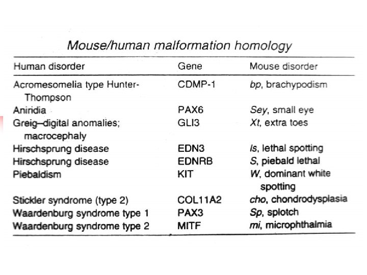
Syndromic hearing loss Over 400 syndromes have been described in which hearing loss is a component part. There are many factors that make specific syndrome diagnosis difficult: * * The rarity of most of these syndromes (lack personal experience) Variability of clinical expression Genetic heterogeneity (a single phenotype may be result of different genes mutations) Pleiotropy (single gene may cause many different phenotypic effects)
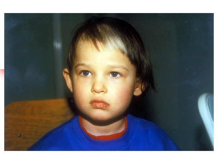
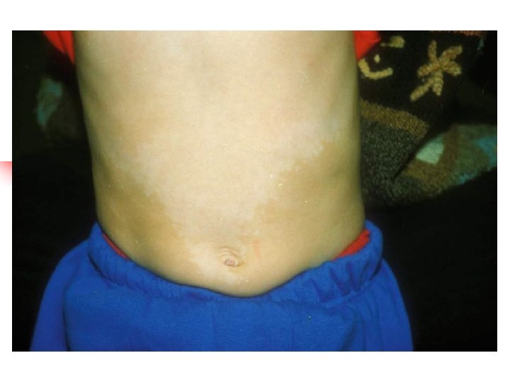
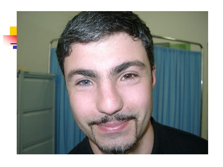
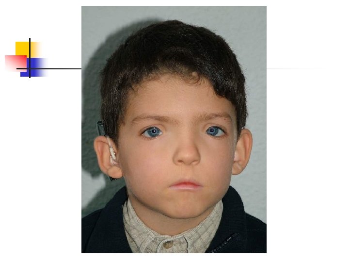
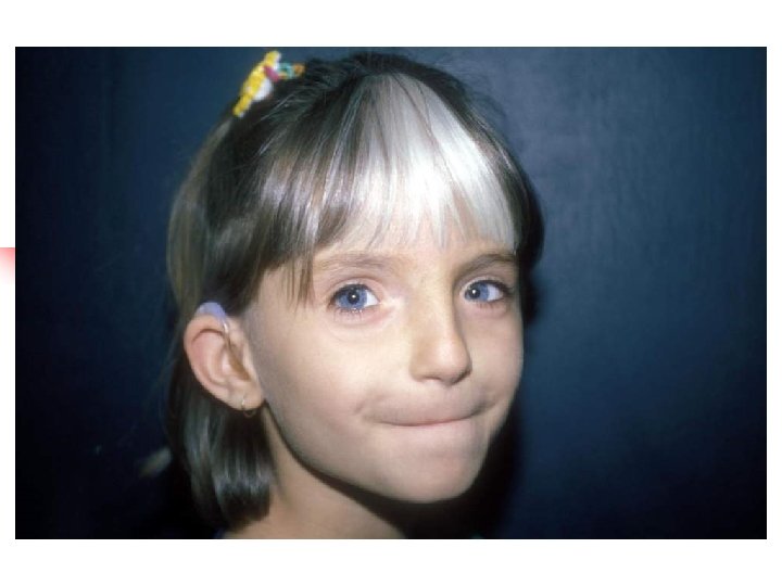
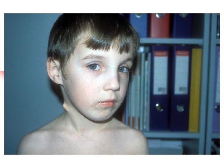
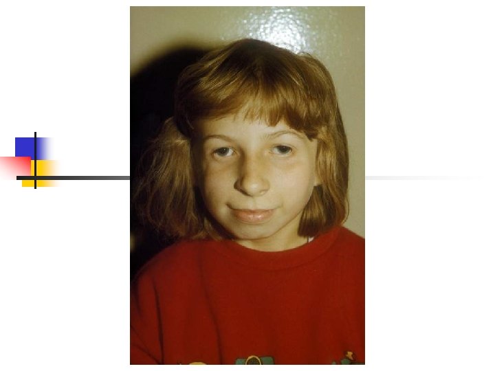
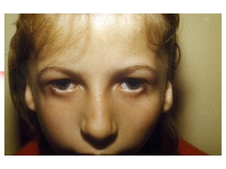
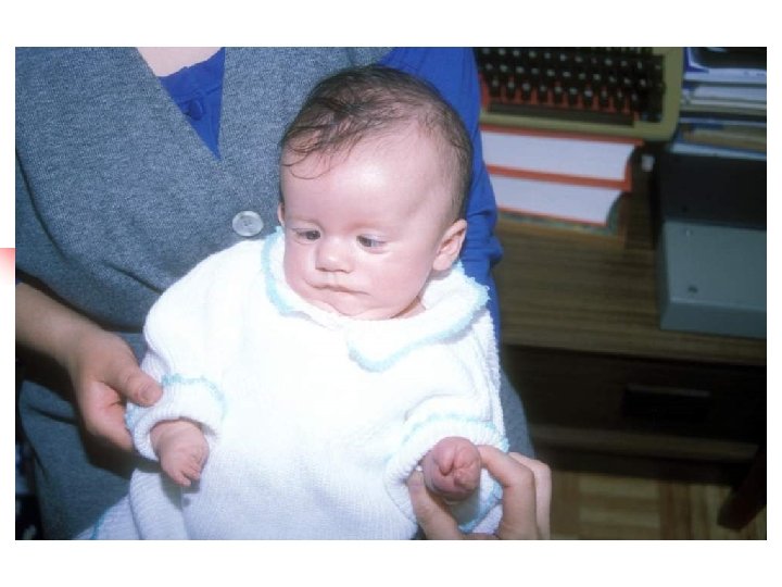
Waardenburg syndromes 1. 2. 3. 4. Bilateral or unilateral sensorineural hearing loss in association with defects in tissues derived from neural crest cells pigmentary abnormalities hair, skin and eyes hearing loss is due to defective migration of melanocytes info the intermediate layer of the stria vascularis genetically heterogeneous; inheritance AD four clinical subtypes
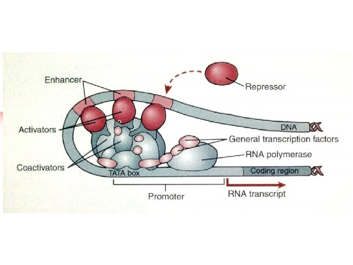
Waardenburg syndromes Type Gene Protein/funct ion Clinical features WS 1 PAX 3 transcription factor Abnormal pigmentation of hair, eyes and skin. Dystopia canthorum, short philtrum, synophrys. Deafness in 20% (unilateral or bilateral) WS 2 MITF transcription factor Abnormal pigmentation of hair, eyes and skin. Deafness in 40% (unilateral or bilateral). No dysmorphic features WS 3 PAX 3 transcription factor Features of WS 1 with limb anomalies WS 4 EDN 3 EDNRB SOX 10 endothelin ligand endothelin receptor transcription factor Abnormal pigmentation of hair, eyes and skin with Hirschprung disease
Transcription factor hierarchy in Waardenburg syndrome: regulation of MITF expression by SOX 10 and PAX 3 EDN 3 EDNRB 3 WS 1 WS 3 PAX 3 SOX 10 transactivation WS 2 MITF transactivation melanocyte tyrosinase WS 4
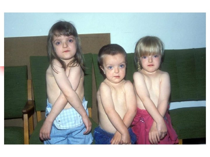
Branchio-oto-renal syndrome Hearing loss conductive, sensorineural or mixed; Branchial cysts and fistulae, external ear malformations, renal dysplasia or hypoplasia. Some patients also eye anomalies Gene EYA 1 on 8 q 13. 3; encoded molecule – transcription factor. Inheritance autosomal dominant. Genetically heterogenous (second BOR locus on 1 p 31)
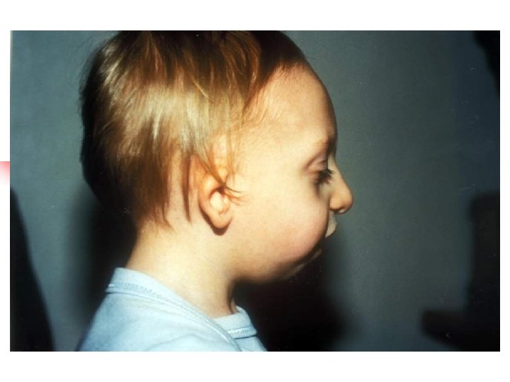
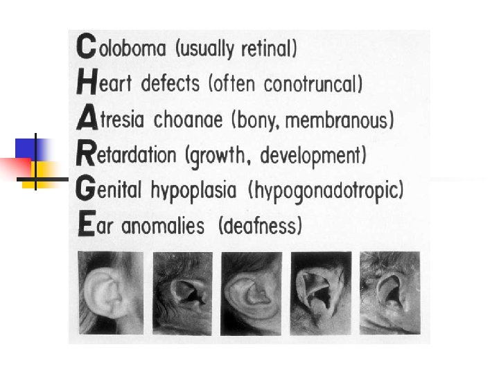
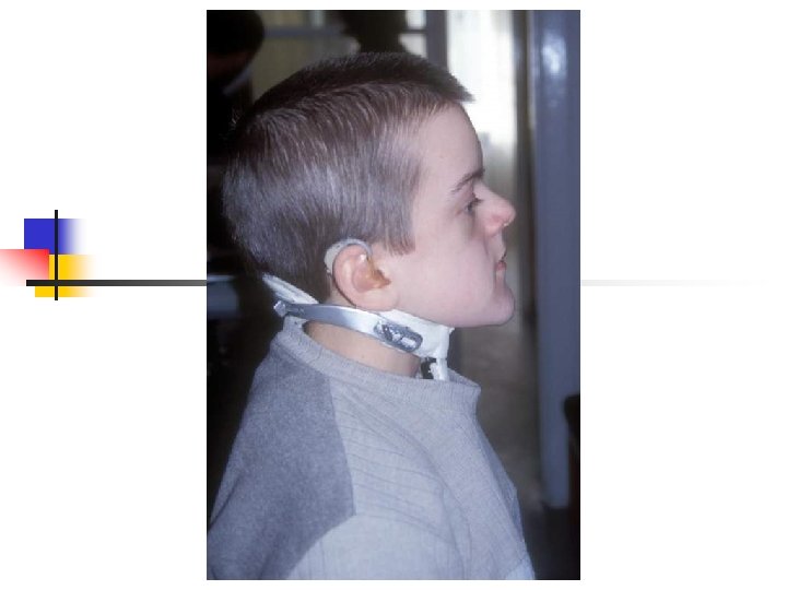
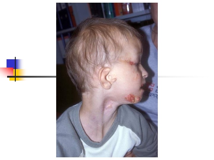
Treacher-Collins syndrome Hearing loss conductive, sensorineural or mixed; Clinical features: down-slanting palpebral fissures, malformation of external and middle ears, sparse lower eyelashes and colobomata of lower eyelids, malar hypoplasia. Gene TCOF; encoded nuclear cytoplasmic transport protein Inheritance autosomal dominant
Usher syndromes 1. 2. 3. Syndromic association of hearing loss with retinitis pigmentosa Accounts 2 -4% of all cases of profound deafness and 50% of the deaf-blind population Inheritance autosomal recessive. Genetic heterogeneity high – more than 12 loci Clinically three main types: TYPE HEARING LOSS VESTIBULAR RESPONSET OF REINITIS PIGM. I Profound from birth Absent 1 st decade II Moderate from birth Normal 1 st or 2 nd decade III Progressive Variable
Usher syndrome Type Locus Gene Protein USH 1 A 14 q 32 - - USH 1 B 11 q 13. 5 MYO 7 A myozyn VIIA USH 1 C 11 p 15. 1 USH 1 C harmonin USH 1 D 10 q 21 CDH 23 cadherina 23 USH 1 E 21 q 21 - - USH 1 F 10 q 21 -22 PCDH 15 protocadherin 15 USH 1 G 17 q 24 -25 USH 1 G SANS USH 2 A 1 q 41 USH 2 A usherin USH 2 B 3 p 23 -24. 2 - - USH 2 C 5 q 14. 3 -21. 3 - - USH 3 A 3 q 21 -25 USH 3 A clarin 1
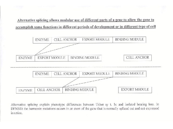
Usher syndromes Usher syndrome type Gene Molecule encoded/function clinical features 1 B MYO 7 A myosin 7 A (motor molecule) profound congenital deafness, retinitis pigmentosa, vestibular areflexia 1 C USH 1 C harmonin 1 D CDH 23 cadherin 23 progfound congenital deafness, variable retinitis pogmentosa and variable vestibular function 1 F PCDH 15 protocadherin 15 profound congenital deafness, retinitis pigmentosa, vestibular dysfunction 2 A USH 2 A usherin (extracellular matrix protein) congenital moderate to severe sensorineural hearing loss (normal vestibular function) retinitis pigmentosa 3 A USH 3 A clarin 1 (transmembrane protein) Progressive sensorineural hearing loss, normal or absent vestibular function, retinitis pigmentosa - " - -"- Nonsyndromic deafness: DFNA 11 (dominant) and DFNB 2 (recessive) results from other alleles of MYO 7 A; DFNB 18 results from different harmonin mutation.
Pendred syndrome Sensorineural deafness, goiter and malformation of the inner ear v Hearing loss is most frequently profound, variable in its onset, rapidly progressive v Goiter results from a specific defect in the organification of iodine (abnormal release of iodine trapped by thyroid after administration of perchlorate) v Malformation of the inner ear in 86% of cases: dilatation of the vestibular aqueduct and endolymphatic sacs, Mondini malformation Inheritance autosomal recessive Mutation of SLC 26 A 4 gene encoding pendrin – protein primarily involved in transport of chloride and iodide ions. Nonsyndromic deafness DFNB 4 also result from mutation in the SLC 26 A 4 gene.
Jervell and Lange-Nielsen syndrome * * * Congenital sensorineural hearing loss and prolongation of the QT interval on electrocardiogram Hearing loss initially involves the high frequencies and progress to become a profound Prolongation of QT reflect a defect in cardiac repolarization. This can lead to recurrent attacks of syncope, ventricular arrhythmia and possible sudden death. Mutation in genes KCNQ 4, KCNE 1 coding potassium chanels (K+ active transport in outer hair cells) Inheritance autosomal recessive
Alport syndrome # # Association of sensorineural high frequency hearing loss with progressive nephritis. Anterior lenticonus, macular flecks, cataracts Gene mutation: COL 4 A 5, COL 4 A 3, COL 4 A 4 coding tissue specific polypeptide subunits of collagen The subunits are expressed in the basilar membrane, spiral ligament and basement membranes of the stria vascularis Genetically heterogeneous. Inheritance X-linked dominant and autosomal recessive
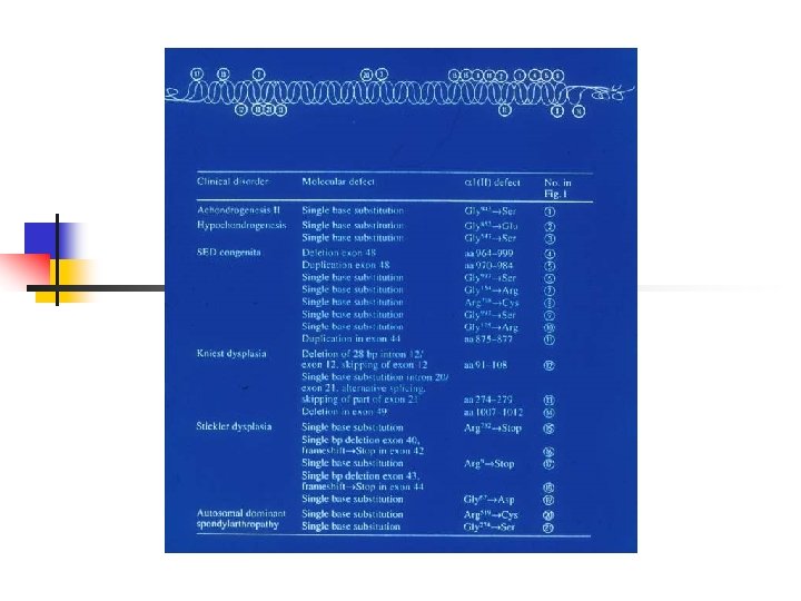
Stickler syndrome – sensorineural hearing loss, high frequency, progressive Myopia, retinal detachment Arthropathy Mid-face hypoplasia, cleft palate, micrognathia Gene defect: COL 2 A 1, COL 11 A 1, – Inheritance autosomal dominant – – COL 11 A 2
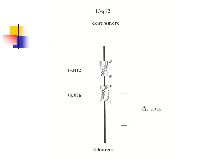
Most important genes involved in non-syndromic hearing loss Chromosomal location Locus /mutation 13 q 11 -12 Gene symbol Inheritance Protein Function DFNB 1/DFNA 3 GJB 2 AR/AD Conexin 26 Gap junction GJB 6 AR/AD Conexin 30 Gap junction 7 q 31 DFNB 4 SLC 26 A 4 AR pendrin Anion transporter 14 q 12 -13 DFNA 9 COCH AD cochlin Extracellular matrix protein mitochondriu m 1555 A>G MTRNR 1 Mitoch. 7445 A>G MTTs 1 12 Sr. RNA t. RNA serine 7472 ins. C 7511 T>C Xq 21. 1 DFN 3 POU 3 F 4 XL domain class 3 Pou Transcription factor 4 p 16. 1 DFNA 6/14/38 WFS 1 AD wolframin ER transmembrane protein
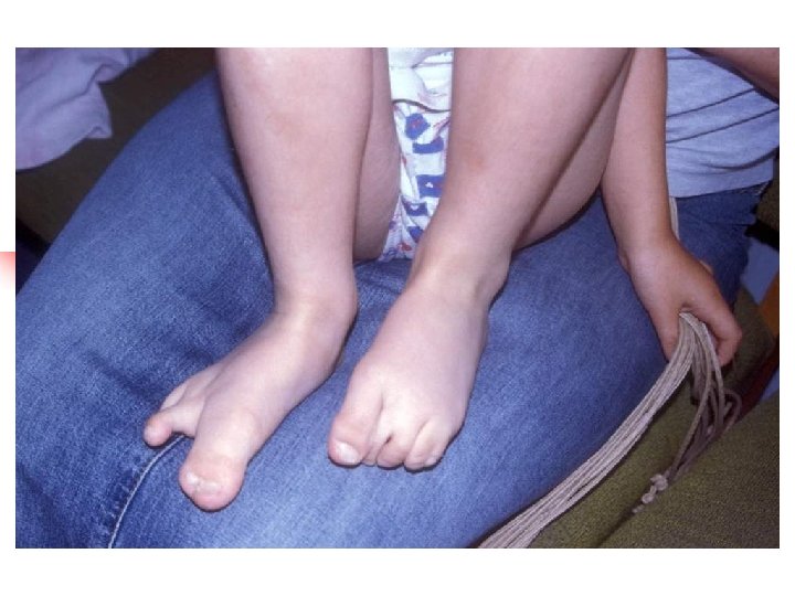
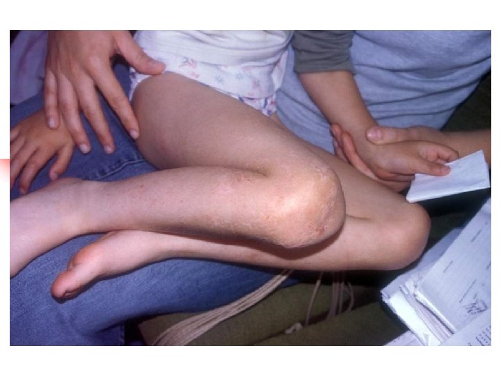
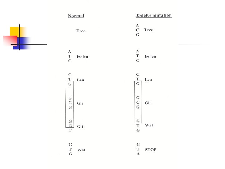
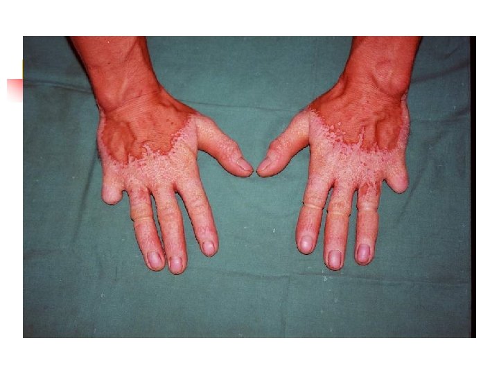
Hearing loss caused by mutation in GJB 2 (connexin deafness) – – – most common cause of hearing loss in many populations deafness usually stable, onset is nearly always prelingual (but not necessarily congenital); hearing may be normal at birth and hearing loss progress rapidly during first few month of life (some babies may pass neonatal hearing screening but become deaf during infancy) GJB 2 encodes a gap junction protein – connexin 26 most common mutation is a deletion of single guanine – 35 del. G (70% mutant alleles, carrier frequency 2 -3%) mutation 35 del. G in thought rather a founder effect not hot-spot deletion GJB 2 mutations may also be a rare cause of autosomal dominant deafness – syndromic and nonsyndromic (DFNA 3). Specific mutation: - hyperkeratosis palmoplantaris - mutilating keratoderma – (Vohwinkel sy. ) - keratoderma – ichthyosis – deafness (KID sy. )
Screening GJB 2 should be offering as part of the routine work-up in the diagnosis of all cases of non-syndromic deafness of unknown cause. Rationale: - common cause of hearing impairment phenotype unremarkable and variable small coding region common mutations in some populations enables accurate genetic information to be given to families disadvantages: counselling difficult with missense and heterozygous mutation
Mitochondrial hearing loss – – – Sensorineural hearing loss is present in 40 -70% patients with mitochondrial disorders and can be syndromic or non-syndromic. Mitochondrial mutations are transmitted exclusively through the maternal line and demonstrate complete (or nearly complete) homoplasmy. Up to 20% patients receiving aminoglycosides experience hearing impairment. 50% of those carry the 12 S ribosomal RNA mutation 1555 A>G. Mitochondrial hearing loss may be syndromic: Kearns-Sayre sy. , MELAS, maternally inherited diabetes and deafness, and others Pathogenesis of mitochondrial hearing loss is based on high ATP requirement in the cochlear hair cells. A reduction of available ATP caused by dysfunction of the mitochondrial oxidative phosphorylation results in disturbances of the ionic gradient in the inner ear.