Peripheral blood Hematopoiesis Part 1 Blood Dr Larry

Peripheral blood & Hematopoiesis Part 1 Blood • Dr. Larry Johnson • Texas A&M University 19761

Objectives of Peripheral blood & Hematopoiesis Part 1 • Examine a blood smear and distinguish each of the formed elements. • Describe the functions of the granulocytes, agranulocytes, platelets, and erythrocytes. • Know the relative cell counts for each of the formed elements in the blood. Part 2 • Describe the normal development of red and white blood cells. Peripheral blood Bone marrow

BLOOD - DIAGNOSTIC VALUE - MOST EXAMINED TYPES OF INFORMATION: IDENTIFY NATURE OF DISEASE VIRAL – T LYMPHOCYTES BACTERIAL – NEUTROPHILS PARASITIC – EOSINOPHILS FOLLOWS THE COURSE OF DISEASE ALLOWS METHOD TO EVALUATE THE EFFECTIVENESS OF TREATMENT
 BUFFY COAT (WHITE BLOOD CELLS) BLOOD PLASMA")
BLOOD COMPOSITION PACKED RED BLOOD CELLS (HEMATOCRIT) BUFFY COAT (WHITE BLOOD CELLS) BLOOD PLASMA

BLOOD PLASMA LIQUID PART OF BLOOD - 55% OF WHOLE BLOOD COMPOSITION : WATER ALBUMIN - OSMOTIC PRESSURE and NUTRALIZES PROTEASES GLOBULIN - ANTIBODIES (Ig. G, Ig. E, ETC. ) TRANSFERRIN - IRON, COPPER, ZINC CHYLOMICRONS - LIPIDS FROM INTESTINE LOW-DENSITY LIPOPROTEIN CHOLESTEROL REMOVAL

BLOOD PLASMA COMPOSITION con’t: PLASMA CLOTTING FACTORS • FIBRINOGEN • PROTHROMBIN • THROMBOPLASTIN • THROMBIN HORMONES - DIRECT ACTIVITY OF CELLS CHEMOTAXIS FACTORS – ATTRACT IMMUNE CELLS

BLOOD SERUM – YELLOW LIQUID FROM CLOTTED BLOOD (e. g. , PLASMA MINUS CLOTTING FACTORS)
 BLOOD - FLUID TISSUE COMPOSED OF ERYTHROCYTES (RBC), LEUKOCYTES (WBC),")
BLOOD (definition and function) BLOOD - FLUID TISSUE COMPOSED OF ERYTHROCYTES (RBC), LEUKOCYTES (WBC), AND PLATELETS SUSPENDED IN BLOOD PLASMA FUNCTION OF BLOOD IS TRANSPORTATION OF CELLS AND FLUID RBC 02 to and C 02 from TISSUE

BLOOD FUNCTION OF BLOOD IS TRANSPORTATION OF CELLS AND FLUID WBC PLASMA IMMUNE DEFENSE OF BODY NUTRIENTS TO TISSUE WASTE FROM TISSUE PROTEINS TO HOLD WATER IN PLASMA HORMONES AND OTHER INFORMATIONAL MEDIATORS PLATELETS PREVENT LOSS OF TRANSPORTATION

Blood Copyright Mc. Graw-Hill Companies Blood summary: Composition and cells Blood plasma is the liquid component of blood that holds blood cells in whole blood vs. blood serum is blood plasma without clotting factors.

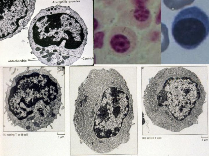
GRANULAR LEUKOCYTES NONGRANULAR LEUKOCYTES
 Lymphocyte Eosinophil Monocyte Neutrophil Basophil (Slide 22) Platelets")
Slide 21: Blood smear (Giemsa stain) Lymphocyte Eosinophil Monocyte Neutrophil Basophil (Slide 22) Platelets

Study Questions % Prevalence in normal blood Distinguishing characteristics Contents of specific granules Life span 54 -62% 3 -5 connected lobes, cytoplasm is granulated 1. Azurophilic primary granules 1. Myeloperoxidase 2. Lysozyme 3. Defensins 2. Specific secondary granules 1 -4 days 25 -33% No granules in cytoplasm, densestaining nucleus surrounded by narrow cytoplasmic rim Agranular Hours to many years 3 -7% Largest agranular leukocyte with horshoe-shaped nucleus Agranular Hours to years 1 -3% Typically bilobed nucleus, cytoplasm has large eosinophilic granules 0 -0. 75% Cytoplasm contains dark blue or brown granules, basophilic nucleus normally obscured by dense cytoplasmic granules Neutrophils Lymphocytes Monocytes Eosinophils Basophils 1. Major basic proteins 2. Others 1. Heparin 2. Histamine 3. Others 1 -2 weeks Several months

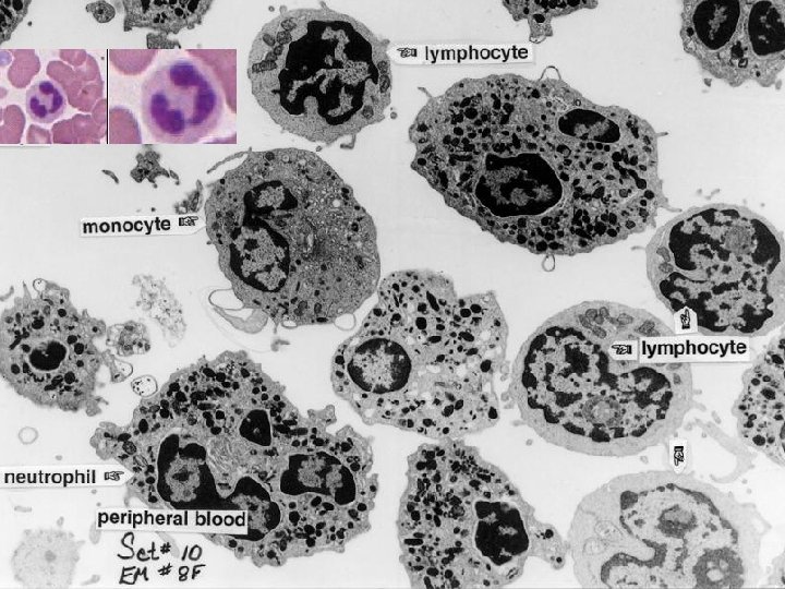
Neutrophils Platelets Lymphocyte Monocyte Erythrocytes Slide 113
 AND FUNCTIONS IN BLOOD CELL TYPE ERYTHROCYTES MAIN FUNCTIONS C")
FORMED ELEMENTS (non-fluid, cellular) AND FUNCTIONS IN BLOOD CELL TYPE ERYTHROCYTES MAIN FUNCTIONS C 02 AND 02 TRANSPORT NEUTROPHILS PHAGOCYTOSIS OF BACTERIA EOSINOPHILS PARASITIC INFECTIONS, INFLAMMATORY PROCESSES RELEASE OF HISTAMINE AND OTHER INFLAMMATION MEDIATORS MONONUCLEAR-PHAGOCYTE SYSTEM become macrophages BASOPHILS MONOCYTES
 AND FUNCTIONS IN BLOOD con’t CELL TYPE B LYMPHOCYTES (CYTOTOXIC")
FORMED ELEMENTS (non-fluid, cellular) AND FUNCTIONS IN BLOOD con’t CELL TYPE B LYMPHOCYTES (CYTOTOXIC T CELL) MAIN FUNCTIONS GENERATION OF ANTIBODY-PRODUCING PLASMA CELLS KILLING OF VIRUSINFECTED CELLS KILLING OF SOME TUMOR AND VIRUS-INFECTED CELLS PLATELETS CLOTTING OF BLOOD T LYMPHOCYTES NATURAL KILLER
 BONE MARROW DERIVED ANUCLEATED CELLS")
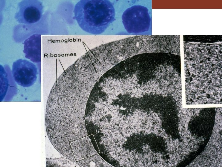
ERYTHROCYTES MINUTE CORPUSCLES - RED COLOR OF BLOOD (iron) BONE MARROW DERIVED ANUCLEATED CELLS (MAMMALS) • NONFUNCTIONAL NUCLEI IN BIRDS, REPTILES, AMPHIBIA, AND FISH 4. 8 TO 5. 4 X 10⁶/MM 3 = 1µL OF BLOOD, 45% OF BLOOD 25 -30 X 1012/PERSON (LARGER AT HIGH ELEVATION)
 basophil, eosinophil, and neutrophils The basic functions of the")
110 Peripheral blood smear (Leishman-Giemsa) basophil, eosinophil, and neutrophils The basic functions of the leukocytes: Neutrophil: bacteria, fungi Eosinophil: parasites, allergic inflammatory responses Basophil: release histamine for inflammatory response Lymphocyte: B cells, T cells, Natural killer cells Monocyte: migrates from blood to tissue and becomes a macrophage
 Monocyte Lymphocyte Monocyte 113")
Peripheral blood smear (May-Grunwald-Giemsa) Monocyte Lymphocyte Monocyte 113

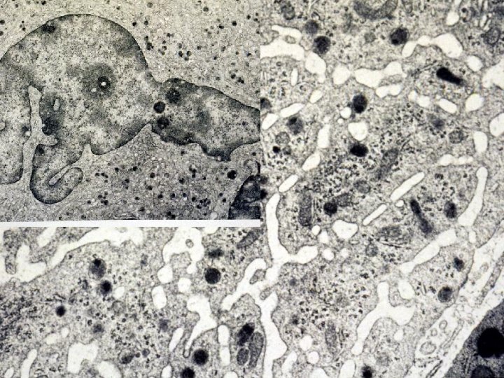
EM 6

Plasma cells

EM of white blood cells EM 8 f

21

Neutrophils
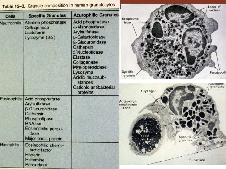
 AZUROPHILIC ACID PHOSPHATASE (HYDROLYTIC ENZYMES) PRIMARY LYSOSOMES SPECIFIC BASIC PROTEIN (PHAGOCYTINS, ANTI-BACTERIAL")
NEUROPHILS (GRANDULES) AZUROPHILIC ACID PHOSPHATASE (HYDROLYTIC ENZYMES) PRIMARY LYSOSOMES SPECIFIC BASIC PROTEIN (PHAGOCYTINS, ANTI-BACTERIAL ACTION)

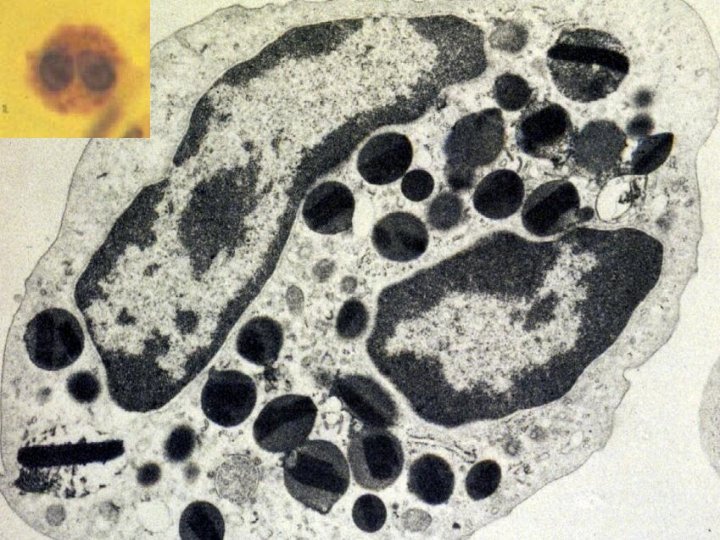
EOSINOPHILS 8 -12 DAY LIFE SPAN TRANSIENT OF BLOOD - 3 TO 4 HRS. SPECIFIC GRANULES - RED/ORANGE, LARGE, CRYSTALLOIDS • MAJOR BASIC PROTEIN (KILLS PARASITIC WORMS) COMMON IN LAMINA PROPRIA (CT UNDER LINING EPITHELIUM) • ALIMENTARY TRACT • RESPIRATORY TRACT ATTRACTED BY CHEMOTACTIC FACTORS GIVEN OFF BY MAST CELLS AND BASOPHILS ROLE • PARASITIC DISEASES • ALLERGY

21
 • METACHROMASIA (HEPARIN in several")
BASOPHILS LEAST NUMEROUS WBC SPECIFIC GRANULES (BLUE, LARGE, HISTAMINE) • METACHROMASIA (HEPARIN in several varied colors) MAY BE INDUCED TO DEGRANULATE (LIKE MAST CELLS) SPECIFIC RECEPTORS FOR Ig. E IMMUNOGLOBULIN • Ig. E USUALLY DILUTES IN BLOOD • 20 FOLD IN PERSONS WITH HAY FEVER, ASTHMA, OR ALLERGIC DERMATITIS

BASOPHILS HISTAMINE - ITCHING AND INCREASED VASCULAR PERMEABILITY – EDEMA CUTANEOUS BASOPHIL HYPERSENSITIVITY REBUCK WINDOW • (BASOPHILS MIGRATE TO DERMIS - CELL MEDIATED IMMUNITY) Note granules above nucleus

EM 8 b BASOPHIL

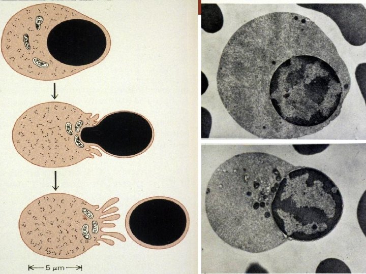
PLATELET FUNCTION - BLOOD CLOTTING 1. PRIMARY AGGREGATION OF PLATELET PLUG - ATTACHMENT TO DAMAGED TISSUE 2. SECONDARY AGGREGATION • ALPHA AND DELTA GRANULES – INDUCE FURTHER AGGREGATION 3. BLOOD COAGULATION - CASCADE OF PLASMA PROTEINS TO FORM BLOOD CLOT OR THROMBUS 4. CLOT RETRACTION - PLATELET ACTIN, MYOSIN AND ATP 5. CLOT REMOVAL - PLASMIN (PROTEOLYTIN ENZYME) AND PLATELET ENZYME CONTAINING GRANULES

PLATELET’S ROLE IN STOPPING BLEEDING PRODUCTION OF SEROTONIN – VASOCONSTRICTION TO STOP BLOOD FLOW PRODUCE THROMBOPLASTIN FIBRIN POLYMERIZES AND PRODUCES THE HEMOSTATIC PLUG

SUMMARY OF PLATELETS - THROMBOPLASTIDS HAVE MANY METABOLIC FUNCTION OF WHOLE CELLS WHEN EXPOSED TO DAMAGED CELLS, STICKY AGGREGATION • STICK TO EACH OTHER

SUMMARY OF PLATELETS - THROMBOPLASTIDS HAVE MANY METABOLIC FUNCTION OF WHOLE CELLS WHEN EXPOSED TO DAMAGED CELLS, STICKY AGGREGATION • STICK TO EACH OTHER RELEASE ADP (FROM ATP INSIDE PLATELETS) WHICH ATTACHES TO OTHER PLATELETS

SUMMARY OF PLATELETS - THROMBOPLASTIDS HAVE MANY METABOLIC FUNCTION OF WHOLE CELLS WHEN EXPOSED TO DAMAGED CELLS, STICKY AGGREGATION • STICK TO EACH OTHER RELEASE ADP (FROM ATP INSIDE PLATELETS) WHICH ATTACHES TO OTHER PLATELETS PRODUCE THROMBOPLASTIC WHICH CATALIZES PROTHROMBIN TO THROMBIN WHICH CATALIZES FIBRINOGEN TO FIBRIN, FORMS NET TO CATCH RBC AND OTHER PLATELETS (BASIS OF THE BLOOD CLOT)
 ZONES • HYDALOMERE – MICROTUBULES AND SMALL VESICLES")
SUMMARY OF PLATELETS THROMBOPLASTIDS ANUCLEATE (MAMMALS) ZONES • HYDALOMERE – MICROTUBULES AND SMALL VESICLES • GRANULOMERE - AZUROPHILIC GRANULES - SOME CONTAIN SEROTONIN VASOCONSTRICTION PLATELETS PRODUCED BY MEGAKARYOCYTES (BONE MARROW) LAST 8 TO 11 DAYS

White blood cells in blood

White blood cells in blood

End of Part 1


Peripheral blood & Hematopoiesis Part 2 Hematopoiesis • Dr. Larry Johnson • Texas A&M University 19761

Objectives of Peripheral blood & Hematopoiesis Part 1 • Examine a blood smear and distinguish each of the formed elements. • Describe the functions of the granulocytes, agranulocytes, platelets, and erythrocytes. • Know the relative cell counts for each of the formed elements in the blood. Part 2 • Describe the normal development of red and white blood cells. Peripheral blood Bone marrow

17

32583

19761

Bone Marrow 19761

Erythropoiesis Mc. Graw-Hill Companies The cell where hemoglobin appears in the developing erythrocytes is the basophilic erythroblasts (early erythroblast) and takes 4 days to form erythrocytes from stem cells in the bone marrow.

Mc. Graw-Hill Companies

Mc. Graw-Hill Companies

Mc. Graw-Hill Companies

Mc. Graw-Hill Companies

Mc. Graw-Hill Companies

Bone marrow Erythrocyte entering blood vessel 32583

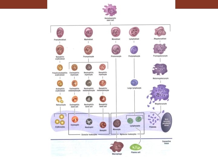
Study Questions 2. Describe the formation of red blood cells, neutrophils and lymphocytes in the bone marrow from their stem cells to their fully differentiated cells. • Erythropoiesis • Takes about 1 week and stimulated by erythropoietin • Originate in bone marrow • Involves major cellular changes • Cell and nuclear volume decrease • Nucleoli diminishes and disappears • Chromatin density increases until nucleus extruded from cell • Decrease in polyribosomes (basophilia) and increase in hemoglobin (eosinophilic) • Mitochondria and other organelles disappear • Steps • 1. Proerythroblast • Large cell with loose, lacy chromatin, nucleoli, and basophilic cytoplasm • 2. Basophilic erythroblast • Strongly basophilic cytoplasm, condensed nucleus, and no visible nucleolus • 3. Polychromatophilic erythroblast • Cell volume is reduced, polysomes decrease, and some cytoplasmic areas fill with hemoglobin producing basophilic and acidophilic regions in cell • 4. Orthochromatic erythroblast • Cell and nuclear volumes continue to condense and no basophilia is evident • 5. Reticulocyte • Cell ejects nucleus to become a reticulocyte, has a small number of polyribosomes • 6. Erythrocyte • Enter blood circulation, lose polyribosomes, and mature as erythrocytes

Bone marrow

EM 12 a marrow

EM 8 a marrow blood

ut 112 Platelets

ut 112 Basophilic erythroblast Platelets Proerythroblast

ut 112 Late polychromatophilic erythroblast Basophilic erythroblast Platelets Polychromatophilic erythroblast Proerythroblast

ut 112 Orthochromatophilic erythroblasts Late polychromatophilic erythroblast Basophilic erythroblast Platelets Polychromatophilic erythroblast Proerythroblast

ut 112 Orthochromatophilic erythroblasts Erythrocytes Late polychromatophilic erythroblast Basophilic erythroblast Platelets Polychromatophilic erythroblast Reticulocyte Proerythroblast

Red Blood Cell Series Cell Name Size Cytoplasm proerythroblast 16 -20 µm basophilic erythroblast (early erythroblast) 12 -16 µm polychromatophilic erythroblast 10 -16 µm orthochromatophilic erythroblast (late erythroblast, normoblast) 8 -10 µm Granules Nucleus deep blue, no hemoglobin none large, round with delicate chromatin, 26 nucleoli tinge of hemoglobin none condensation of chromatin, no nucleoli muddy color none checkerboard pattern no basophilia none dense

Granulopoiesis Mc. Graw-Hill Companies Specific granules first appear in the myelocyte. Granulopoiesis typically takes 10 to 14 days.

White Blood Cell Series Cell Name Size Cytoplasm Granules Nucleus myeloblast 15 -20 µm sky blue no granules red staining, round, delicate chromatin sky blue non-specific, 1 -40 azurophilic granules round to oval, delicate chromatin promyelocyte 15 -20 µm neutrophilic myelocyte 17 -25 µm some basophilia, some pink few fine neutrophilic granules round to oval, delicate chomatin neutrophilic metamyelocyte 12 -16 µm no basophilia, becoming pink many fine neutrophilic granules bean shaped, moderately dense chromatin neutrophilic band or stab 12 -16 µm no basophilia, pink many fine neutrophilic granules band or S-shaped eosinophilic myelocyte 13 -17 µm some basophilia, specific granules few large eosinophilic granules round to oval, delicate chromatin eosinophilic metamyelocyte 12 -16 µm no basophilia, specific granules many large eosinophilic granules bean-shaped, moderately dense chromatin eosinophilic band or stab 12 -16 µm no basophilia, specific granules many large eosinophilic granules band or S-shaped

EM 12 b Neutrophilic myelocyte Neutrophi Reticulocytes

ut 112 Platelets Eosinophil or basophil Neutrophilic myelocytes

ut 112 Neutrophilic metamyelocytes Platelets Eosinophil or basophil Neutrophilic myelocytes

ut 112 Neutrophilic metamyelocytes Platelets Eosinophil or basophil Neutrophilic myelocytes

Granulopoiesis. Takes about 10 to 14 days • Majority of process is dominated by the synthesis of proteins for cytoplasmic granules • Steps 1. Myeloblast • Most immature, finely dispersed chromatin, faint nuclei, and no cytoplasmic 2. Promyelocyte • Basophilic cytoplasm and azurophilic granules being secreted in Golgi apparatus 3. Neutrophilic myelocyte • Specific granules produced in Golgi, moderate number of azurophilic granuloes 4. Neutrophilic metamyelocyte • Specific granules occupy most of cytoplasm, dispersed azurophilic granules, Golgi reduced 5. Neutrophilic band cell • Nucleus is elongated but not yet polymorphic 6. Neutrophil • 3 -5 connected lobes, cytoplasm is granulated
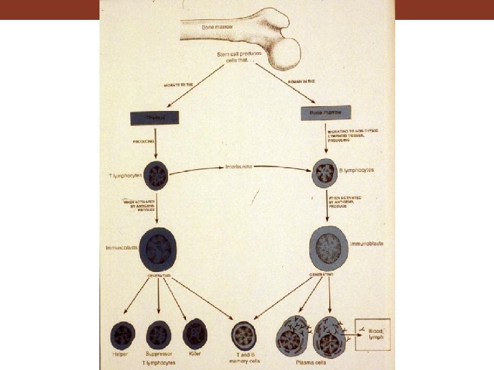

Maturation of agranulocytes. • All lymphocyte progenitor cells originate in bone marrow and migrate to the thymus or other lymphoid organ (lymph node, spleen, etc. ) to proliferate. • Some lymphocytes migrate to thymus, and acquire properties to be T lymphocytes. • Some lymphocytes differentiate into B lymphocytes in the bone marrow and then migrate to peripheral lymphoid organs • Steps • 1. Lymphoblast • Large cells capable of dividing 2 -3 X to form lymphocytes • 2. Lymphocyte • Maturation= nuclei becomes smaller, nucleoli become less visible, cells decrease in size • Bone marrow= B lymphocyte specialization • Thymus= T lymphocyte specialization • 3. Mature lymphocyte • Larger than newly formed lymphocytes • Acquire distinctive cell surface and other proteins

140 Cardiac stomach w/chronic infection

140 Cardiac stomach w/chronic infection

432 Lung with bronchus and lung – macrophages

EM 8 b • macrophage

EM 12 c
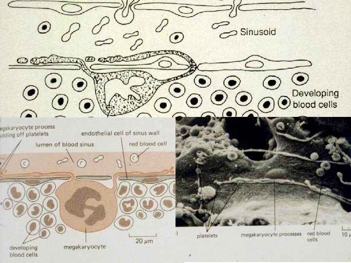

Slide 17: in situ bone marrow Megakaryocyte Adipocyte

Slide 18: in situ bone marrow Megakaryocytes Adipocytes

ne marrow 32583 Developing granulated cells Megakaryocytes

Lymphocytes Unknown nuclei Lymphocytes

Promyelocyte

Neutrophilic myelocyte Promyelocyte Neutrophilic myelocytes

Neutrophilic metamyelocytes Neutrophilic myelocyte Promyelocyte Neutrophilic myelocytes Neutrophilic metamyelocytes

Neutrophilic metamyelocytes Neutrophilic myelocyte Promyelocyte Neutrophilic band cells Neutrophilic myelocytes Neutrophilic band cells Neutrophilic metamyelocytes Neutrophilic band cell

Basophilic erythroblast

Orthochromatophilic erythroblast Basophilic erythroblast Orthochromatophilic erythroblast

Orthochromatophilic erythroblast Reticulocytes Basophilic erythroblast Orthochromatophilic erythroblast

Orthochromatophilic erythroblast Reticulocytes Basophilic erythroblast Erythrocytes Orthochromatophilic erythroblast

Neutrophilic metamyelocyte Lymphocytes Neutrophilic myelocyte Promyelocyte Unknown nuclei Orthochromatophilic erythroblast Neutrophilic myelocyte Lymphocytes Neutrophilic band Reticulocytes Neutrophilic metamyelocyte Erythrocytes Basophilic erythroblast Orthochromatophilic erythroblast Neutrophilic band

Neutrophilic metamyelocyte Unknown nuclei Neutrophilic myelocyte Neutrophilic band Neutrophilic metamyelocyte Proerythroblast Orthochromatophilic erythroblast Eosinophils or basophils Orthochromatophilic erythroblast
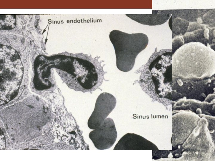

32583 Bone marrow White blood cell entering a blood vessel

Many illustrations in these VIBS Histology You. Tube videos were modified from the following books and sources: Many thanks to original sources! • Bruce Alberts, et al. 1983. Molecular Biology of the Cell. Garland Publishing, Inc. , New York, NY. • Bruce Alberts, et al. 1994. Molecular Biology of the Cell. Garland Publishing, Inc. , New York, NY. • William J. Banks, 1981. Applied Veterinary Histology. Williams and Wilkins, Los Angeles, CA. • Hans Elias, et al. 1978. Histology and Human Microanatomy. John Wiley and Sons, New York, NY. • Don W. Fawcett. 1986. Bloom and Fawcett. A textbook of histology. W. B. Saunders Company, Philadelphia, PA. • Don W. Fawcett. 1994. Bloom and Fawcett. A textbook of histology. Chapman and Hall, New York, NY. • Arthur W. Ham and David H. Cormack. 1979. Histology. J. S. Lippincott Company, Philadelphia, PA. • Luis C. Junqueira, et al. 1983. Basic Histology. Lange Medical Publications, Los Altos, CA. • L. Carlos Junqueira, et al. 1995. Basic Histology. Appleton and Lange, Norwalk, CT. • L. L. Langley, et al. 1974. Dynamic Anatomy and Physiology. Mc. Graw-Hill Book Company, New York, NY. • W. W. Tuttle and Byron A. Schottelius. 1969. Textbook of Physiology. The C. V. Mosby Company, St. Louis, MO. • Leon Weiss. 1977. Histology Cell and Tissue Biology. Elsevier Biomedical, New York, NY. • Leon Weiss and Roy O. Greep. 1977. Histology. Mc. Graw-Hill Book Company, New York, NY. • Nature (http: //www. nature. com), Vol. 414: 88, 2001. • A. L. Mescher 2013 Junqueira’s Basis Histology text and atlas, 13 th ed. Mc. Graw • Douglas P. Dohrman and TAMHSC Faculty 2012 Structure and Function of Human Organ Systems, Histology Laboratory Manual - Slide selections were largely based on this manual for first year medical students at TAMHSC

The End!
- Slides: 104